
Search Count: 384
 |
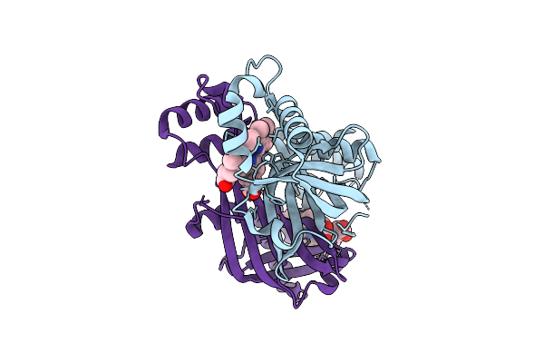
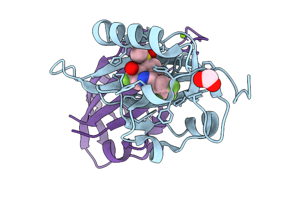
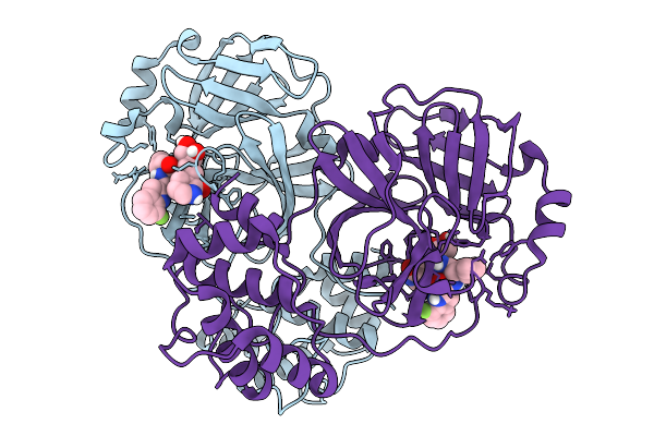
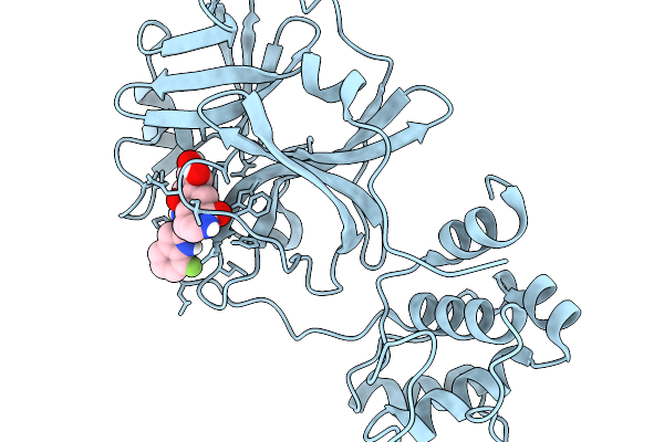
Crystal Structure Of Plcg1 Sh2(C) Domain With A Triple Tyr Phosphorylated Peptide Derived From Ntrk1
Organism: Homo sapiens
Method: X-RAY DIFFRACTION Resolution:2.40 Å Release Date: 2026-06-17 Classification: ONCOPROTEIN Ligands: CL, PO4 |
 |
Organism: Mus musculus
Method: ELECTRON MICROSCOPY Release Date: 2026-05-27 Classification: STRUCTURAL PROTEIN Ligands: GTP, MG, GDP, ATP |
 |
Organism: Haloarcula taiwanensis
Method: ELECTRON MICROSCOPY Resolution:4.90 Å Release Date: 2026-04-22 Classification: SIGNALING PROTEIN Ligands: RET |
 |
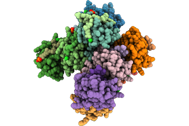
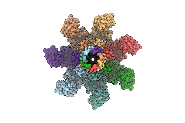
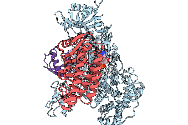
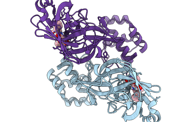
Structural Basis For The Assembly And Translocation Of The Vip1-Vip2 Insecticidal Binary Toxin From Bacillus Thuringiensis
Organism: Bacillus thuringiensis
Method: ELECTRON MICROSCOPY Resolution:3.05 Å Release Date: 2026-04-15 Classification: TOXIN Ligands: CA |
 |
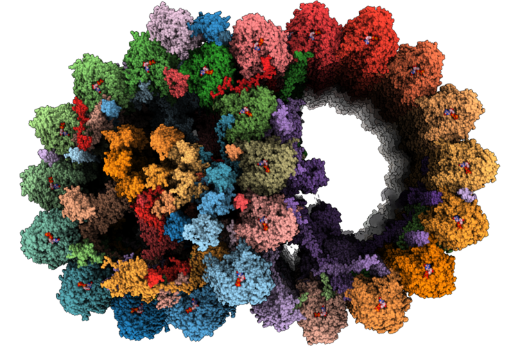
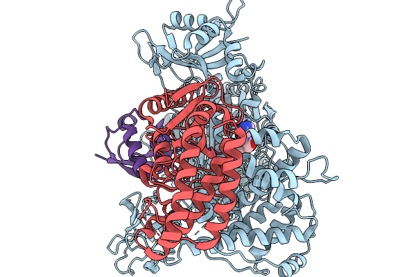
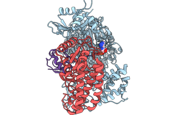
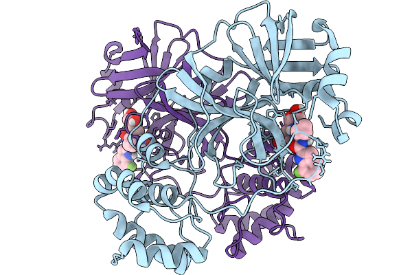
Structural Basis For The Assembly And Translocation Of The Vip1-Vip2 Insecticidal Binary Toxin From Bacillus Thuringiensis
Organism: Bacillus thuringiensis
Method: ELECTRON MICROSCOPY Resolution:2.40 Å Release Date: 2026-04-15 Classification: TOXIN Ligands: CA |
 |
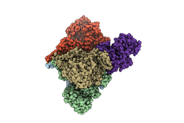
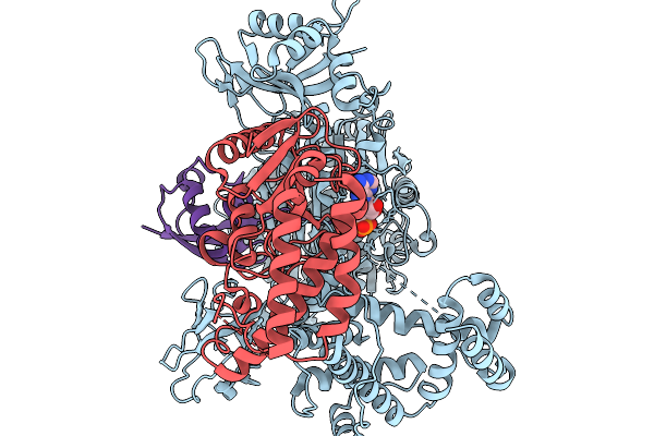
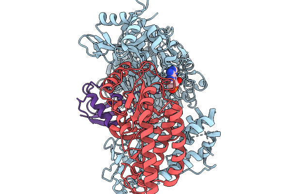
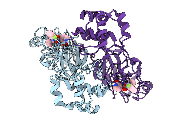
Structural Basis For The Assembly And Translocation Of The Vip1-Vip2 Insecticidal Binary Toxin From Bacillus Thuringiensis
Organism: Bacillus thuringiensis
Method: ELECTRON MICROSCOPY Resolution:3.31 Å Release Date: 2026-04-15 Classification: TOXIN Ligands: CA |
 |
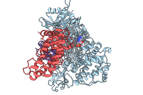
The Crystal Structure Of Paib From Bacillus Stearothermophilus Bound To Hem
Organism: Geobacillus kaustophilus hta426
Method: X-RAY DIFFRACTION Resolution:2.42 Å Release Date: 2026-04-01 Classification: TRANSCRIPTION Ligands: HEM |
 |
Organism: Homo sapiens
Method: ELECTRON MICROSCOPY Release Date: 2026-03-25 Classification: CYTOSOLIC PROTEIN Ligands: AMP |
 |
Organism: Homo sapiens
Method: ELECTRON MICROSCOPY Release Date: 2026-03-25 Classification: CYTOSOLIC PROTEIN Ligands: AMP |
 |
Organism: Homo sapiens
Method: ELECTRON MICROSCOPY Release Date: 2026-03-25 Classification: CYTOSOLIC PROTEIN Ligands: AMP |
 |
Organism: Homo sapiens
Method: ELECTRON MICROSCOPY Release Date: 2026-03-25 Classification: CYTOSOLIC PROTEIN Ligands: AMP |
 |
Organism: Homo sapiens
Method: ELECTRON MICROSCOPY Release Date: 2026-03-25 Classification: CYTOSOLIC PROTEIN Ligands: AMP |
 |
Organism: Homo sapiens
Method: ELECTRON MICROSCOPY Release Date: 2026-03-25 Classification: CYTOSOLIC PROTEIN Ligands: AMP |
 |
Organism: Homo sapiens
Method: X-RAY DIFFRACTION Resolution:1.56 Å Release Date: 2026-02-18 Classification: TRANSCRIPTION Ligands: A1I0H, FMT, MG |
 |
Organism: Tylonycteris bat coronavirus hku4
Method: X-RAY DIFFRACTION Resolution:2.05 Å Release Date: 2026-02-18 Classification: VIRUS Ligands: A1BWH |
 |
Organism: Severe acute respiratory syndrome coronavirus 2
Method: X-RAY DIFFRACTION Resolution:2.00 Å Release Date: 2026-02-18 Classification: VIRAL PROTEIN Ligands: A1BWH |
 |
Organism: Porcine epidemic diarrhea virus
Method: X-RAY DIFFRACTION Resolution:1.50 Å Release Date: 2026-02-18 Classification: VIRAL PROTEIN Ligands: A1BWH |
 |
Organism: Camel alphacoronavirus
Method: X-RAY DIFFRACTION Resolution:2.30 Å Release Date: 2026-02-18 Classification: VIRAL PROTEIN Ligands: A1BWH, PEG |
 |
Organism: Middle east respiratory syndrome-related coronavirus
Method: X-RAY DIFFRACTION Resolution:2.50 Å Release Date: 2026-02-18 Classification: VIRAL PROTEIN Ligands: A1BWH |
 |
Organism: Miniopterus bat coronavirus hku8
Method: X-RAY DIFFRACTION Resolution:1.80 Å Release Date: 2026-02-18 Classification: VIRAL PROTEIN Ligands: A1BWH |