

Search Count: 16
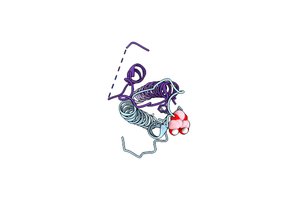 |
Structure Of The N-Terminal Domain Of Wag31
Organism: Mycobacterium tuberculosis (strain atcc 25618 / h37rv)
Method: X-RAY DIFFRACTION Resolution:2.30 Å Release Date: 2020-07-15 Classification: LIPID BINDING PROTEIN Ligands: PGE |
Organism: Mycobacterium tuberculosis (strain atcc 25618 / h37rv)
Method: X-RAY DIFFRACTION
Release Date: 2020-07-15
Ligands: PGE
 |
Crystal Structure Of Imidazole Glycerol Phosphate Dehydratase
Organism: Filobasidiella neoformans
Method: X-RAY DIFFRACTION Resolution:2.30 Å Release Date: 2004-05-04 Classification: LYASE Ligands: HG, EMC, SO4, GOL, ACY |
Organism: Filobasidiella neoformans
Method: X-RAY DIFFRACTION
Release Date: 2004-05-04
Ligands: HG, EMC, SO4, GOL, ACY
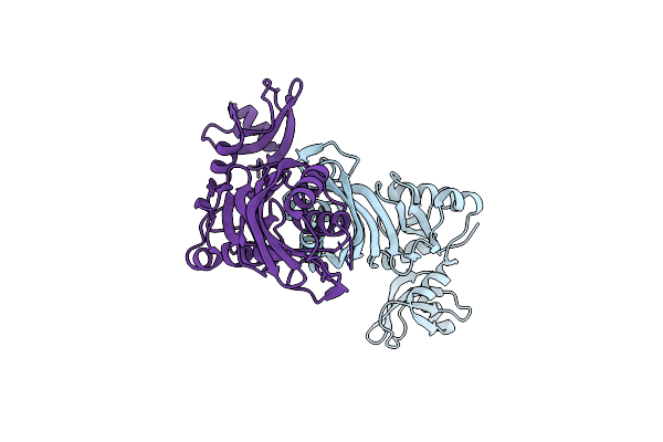 |
Structure Of Trna Psi55 Pseudouridine Synthase (Trub)
Organism: Mycobacterium tuberculosis
Method: X-RAY DIFFRACTION Resolution:1.90 Å Release Date: 2004-03-02 Classification: LYASE |
Organism: Mycobacterium tuberculosis
Method: X-RAY DIFFRACTION
Release Date: 2004-03-02
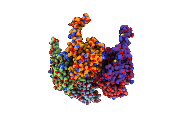 |
The Crystal Structure Of The First Enzyme Of Pantothenate Biosynthetic Pathway, Ketopantoate Hydroxymethyltransferase From Mycobacterium Tuberculosis Shows A Decameric Assembly And Terminal Helix-Swapping
Organism: Mycobacterium tuberculosis
Method: X-RAY DIFFRACTION Resolution:2.80 Å Release Date: 2003-07-15 Classification: TRANSFERASE Ligands: MG |
Organism: Mycobacterium tuberculosis
Method: X-RAY DIFFRACTION
Release Date: 2003-07-15
Ligands: MG
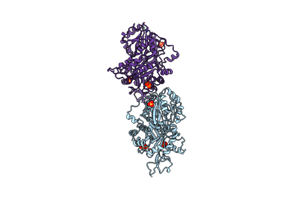 |
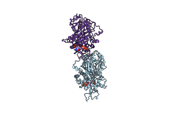
Towards Understanding The Mechanism Of The Complex Cyclization Reaction Catalyzed By Imidazole Glycerophosphate Synthase
Organism: Saccharomyces cerevisiae
Method: X-RAY DIFFRACTION Resolution:2.50 Å Release Date: 2003-06-17 Classification: TRANSFERASE, LYASE Ligands: SO4, NI, POP |
Organism: Saccharomyces cerevisiae
Method: X-RAY DIFFRACTION
Release Date: 2003-06-17
Ligands: SO4, NI, POP
 |
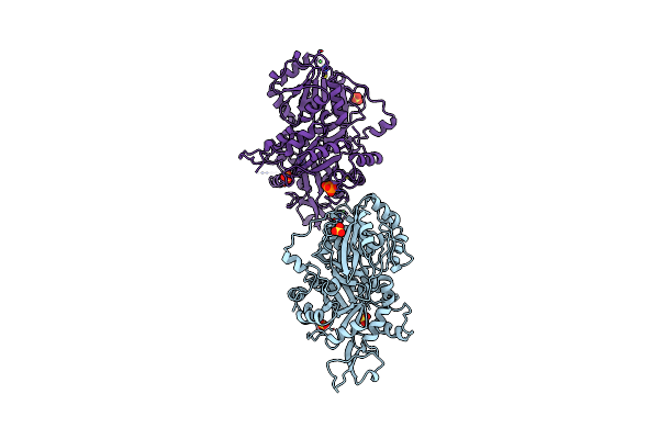
Towards Understanding The Mechanism Of The Complex Cyclization Reaction Catalyzed By Imidazole Glycerophosphate Synthase
Organism: Saccharomyces cerevisiae
Method: X-RAY DIFFRACTION Resolution:2.50 Å Release Date: 2003-06-17 Classification: TRANSFERASE, LYASE Ligands: NI, 1PR |
Organism: Saccharomyces cerevisiae
Method: X-RAY DIFFRACTION
Release Date: 2003-06-17
Ligands: NI, 1PR
 |
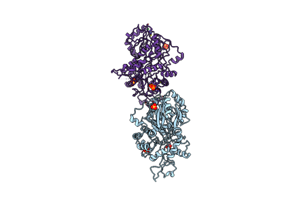
Towards Understanding The Mechanism Of The Complex Cyclization Reaction Catalyzed By Imidazole Glycerophosphate Synthase
Organism: Saccharomyces cerevisiae
Method: X-RAY DIFFRACTION Resolution:2.40 Å Release Date: 2003-06-17 Classification: TRANSFERASE, LYASE Ligands: NI, SO4, POP |
Organism: Saccharomyces cerevisiae
Method: X-RAY DIFFRACTION
Release Date: 2003-06-17
Ligands: NI, SO4, POP
 |
Hinge-Bending Motion Of D-Allose Binding Protein From Escherichia Coli: Three Open Conformations
Organism: Escherichia coli
Method: X-RAY DIFFRACTION Resolution:3.10 Å Release Date: 2003-03-06 Classification: SUGAR BINDING PROTEIN Ligands: NI |
Organism: Escherichia coli
Method: X-RAY DIFFRACTION
Release Date: 2003-03-06
Ligands: NI
 |
Hinge-Bending Motion Of D-Allose Binding Protein From Escherichia Coli: Three Open Conformations
Organism: Escherichia coli
Method: X-RAY DIFFRACTION Resolution:1.71 Å Release Date: 2003-03-06 Classification: PERIPLASMIC BINDING PROTEIN Ligands: ZN |
Organism: Escherichia coli
Method: X-RAY DIFFRACTION
Release Date: 2003-03-06
Ligands: ZN
 |
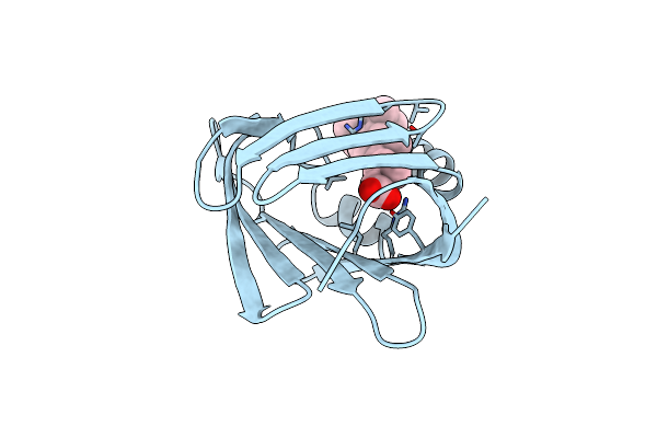
Crystal Structure Of Imidazole Glycerol Phosphate Synthase: A Tunnel Through A (Beta/Alpha)8 Barrel Joins Two Active Sites
Organism: Saccharomyces cerevisiae
Method: X-RAY DIFFRACTION Resolution:2.10 Å Release Date: 2001-10-12 Classification: TRANSFERASE Ligands: NI, SO4, POP |
Organism: Saccharomyces cerevisiae
Method: X-RAY DIFFRACTION
Release Date: 2001-10-12
Ligands: NI, SO4, POP
 |
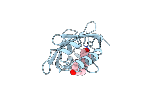
Cellular Retinoic Acid Binding Protein Ii In Complex With A Synthetic Retinoic Acid (Ro-13 6307)
Organism: Homo sapiens
Method: X-RAY DIFFRACTION Resolution:2.10 Å Release Date: 1999-12-22 Classification: TRANSPORT PROTEIN Ligands: R13 |
Organism: Homo sapiens
Method: X-RAY DIFFRACTION
Release Date: 1999-12-22
Ligands: R13
 |
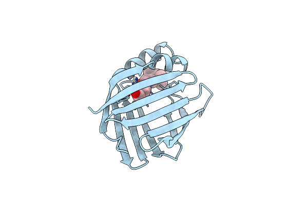
Cellular Retinoic Acid Binding Protein Ii In Complex With A Synthetic Retinoic Acid (Ro-12 7310)
Organism: Homo sapiens
Method: X-RAY DIFFRACTION Resolution:2.00 Å Release Date: 1999-12-22 Classification: TRANSPORT PROTEIN Ligands: R12 |
Organism: Homo sapiens
Method: X-RAY DIFFRACTION
Release Date: 1999-12-22
Ligands: R12
 |
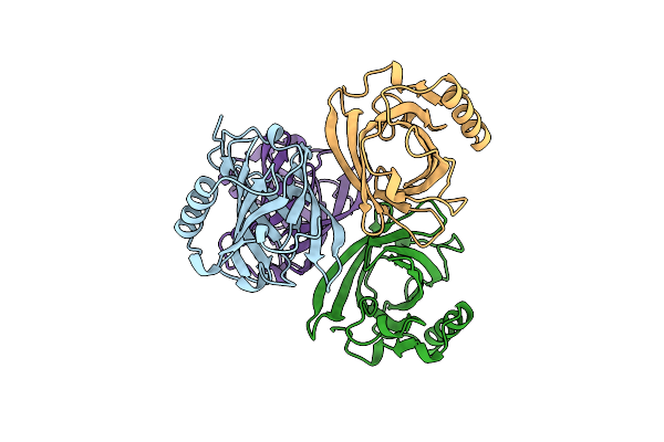
Cellular Retinoic Acid Binding Protein I In Complex With A Retinobenzoic Acid (Am80)
Organism: Bos taurus
Method: X-RAY DIFFRACTION Resolution:2.80 Å Release Date: 1999-12-21 Classification: TRANSPORT PROTEIN Ligands: A80 |
Organism: Bos taurus
Method: X-RAY DIFFRACTION
Release Date: 1999-12-21
Ligands: A80
 |
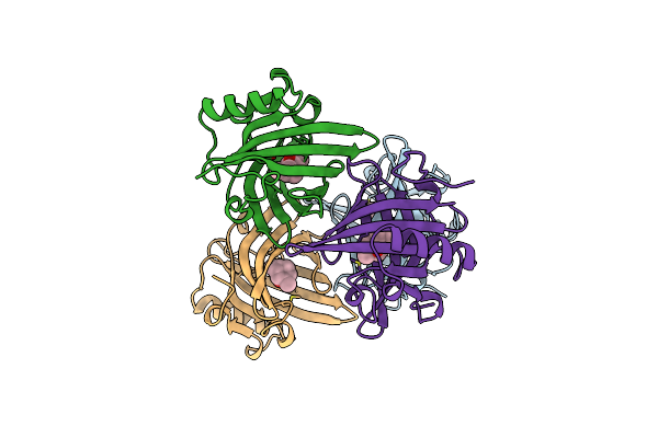
The Crystal Structures Of A2U-Globulin And Its Complex With A Hyaline Droplet Inducer.
Organism: Rattus norvegicus
Method: X-RAY DIFFRACTION Resolution:2.50 Å Release Date: 1999-08-26 Classification: LIPID BINDING PROTEIN |
Organism: Rattus norvegicus
Method: X-RAY DIFFRACTION
Release Date: 1999-08-26
 |
The Crystal Structures Of A2U-Globulin And Its Complex With A Hyaline Droplet Inducer.
Organism: Rattus norvegicus
Method: X-RAY DIFFRACTION Resolution:2.90 Å Release Date: 1999-08-13 Classification: LIPID BINDING PROTEIN Ligands: LEO |
Organism: Rattus norvegicus
Method: X-RAY DIFFRACTION
Release Date: 1999-08-13
Ligands: LEO
 |
Crystal Structure Of D-Allose Binding Protein From Escherichia Coli
Organism: Escherichia coli k12
Method: X-RAY DIFFRACTION Resolution:1.80 Å Release Date: 1999-02-16 Classification: PERIPLASMIC SUGAR RECEPTOR Ligands: ZN, SO4, ALL |
Organism: Escherichia coli k12
Method: X-RAY DIFFRACTION
Release Date: 1999-02-16
Ligands: ZN, SO4, ALL