

Search Count: 27
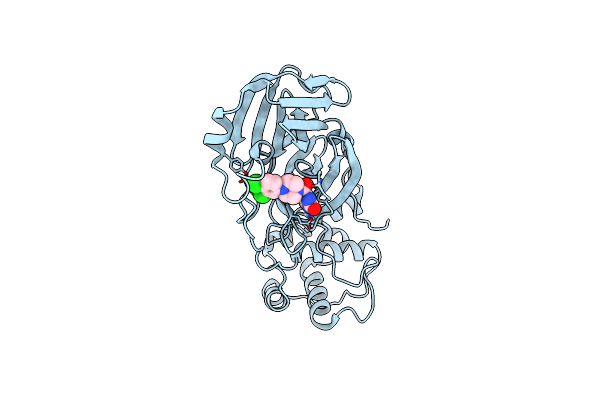 |
Room-Temperature X-Ray Structure Of Sars-Cov-2 Main Protease (3Cl Mpro) In Complex With A Non-Covalent Inhibitor Mcule-5948770040
Organism: Severe acute respiratory syndrome coronavirus 2
Method: X-RAY DIFFRACTION Resolution:1.80 Å Release Date: 2021-03-03 Classification: HYDROLASE/HYDROLASE INHIBITOR Ligands: YD1 |
Organism: Severe acute respiratory syndrome coronavirus 2
Method: X-RAY DIFFRACTION
Release Date: 2021-03-03
Ligands: YD1
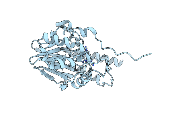 |
Crystal Structure Of The Metallo Beta Lactamase L1 From Stenotrophomonas Maltophilia Determined By Serial Crystallography
Organism: Stenotrophomonas maltophilia (strain k279a)
Method: X-RAY DIFFRACTION Resolution:1.85 Å Release Date: 2020-12-30 Classification: HYDROLASE Ligands: ZN |
Organism: Stenotrophomonas maltophilia (strain k279a)
Method: X-RAY DIFFRACTION
Release Date: 2020-12-30
Ligands: ZN
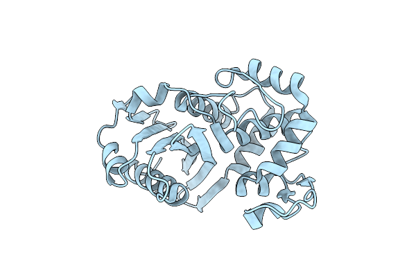 |
Crystal Structure Of The Beta Lactamase Class D From Chitinophaga Pinensis By Serial Crystallography
Organism: Chitinophaga pinensis dsm 2588
Method: X-RAY DIFFRACTION Resolution:1.80 Å Release Date: 2020-09-23 Classification: HYDROLASE |
Organism: Chitinophaga pinensis dsm 2588
Method: X-RAY DIFFRACTION
Release Date: 2020-09-23
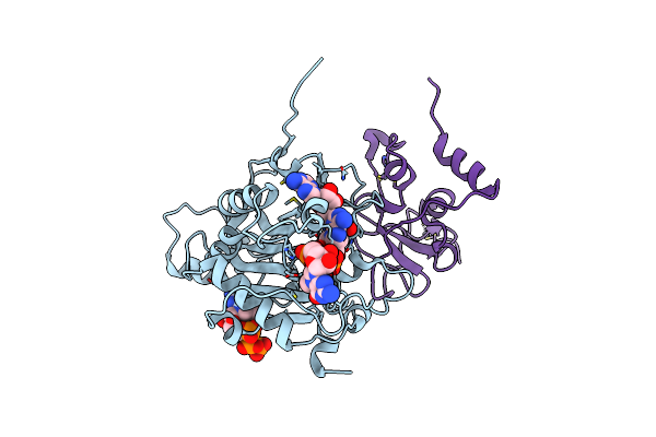 |
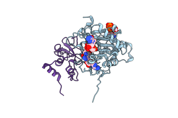
Room Temperature Structure Of Sars-Cov-2 Nsp10/Nsp16 Methyltransferase In A Complex With 2'-O-Methylated M7Gpppa Cap-1 And Sah Determined By Fixed-Target Serial Crystallography
Organism: Severe acute respiratory syndrome coronavirus 2
Method: X-RAY DIFFRACTION Resolution:2.25 Å Release Date: 2020-08-26 Classification: VIRAL PROTEIN Ligands: CL, SAH, V9G, MGP, ZN |
Organism: Severe acute respiratory syndrome coronavirus 2
Method: X-RAY DIFFRACTION
Release Date: 2020-08-26
Ligands: CL, SAH, V9G, MGP, ZN
 |
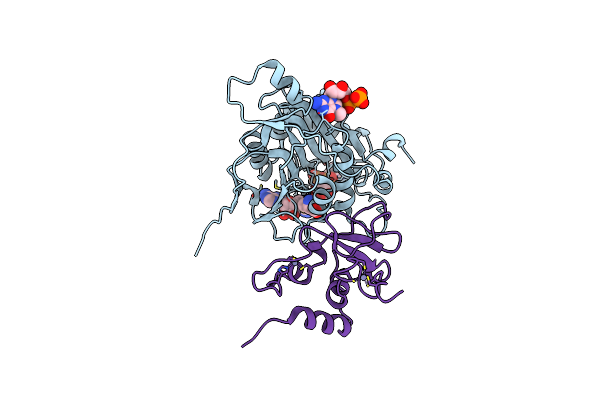
Room Temperature Crystal Structure Of Nsp10/Nsp16 From Sars-Cov-2 With Substrates And Products Of 2'-O-Methylation Of The Cap-1
Organism: Severe acute respiratory syndrome coronavirus 2
Method: X-RAY DIFFRACTION Resolution:2.65 Å Release Date: 2020-08-26 Classification: VIRAL PROTEIN Ligands: CL, SAM, SAH, GTA, V9G, MGP, ZN |
Organism: Severe acute respiratory syndrome coronavirus 2
Method: X-RAY DIFFRACTION
Release Date: 2020-08-26
Ligands: CL, SAM, SAH, GTA, V9G, MGP, ZN
 |
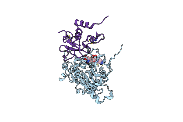
Room Temperature Structure Of Sars-Cov-2 Nsp10/Nsp16 Methyltransferase In A Complex With M7Gpppa Cap-0 And Sam Determined By Fixed-Target Serial Crystallography
Organism: Severe acute respiratory syndrome coronavirus 2
Method: X-RAY DIFFRACTION Resolution:2.18 Å Release Date: 2020-08-26 Classification: VIRAL PROTEIN Ligands: GTA, SAM, M7G, ZN |
Organism: Severe acute respiratory syndrome coronavirus 2
Method: X-RAY DIFFRACTION
Release Date: 2020-08-26
Ligands: GTA, SAM, M7G, ZN
 |
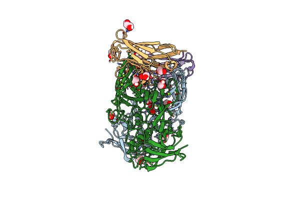
Room Temperature Structure Of Sars-Cov-2 Nsp10/Nsp16 Methyltransferase In A Complex With Sam Determined By Fixed-Target Serial Crystallography
Organism: Severe acute respiratory syndrome coronavirus 2
Method: X-RAY DIFFRACTION Resolution:2.25 Å Release Date: 2020-07-08 Classification: VIRAL PROTEIN Ligands: CL, SAM, ZN |
Organism: Severe acute respiratory syndrome coronavirus 2
Method: X-RAY DIFFRACTION
Release Date: 2020-07-08
Ligands: CL, SAM, ZN
 |
Crystal Structure Of The Murine Norovirus Vp1 P Domain In Complex With The Cd300Lf Receptor
Organism: Murine norovirus 1, Mus musculus
Method: X-RAY DIFFRACTION Resolution:2.00 Å Release Date: 2018-09-12 Classification: VIRAL PROTEIN/LIPID BINDING PROTEIN Ligands: EDO, CL, MG |
Organism: Murine norovirus 1, Mus musculus
Method: X-RAY DIFFRACTION
Release Date: 2018-09-12
Ligands: EDO, CL, MG
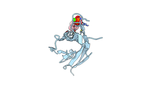 |
Crystal Structure Of Murine Cd300Lf In Complex With Phosphocholine
Organism: Mus musculus
Method: X-RAY DIFFRACTION Resolution:1.36 Å Release Date: 2018-09-12 Classification: LIPID BINDING PROTEIN Ligands: PC, CA |
Organism: Mus musculus
Method: X-RAY DIFFRACTION
Release Date: 2018-09-12
Ligands: PC, CA
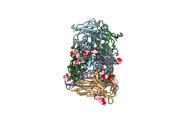 |
Crystal Structure Of The Murine Norovirus Vp1 P Domain In Complex With The Cd300Lf Receptor And Glycochenodeoxycholic Acid
Organism: Murine norovirus 1, Mus musculus
Method: X-RAY DIFFRACTION Resolution:1.95 Å Release Date: 2018-09-12 Classification: VIRAL PROTEIN Ligands: MG, EDO, CHO |
Organism: Murine norovirus 1, Mus musculus
Method: X-RAY DIFFRACTION
Release Date: 2018-09-12
Ligands: MG, EDO, CHO
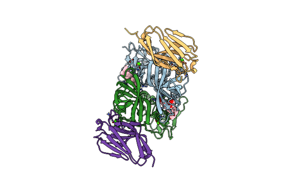 |
Crystal Structure Of The Murine Norovirus Vp1 P Domain In Complex With The Cd300Lf Receptor And Lithocholic Acid
Organism: Murine norovirus 1, Mus musculus
Method: X-RAY DIFFRACTION Resolution:1.80 Å Release Date: 2018-09-12 Classification: VIRAL PROTEIN Ligands: 4OA, CA, EDO |
Organism: Murine norovirus 1, Mus musculus
Method: X-RAY DIFFRACTION
Release Date: 2018-09-12
Ligands: 4OA, CA, EDO
 |
Mouse Norovirus Model Using The Crystal Structure Of Mnv P Domain And The Norwalkvirus Shell Domain
Organism: Norwalk virus, Murine norovirus 1
Method: ELECTRON MICROSCOPY Release Date: 2018-04-04 Classification: VIRUS |
Organism: Norwalk virus, Murine norovirus 1
Method: ELECTRON MICROSCOPY
Release Date: 2018-04-04
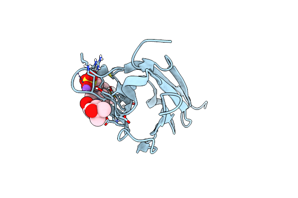 |
Crystal Structure Of Mouse Cd300Lf At 1.6 Angstroms Resolution.
Organism: Mus musculus
Method: X-RAY DIFFRACTION Resolution:1.60 Å Release Date: 2016-08-31 Classification: IMMUNE SYSTEM Ligands: EPE, NA, MPD |
Organism: Mus musculus
Method: X-RAY DIFFRACTION
Release Date: 2016-08-31
Ligands: EPE, NA, MPD
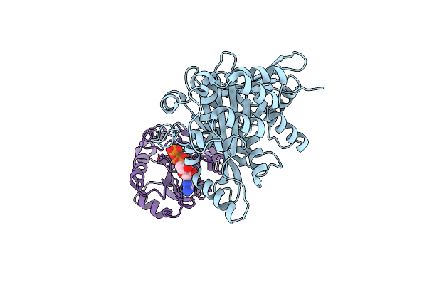 |
Structure Of Espg-Arf6 Complex
Organism: Escherichia coli, Homo sapiens
Method: X-RAY DIFFRACTION Resolution:2.50 Å Release Date: 2011-01-05 Classification: PROTEIN TRANSPORT Ligands: GTP, MG |
Organism: Escherichia coli, Homo sapiens
Method: X-RAY DIFFRACTION
Release Date: 2011-01-05
Ligands: GTP, MG
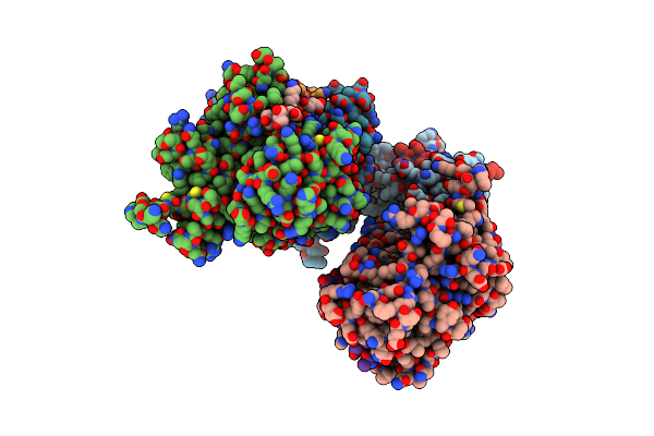 |
Structure Of Espg-Pak2 Autoinhibitory Ialpha3 Helix Complex
Organism: Escherichia coli, Homo sapiens
Method: X-RAY DIFFRACTION Resolution:2.86 Å Release Date: 2011-01-05 Classification: PROTEIN TRANSPORT/TRANSFERASE |
Organism: Escherichia coli, Homo sapiens
Method: X-RAY DIFFRACTION
Release Date: 2011-01-05
 |
Crystal Structure Of Map And Cdc42 Complex
Organism: Homo sapiens, Escherichia coli
Method: X-RAY DIFFRACTION Resolution:2.30 Å Release Date: 2009-07-21 Classification: SIGNALING PROTEIN/TRANSCRIPTION |
Organism: Homo sapiens, Escherichia coli
Method: X-RAY DIFFRACTION
Release Date: 2009-07-21
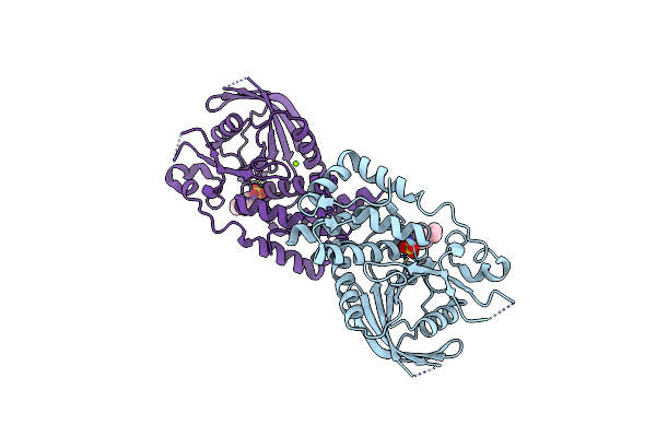 |
Engineering The Ptpbeta Catalytic Domain With Improved Crystallization Properties
Organism: Homo sapiens
Method: X-RAY DIFFRACTION Resolution:1.70 Å Release Date: 2006-09-05 Classification: HYDROLASE Ligands: CL, UA5, MG |
Organism: Homo sapiens
Method: X-RAY DIFFRACTION
Release Date: 2006-09-05
Ligands: CL, UA5, MG
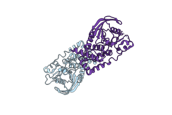 |
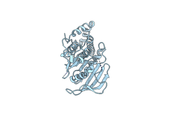
Engineered Catalytic Domain Of Protein Tyrosine Phosphatase Hptpbeta
Organism: Homo sapiens
Method: X-RAY DIFFRACTION Resolution:1.85 Å Release Date: 2006-08-29 Classification: HYDROLASE Ligands: CL |
Organism: Homo sapiens
Method: X-RAY DIFFRACTION
Release Date: 2006-08-29
Ligands: CL
 |
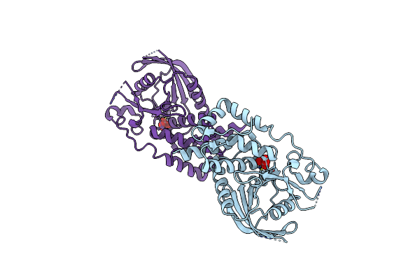
Structural Studies Of Protein Tyrosine Phosphatase Beta Catalytic Domain In Complex With Inhibitors
Organism: Homo sapiens
Method: X-RAY DIFFRACTION Resolution:1.85 Å Release Date: 2006-08-29 Classification: HYDROLASE |
Organism: Homo sapiens
Method: X-RAY DIFFRACTION
Release Date: 2006-08-29
 |
Structural Studies Of Protein Tyrosine Phosphatase Beta Catalytic Domain In Complex With Inhibitors
Organism: Homo sapiens
Method: X-RAY DIFFRACTION Resolution:1.75 Å Release Date: 2006-08-29 Classification: HYDROLASE Ligands: VO4 |
Organism: Homo sapiens
Method: X-RAY DIFFRACTION
Release Date: 2006-08-29
Ligands: VO4