

Search Count: 21
 |
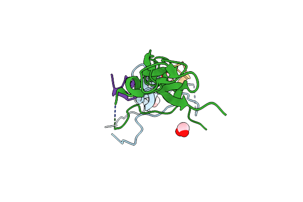
Crystal Structure Of Human Vitamin D Receptor Ligand Binding Domain In Complex With A Vdrm
Organism: Homo sapiens
Method: X-RAY DIFFRACTION Resolution:2.20 Å Release Date: 2018-03-14 Classification: PROTEIN BINDING Ligands: 8VM |
Organism: Homo sapiens
Method: X-RAY DIFFRACTION
Release Date: 2018-03-14
Ligands: 8VM
 |
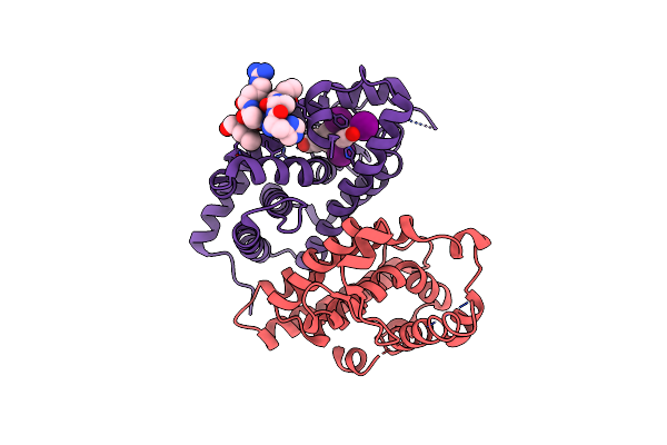
Il-17A In Complex With Peptide
Organism: Homo sapiens, Synthetic construct
Method: X-RAY DIFFRACTION Resolution:1.70 Å Release Date: 2018-02-14 Classification: IMMUNE SYSTEM / INHIBITOR Ligands: EDO, CL |
Organism: Homo sapiens, Synthetic construct
Method: X-RAY DIFFRACTION
Release Date: 2018-02-14
Ligands: EDO, CL
 |
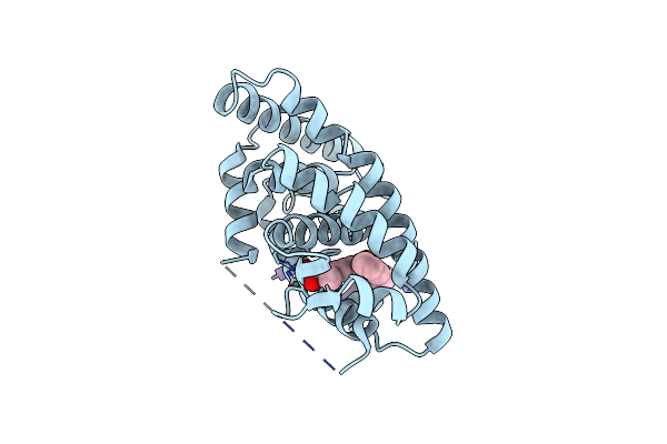
Crystal Structure Of The T3-Bound Tr-Beta Ligand-Binding Domain In Complex With Rxr-Alpha
Method: X-RAY DIFFRACTION
Resolution:3.22 Å Release Date: 2015-09-02 Classification: PROTEIN BINDING Ligands: T3 |
Method: X-RAY DIFFRACTION
Release Date: 2015-09-02
Ligands: T3
 |
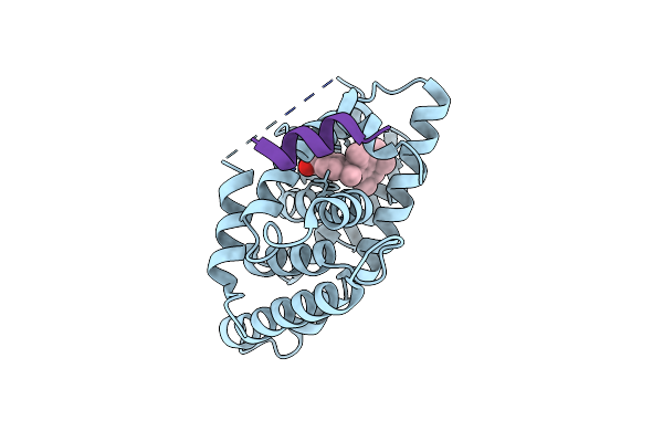
Crystal Structure Of Human Retinoid X Receptor Alpha-Ligand Binding Domain Complex With 9Cuab30 And The Coactivator Peptide Grip-1
Organism: Homo sapiens
Method: X-RAY DIFFRACTION Resolution:2.00 Å Release Date: 2013-11-13 Classification: TRANSCRIPTION Ligands: 1O8 |
Organism: Homo sapiens
Method: X-RAY DIFFRACTION
Release Date: 2013-11-13
Ligands: 1O8
 |
Crystal Structure Of Human Retinoid X Receptor Alpha-Ligand Binding Domain Complex With Targretin And The Coactivator Peptide Grip-1
Organism: Homo sapiens
Method: X-RAY DIFFRACTION Resolution:2.10 Å Release Date: 2013-11-13 Classification: TRANSCRIPTION Ligands: 9RA |
Organism: Homo sapiens
Method: X-RAY DIFFRACTION
Release Date: 2013-11-13
Ligands: 9RA
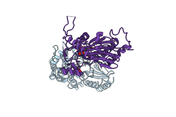 |
Crystal Structure Of Snrk2.6 In Complex With Hab1
Organism: Arabidopsis thaliana
Method: X-RAY DIFFRACTION Resolution:2.60 Å Release Date: 2012-02-15 Classification: SIGNALING PROTEIN Ligands: SO4, MG |
Organism: Arabidopsis thaliana
Method: X-RAY DIFFRACTION
Release Date: 2012-02-15
Ligands: SO4, MG
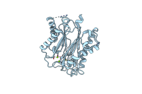 |
Crystal Structure Of Protein Phosphatase Abi2
Organism: Arabidopsis thaliana
Method: X-RAY DIFFRACTION Resolution:1.90 Å Release Date: 2012-02-15 Classification: HYDROLASE Ligands: MG |
Organism: Arabidopsis thaliana
Method: X-RAY DIFFRACTION
Release Date: 2012-02-15
Ligands: MG
 |
Crystal Structure Of Abscisic Acid Bound Pyl2 In Complex With Type 2C Protein Phosphatase Abi2
Organism: Arabidopsis thaliana
Method: X-RAY DIFFRACTION Resolution:2.50 Å Release Date: 2012-02-15 Classification: SIGNALING PROTEIN Ligands: A8S, MG |
Organism: Arabidopsis thaliana
Method: X-RAY DIFFRACTION
Release Date: 2012-02-15
Ligands: A8S, MG
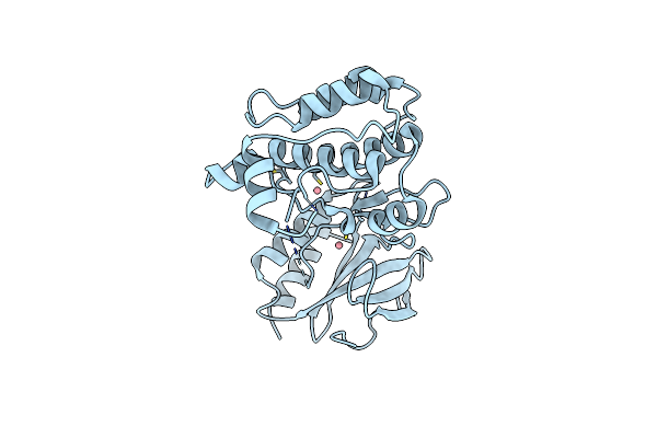 |
The Crystal Structure Of Snf1-Related Kinase 2.3
Organism: Arabidopsis thaliana
Method: X-RAY DIFFRACTION Resolution:1.90 Å Release Date: 2011-12-14 Classification: TRANSFERASE Ligands: CO |
Organism: Arabidopsis thaliana
Method: X-RAY DIFFRACTION
Release Date: 2011-12-14
Ligands: CO
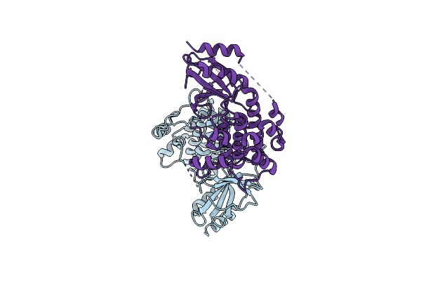 |
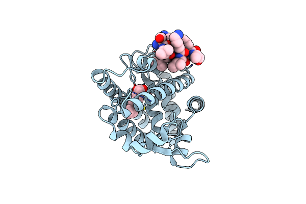
The Crystal Structure Of Snf1-Related Kinase 2.6
Organism: Arabidopsis thaliana
Method: X-RAY DIFFRACTION Resolution:2.30 Å Release Date: 2011-12-14 Classification: TRANSFERASE |
Organism: Arabidopsis thaliana
Method: X-RAY DIFFRACTION
Release Date: 2011-12-14
 |
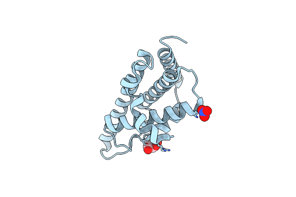
Ligand Binding Domain Of Ppargamma Complexed With Decanoic Acid And Pgc-1A Peptide
Organism: Homo sapiens
Method: X-RAY DIFFRACTION Resolution:1.52 Å Release Date: 2011-11-09 Classification: TRANSCRIPTION Ligands: DKA |
Organism: Homo sapiens
Method: X-RAY DIFFRACTION
Release Date: 2011-11-09
Ligands: DKA
 |
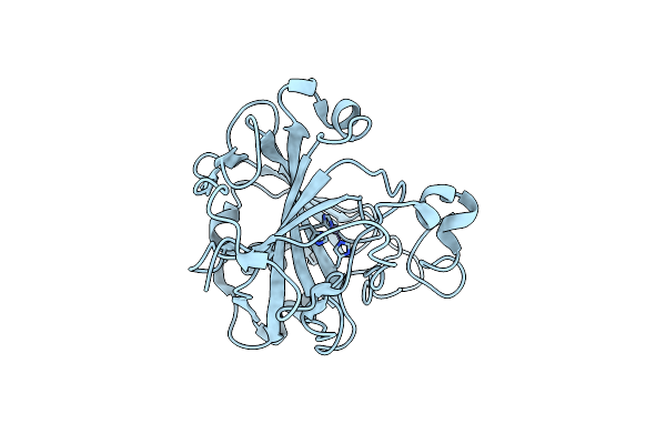
The Structural Basis For Recognition Of J-Base Containing Dna By A Novel Dna-Binding Domain In Jbp1
Organism: Leishmania tarentolae
Method: X-RAY DIFFRACTION Resolution:1.90 Å Release Date: 2011-03-30 Classification: OXIDOREDUCTASE Ligands: NO3, GOL |
Organism: Leishmania tarentolae
Method: X-RAY DIFFRACTION
Release Date: 2011-03-30
Ligands: NO3, GOL
 |
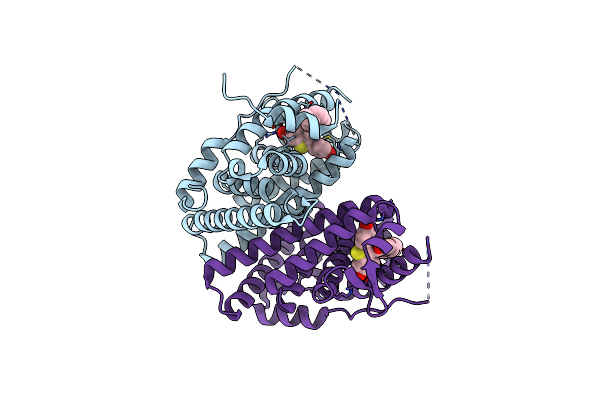
Apo-Human Carbonic Anhydrase Ii Revisited: Implications Of The Loss Of A Metal In Protein Structure, Stability And Solvent Network
Organism: Homo sapiens
Method: X-RAY DIFFRACTION Resolution:1.26 Å Release Date: 2009-07-21 Classification: LYASE Ligands: ZN |
Organism: Homo sapiens
Method: X-RAY DIFFRACTION
Release Date: 2009-07-21
Ligands: ZN
 |
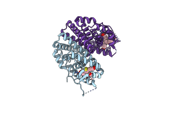
Estrogen Receptor Alpha Ligand-Binding Domain Complexed To A Serm
Organism: Homo sapiens
Method: X-RAY DIFFRACTION Resolution:2.00 Å Release Date: 2008-04-08 Classification: TRANSCRIPTION Ligands: LLB |
Organism: Homo sapiens
Method: X-RAY DIFFRACTION
Release Date: 2008-04-08
Ligands: LLB
 |
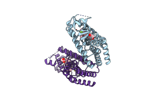
Estrogen Receptor Alpha Ligand-Binding Domain In Complex With A Serm
Organism: Homo sapiens
Method: X-RAY DIFFRACTION Resolution:2.00 Å Release Date: 2008-04-08 Classification: TRANSCRIPTION Ligands: LLC |
Organism: Homo sapiens
Method: X-RAY DIFFRACTION
Release Date: 2008-04-08
Ligands: LLC
 |
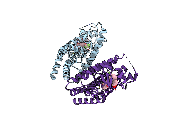
Crystal Structure Of Ppargamma Lbd Bound To Full Agonist Mrl20
Organism: Homo sapiens
Method: X-RAY DIFFRACTION Resolution:2.20 Å Release Date: 2007-10-23 Classification: Ligand binding protein Ligands: 240 |
Organism: Homo sapiens
Method: X-RAY DIFFRACTION
Release Date: 2007-10-23
Ligands: 240
 |
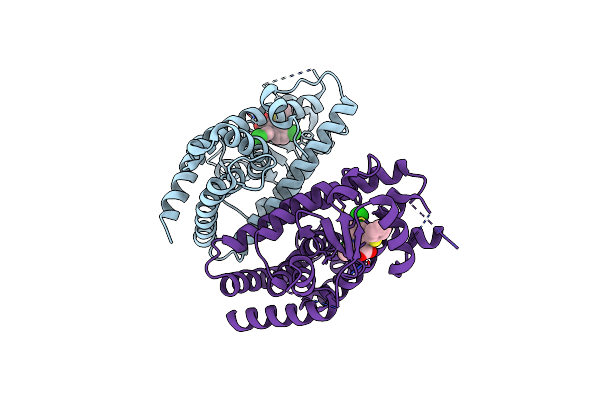
Crystal Structure Of Ppargamma Bound To Partial Agonist Mrl24
Organism: Homo sapiens
Method: X-RAY DIFFRACTION Resolution:2.30 Å Release Date: 2007-10-23 Classification: Ligand binding protein Ligands: 241 |
Organism: Homo sapiens
Method: X-RAY DIFFRACTION
Release Date: 2007-10-23
Ligands: 241
 |
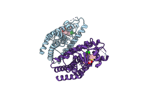
Crystal Structure Of Ppargamma Bound To Partial Agonist Ntzdpa
Organism: Homo sapiens
Method: X-RAY DIFFRACTION Resolution:2.05 Å Release Date: 2007-10-23 Classification: Ligand binding protein Ligands: NZA |
Organism: Homo sapiens
Method: X-RAY DIFFRACTION
Release Date: 2007-10-23
Ligands: NZA
 |
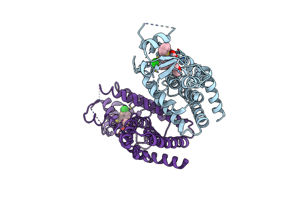
Crystal Structure Of Ppargamma Ligand Binding Domain Bound To Partial Agonist Sr145
Organism: Homo sapiens
Method: X-RAY DIFFRACTION Resolution:2.20 Å Release Date: 2007-10-23 Classification: Ligand binding protein Ligands: SF1 |
Organism: Homo sapiens
Method: X-RAY DIFFRACTION
Release Date: 2007-10-23
Ligands: SF1
 |
Crystal Structure Of Ppar Gamma Complexed With Partial Agonist Sf147
Organism: Homo sapiens
Method: X-RAY DIFFRACTION Resolution:2.41 Å Release Date: 2007-10-23 Classification: Ligand binding protein Ligands: SF2 |
Organism: Homo sapiens
Method: X-RAY DIFFRACTION
Release Date: 2007-10-23
Ligands: SF2