

Search Count: 17
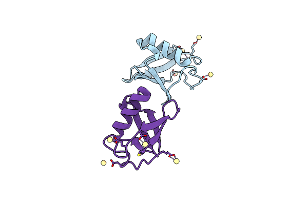 |
Structure Of D-Thr53 Ubiquitin
Organism: Homo sapiens
Method: X-RAY DIFFRACTION Resolution:2.60 Å Release Date: 2022-05-18 Classification: PROTEIN BINDING Ligands: CD |
Organism: Homo sapiens
Method: X-RAY DIFFRACTION
Release Date: 2022-05-18
Ligands: CD
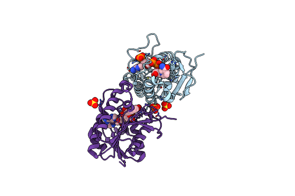 |
Crystal Structure Of Human Biliverdin Ix-Beta Reductase B
Organism: Homo sapiens
Method: X-RAY DIFFRACTION Resolution:1.60 Å Release Date: 2022-01-19 Classification: OXIDOREDUCTASE Ligands: NAP, SO4 |
Organism: Homo sapiens
Method: X-RAY DIFFRACTION
Release Date: 2022-01-19
Ligands: NAP, SO4
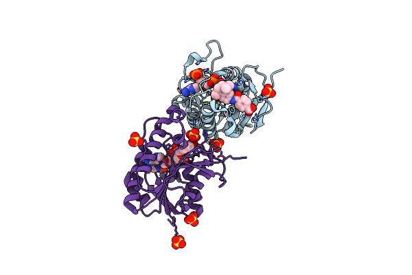 |
Crystal Structure Of Hyman Biliverdin Ix-Beta Reductase B With Tamibarotene (A80)
Organism: Homo sapiens
Method: X-RAY DIFFRACTION Resolution:1.70 Å Release Date: 2022-01-19 Classification: OXIDOREDUCTASE Ligands: NAP, A80, SO4 |
Organism: Homo sapiens
Method: X-RAY DIFFRACTION
Release Date: 2022-01-19
Ligands: NAP, A80, SO4
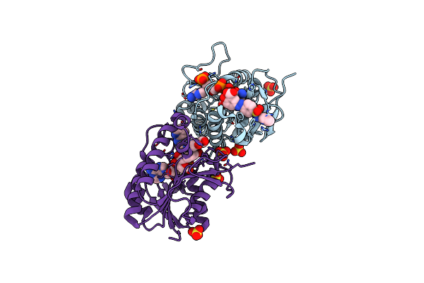 |
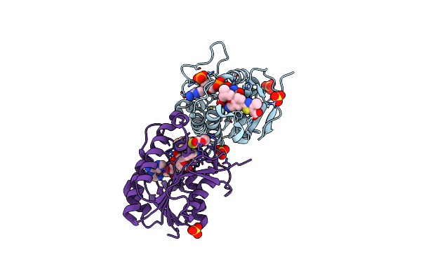
Crystal Structure Of Human Biliverdin Ix-Beta Reductase B With Sulfasalazine (Sas)
Organism: Homo sapiens
Method: X-RAY DIFFRACTION Resolution:1.45 Å Release Date: 2022-01-19 Classification: OXIDOREDUCTASE Ligands: NAP, SAS, SO4 |
Organism: Homo sapiens
Method: X-RAY DIFFRACTION
Release Date: 2022-01-19
Ligands: NAP, SAS, SO4
 |
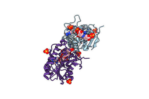
Crystal Structure Of Human Biliverdin Ix-Beta Reductase B With Febuxostat (Tei)
Organism: Homo sapiens
Method: X-RAY DIFFRACTION Resolution:1.45 Å Release Date: 2022-01-19 Classification: OXIDOREDUCTASE Ligands: NAP, TEI, SO4 |
Organism: Homo sapiens
Method: X-RAY DIFFRACTION
Release Date: 2022-01-19
Ligands: NAP, TEI, SO4
 |
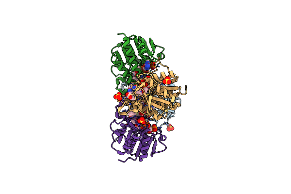
Crystal Structure Of Human Biliverdin Ix-Beta Reductase B With Olsalazine Sodium (Oss)
Organism: Homo sapiens
Method: X-RAY DIFFRACTION Resolution:1.35 Å Release Date: 2022-01-19 Classification: OXIDOREDUCTASE Ligands: NAP, JBC, SO4 |
Organism: Homo sapiens
Method: X-RAY DIFFRACTION
Release Date: 2022-01-19
Ligands: NAP, JBC, SO4
 |
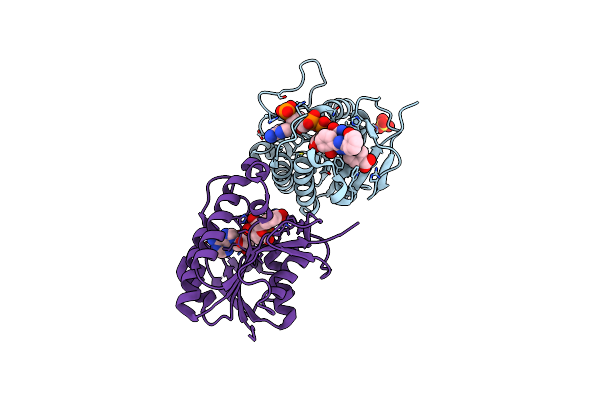
Crystal Structure Of Human Biliverdin Ix-Beta Reductase B With Ataluren (Ptc)
Organism: Homo sapiens
Method: X-RAY DIFFRACTION Resolution:1.50 Å Release Date: 2022-01-19 Classification: OXIDOREDUCTASE Ligands: NAP, JBF, SO4 |
Organism: Homo sapiens
Method: X-RAY DIFFRACTION
Release Date: 2022-01-19
Ligands: NAP, JBF, SO4
 |
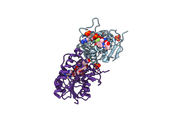
Crystal Structure Of Human Biliverdin Ix-Beta Reductase B With Deferasirox (Icl)
Organism: Homo sapiens
Method: X-RAY DIFFRACTION Resolution:1.50 Å Release Date: 2022-01-19 Classification: OXIDOREDUCTASE Ligands: NAP, JBL, SO4 |
Organism: Homo sapiens
Method: X-RAY DIFFRACTION
Release Date: 2022-01-19
Ligands: NAP, JBL, SO4
 |
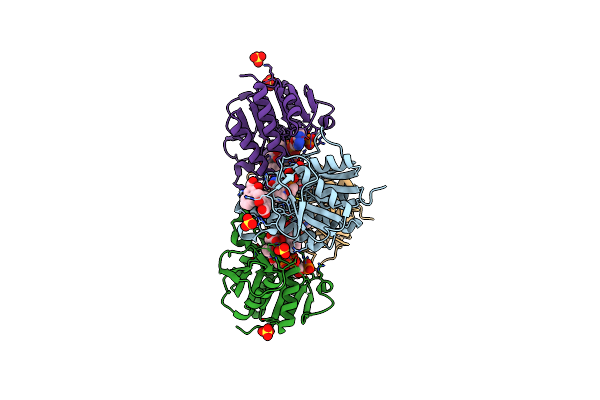
Crystal Structure Of Human Biliverdin Ix-Beta Reductase B With Flunixin Meglumin (Fmg)
Organism: Homo sapiens
Method: X-RAY DIFFRACTION Resolution:2.00 Å Release Date: 2022-01-19 Classification: OXIDOREDUCTASE Ligands: NAP, JBO, SO4 |
Organism: Homo sapiens
Method: X-RAY DIFFRACTION
Release Date: 2022-01-19
Ligands: NAP, JBO, SO4
 |
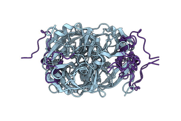
Crystal Structure Of Human Biliverdin Ix-Beta Reductase B With Pyrantel Pamoate (Ppa)
Organism: Homo sapiens
Method: X-RAY DIFFRACTION Resolution:1.60 Å Release Date: 2022-01-19 Classification: OXIDOREDUCTASE Ligands: NAP, JBR, SO4 |
Organism: Homo sapiens
Method: X-RAY DIFFRACTION
Release Date: 2022-01-19
Ligands: NAP, JBR, SO4
 |
Aromatic Interactions Drive The Coupled Folding And Binding Of The Intrinsically Disordered Sesbania Mosaic Virus Vpg Protein
Organism: Sesbania mosaic virus
Method: SOLUTION NMR Release Date: 2021-01-27 Classification: HYDROLASE |
Organism: Sesbania mosaic virus
Method: SOLUTION NMR
Release Date: 2021-01-27
 |
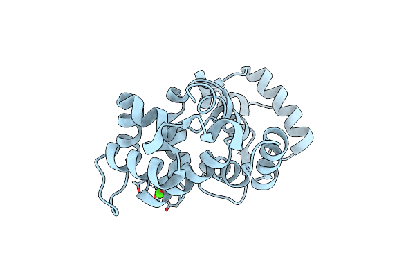
Aromatic Interactions Drive The Coupled Folding And Binding Of The Intrinsically Disordered Sesbania Mosaic Virus Vpg Protein.
Organism: Sesbania mosaic virus
Method: SOLUTION NMR Release Date: 2021-01-20 Classification: PLANT VIRAL PROTEIN |
Organism: Sesbania mosaic virus
Method: SOLUTION NMR
Release Date: 2021-01-20
 |
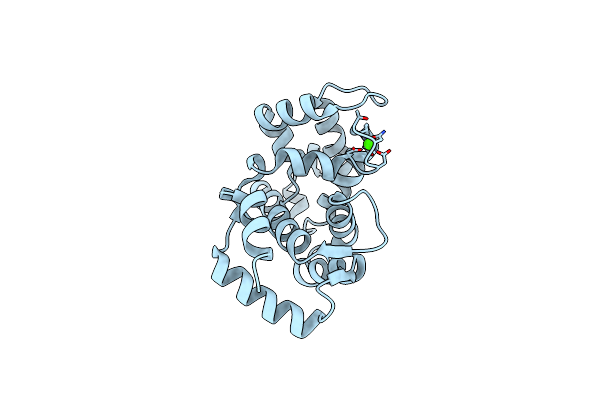
Crystal Structure Of A Non-Myristoylated C39A Recoverin Mutant With One Calcium Ion Bound To Ef-Hand 3
Organism: Bos taurus
Method: X-RAY DIFFRACTION Resolution:1.50 Å Release Date: 2013-11-13 Classification: METAL BINDING PROTEIN Ligands: CA |
Organism: Bos taurus
Method: X-RAY DIFFRACTION
Release Date: 2013-11-13
Ligands: CA
 |
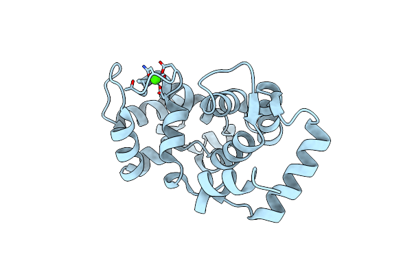
Crystal Structure Of A Non-Myristoylated C39D Recoverin Mutant With One Calcium Ion Bound To Ef-Hand 3
Organism: Bos taurus
Method: X-RAY DIFFRACTION Resolution:1.45 Å Release Date: 2013-11-13 Classification: METAL BINDING PROTEIN Ligands: CA |
Organism: Bos taurus
Method: X-RAY DIFFRACTION
Release Date: 2013-11-13
Ligands: CA
 |
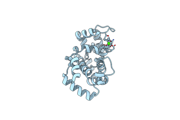
Crystal Structure Of Non-Myristoylated Recoverin With Cysteine-39 Oxidized To Sulfenic Acid
Organism: Bos taurus
Method: X-RAY DIFFRACTION Resolution:1.90 Å Release Date: 2013-11-13 Classification: METAL BINDING PROTEIN Ligands: CA |
Organism: Bos taurus
Method: X-RAY DIFFRACTION
Release Date: 2013-11-13
Ligands: CA
 |
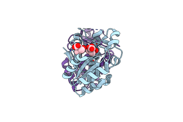
Crystal Structure Of Non-Myristoylated Recoverin At 1.45 A Resolution With Calcium Bound To Ef-Hand 3
Organism: Bos taurus
Method: X-RAY DIFFRACTION Resolution:1.45 Å Release Date: 2013-11-13 Classification: METAL BINDING PROTEIN Ligands: CA |
Organism: Bos taurus
Method: X-RAY DIFFRACTION
Release Date: 2013-11-13
Ligands: CA
 |
Structural And Substrate-Binding Studies Of Pantothenate Synthenate (Ps)Provide Insights Into Homotropic Inhibition By Pantoate In Ps'S
Organism: Escherichia coli
Method: X-RAY DIFFRACTION Resolution:1.67 Å Release Date: 2010-02-09 Classification: LIGASE Ligands: PAF |
Organism: Escherichia coli
Method: X-RAY DIFFRACTION
Release Date: 2010-02-09
Ligands: PAF