
Search Count: 35
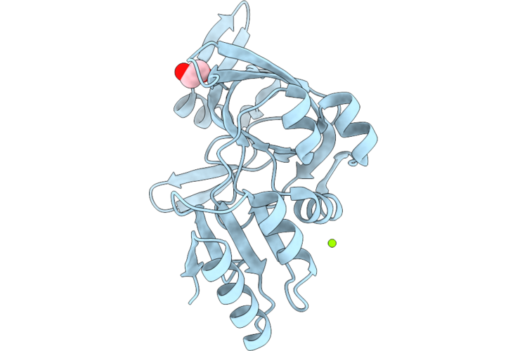 |
Nucleotide Binding Domain (Residues 475-720) Of Abc3 Transporter Permease From Clostridioides Difficile Strain 630
Organism: Clostridioides difficile 630
Method: X-RAY DIFFRACTION Resolution:2.00 Å Release Date: 2026-06-24 Classification: TRANSPORT PROTEIN Ligands: MG, CL, EDO |
 |
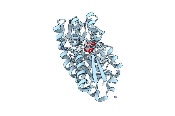
Crystal Structure Of The Ggdef Domain (Residues 31-260) Of Diguanylate Cyclase From Vibrio Cholerae Serotype O1
Organism: Vibrio cholerae o1 biovar el tor str. n16961
Method: X-RAY DIFFRACTION Resolution:2.00 Å Release Date: 2026-06-24 Classification: OXIDOREDUCTASE Ligands: MG, CL |
 |
Crystal Structure Of Duf4097 Domain-Containing Protein From Clostridium Difficile Strain 630
Organism: Clostridioides difficile 630
Method: X-RAY DIFFRACTION Resolution:2.00 Å Release Date: 2026-06-24 Classification: UNKNOWN FUNCTION Ligands: EDO |
 |
Crystal Structure Of The Ggdef Domain Of The Diguanylate Cyclase From Vibrio Vulnificus Cmcp6
Organism: Vibrio vulnificus cmcp6
Method: X-RAY DIFFRACTION Resolution:2.60 Å Release Date: 2026-06-24 Classification: TRANSFERASE |
 |
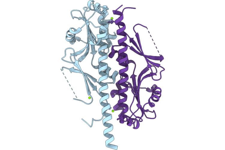
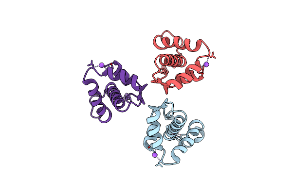
Crystal Structure Of Flagellar Assembly Protein Fliw From Campylobacter Jejuni Subsp. Jejuni 81-176-Drh212
Organism: Campylobacter jejuni subsp. jejuni 81-176-drh212
Method: X-RAY DIFFRACTION Resolution:2.95 Å Release Date: 2026-06-17 Classification: TRANSLATION Ligands: CL |
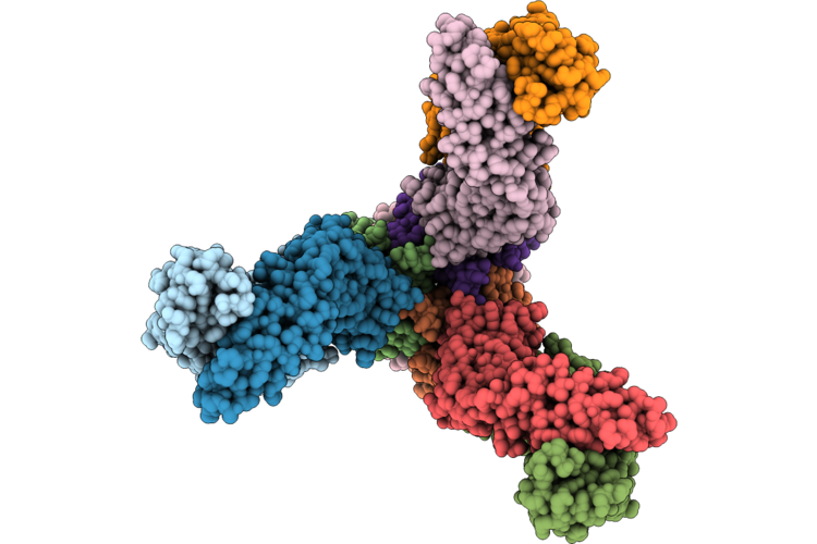 |
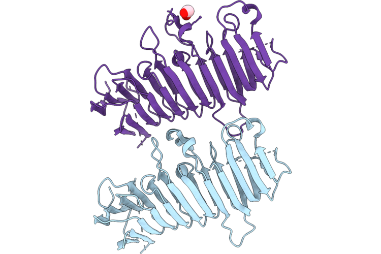
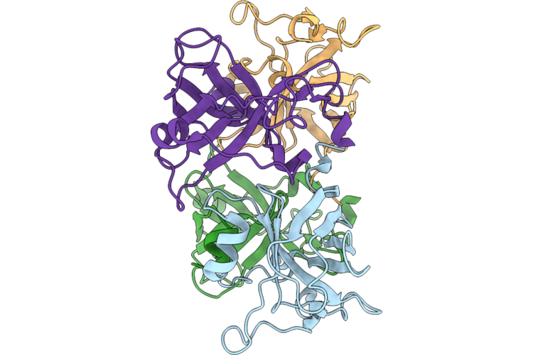
Chlamydia Muridarum Major Outer Membrane Protein Bound To Mus Musculus Mab-18B Fab
Organism: Mus musculus, Chlamydia muridarum
Method: ELECTRON MICROSCOPY Resolution:3.36 Å Release Date: 2026-05-27 Classification: MEMBRANE PROTEIN/IMMUNE SYSTEM Ligands: PEX, DAO, NAG, FO4 |
 |
Crystal Structure Of Putative L-Amino Acid N-Acyltransferase Mnat From Pseudomonas Aeruginosa
Organism: Pseudomonas aeruginosa pao1
Method: X-RAY DIFFRACTION Resolution:1.80 Å Release Date: 2026-04-15 Classification: VIRAL PROTEIN Ligands: EDO |
 |
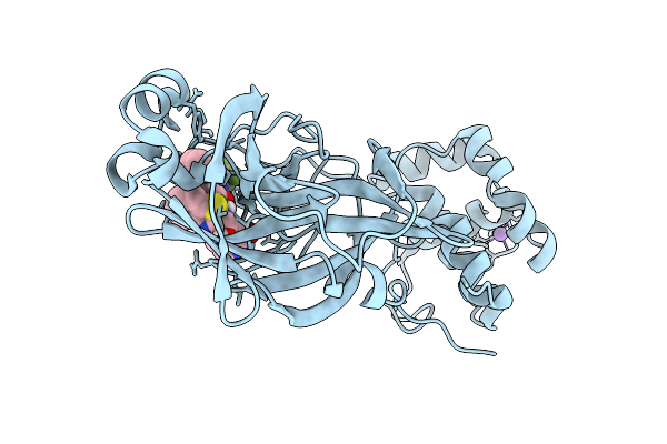
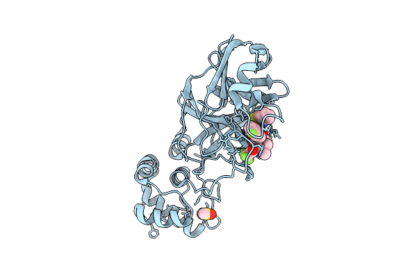
X-Ray Structure Of Sars-Cov-2 Main Protease Covalently Bound To Inhibitor Grl-050-22 At 1.16 A
Organism: Severe acute respiratory syndrome coronavirus 2
Method: X-RAY DIFFRACTION Resolution:1.16 Å Release Date: 2025-12-24 Classification: VIRAL PROTEIN, HYDROLASE/INHIBITOR Ligands: A1C3L |
 |
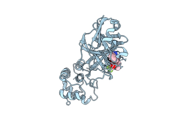
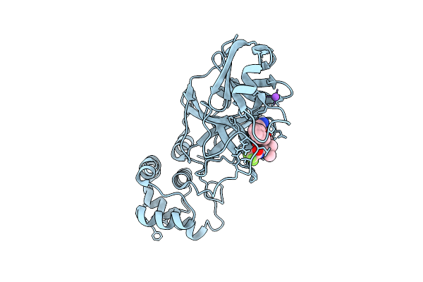
X-Ray Structure Of Sars-Cov-2 Main Protease Covalently Bound To Inhibitor Grl-062-22 At 1.65 A
Organism: Severe acute respiratory syndrome coronavirus 2
Method: X-RAY DIFFRACTION Resolution:1.65 Å Release Date: 2025-12-24 Classification: VIRAL PROTEIN, HYDROLASE/INHIBITOR Ligands: A1C3M, NA |
 |
X-Ray Structure Of Sars-Cov-2 Main Protease T190I Covalently Bound To Compound Grl-051-22 At 1.5 A
Organism: Severe acute respiratory syndrome coronavirus
Method: X-RAY DIFFRACTION Resolution:1.50 Å Release Date: 2025-11-26 Classification: VIRAL PROTEIN, HYDROLASE Ligands: A1BFE, NA |
 |
X-Ray Structure Of Sars-Cov-2 Main Protease Covalently Bound To Compound Grl-050-23 At 1.6 A
Organism: Severe acute respiratory syndrome coronavirus 2
Method: X-RAY DIFFRACTION Resolution:1.60 Å Release Date: 2025-11-26 Classification: VIRAL PROTEIN Ligands: DMS, A1BMU, NA |
 |
X-Ray Structure Of Sars-Cov-2 Main Protease M165I Covalently Bound To Inhibitor Grl-051-22 At 1.90 A
Organism: Severe acute respiratory syndrome coronavirus 2
Method: X-RAY DIFFRACTION Resolution:1.90 Å Release Date: 2025-11-26 Classification: VIRAL PROTEIN Ligands: A1BFE, NA |
 |
X-Ray Structure Of Sars-Cov-2 Main Protease V186G Covalently Bound To Inhibitor Nirmatrelvir At 1.81 A
Organism: Severe acute respiratory syndrome coronavirus 2
Method: X-RAY DIFFRACTION Resolution:1.81 Å Release Date: 2025-11-26 Classification: VIRAL PROTEIN, HYDROLASE/INHIBITOR Ligands: 4WI |
 |
Crystal Structure Of C4-Dicarboxylate-Binding Protein (Pa0884) Of Tripartite Atp-Independent Periplasmic Transporter Family From Pseudomonas Aeruginosa Pao1 In Complex With L-Malate
Organism: Pseudomonas aeruginosa
Method: X-RAY DIFFRACTION Resolution:1.50 Å Release Date: 2025-07-09 Classification: TRANSPORT PROTEIN Ligands: LMR |
 |
Crystal Structure Of C4-Dicarboxylate-Binding Protein (Pa0884) Of Tripartite Atp-Independent Periplasmic Transporter Family From Pseudomonas Aeruginosa Pao1 In Complex With Mesaconic Acid
Organism: Pseudomonas aeruginosa
Method: X-RAY DIFFRACTION Resolution:1.32 Å Release Date: 2025-07-09 Classification: TRANSPORT PROTEIN Ligands: MEZ, MG, CL |
 |
Crystal Structure Of C4-Dicarboxylate-Binding Periplasmic Protein (Pa5167) Of Tripartite Atp-Independent Periplasmic Transporter Family From Pseudomonas Aeruginosa Pao1 In Complex With L-Malate
Organism: Pseudomonas aeruginosa
Method: X-RAY DIFFRACTION Resolution:1.30 Å Release Date: 2025-07-09 Classification: TRANSPORT PROTEIN Ligands: LMR, ZN |
 |
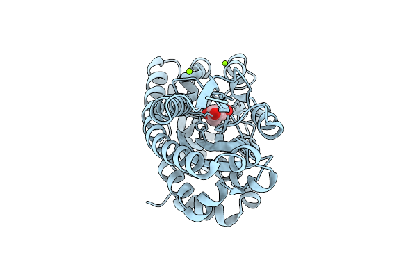
Crystal Structure Of Acyl-Coa Lyase Subunit Beta From Pseudomonas Aeruginosa Pao1
Organism: Pseudomonas aeruginosa
Method: X-RAY DIFFRACTION Resolution:1.85 Å Release Date: 2025-07-09 Classification: LYASE |
 |
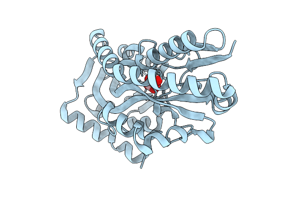
Crystal Structure Of The Sars-Cov-2 2'-O-Methyltransferase With (M7Gpppa)Pupu (Cap-0) And S-Adenosyl-L-Homocysteine (Sah).
Organism: Severe acute respiratory syndrome coronavirus 2
Method: X-RAY DIFFRACTION Resolution:1.95 Å Release Date: 2025-07-02 Classification: TRANSFERASE, Viral Protein Ligands: SAH, CL, SO4, ZN, MGT |
 |
Crystal Structure Of The C-Terminal Cytoplasmic Domain Of Nsp4 From Sars-Cov-2
Organism: Severe acute respiratory syndrome coronavirus 2
Method: X-RAY DIFFRACTION Resolution:2.35 Å Release Date: 2025-06-25 Classification: VIRAL PROTEIN |
 |
Crystal Structure Of The Surface Protein (Cd630_07380) From Clostridium Difficile Strain 630
Organism: Clostridioides difficile 630
Method: X-RAY DIFFRACTION Resolution:1.85 Å Release Date: 2025-06-18 Classification: MEMBRANE PROTEIN |