

Search Count: 21
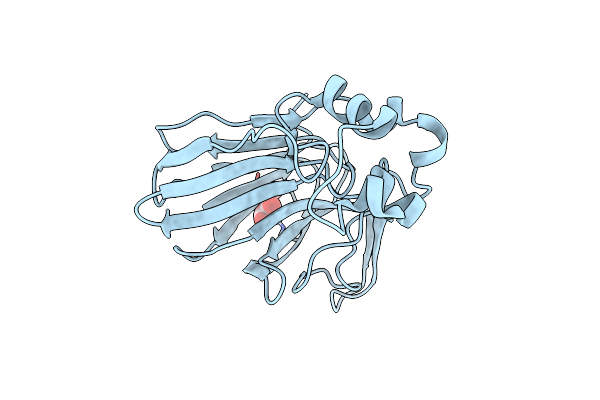 |
Serial Microseconds Crystallography At Id29 Using Sacla Extruder: Thaumatin
Organism: Thaumatococcus daniellii
Method: X-RAY DIFFRACTION Resolution:1.75 Å Release Date: 2025-01-22 Classification: PLANT PROTEIN Ligands: TLA |
Organism: Thaumatococcus daniellii
Method: X-RAY DIFFRACTION
Release Date: 2025-01-22
Ligands: TLA
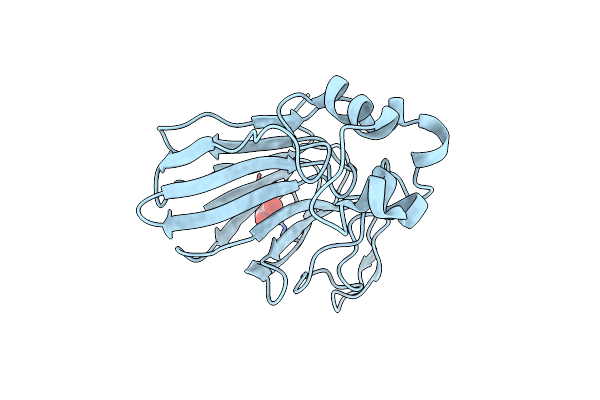 |
Serial Microseconds Crystallography At Id29 Using Asu Extruder: Thaumatin
Organism: Thaumatococcus daniellii
Method: X-RAY DIFFRACTION Resolution:1.75 Å Release Date: 2025-01-22 Classification: PLANT PROTEIN Ligands: TLA |
Organism: Thaumatococcus daniellii
Method: X-RAY DIFFRACTION
Release Date: 2025-01-22
Ligands: TLA
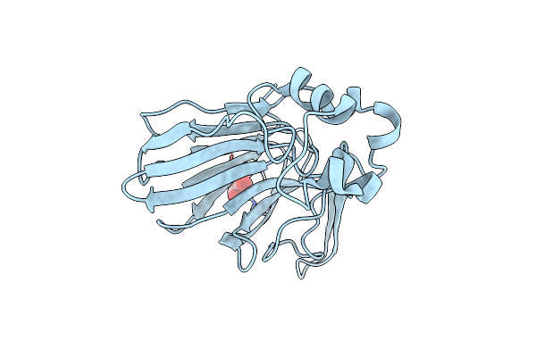 |
Serial Microseconds Crystallography At Id29 Using Mpi Extruder: Thaumatin
Organism: Thaumatococcus daniellii
Method: X-RAY DIFFRACTION Resolution:1.75 Å Release Date: 2025-01-22 Classification: PLANT PROTEIN Ligands: TLA |
Organism: Thaumatococcus daniellii
Method: X-RAY DIFFRACTION
Release Date: 2025-01-22
Ligands: TLA
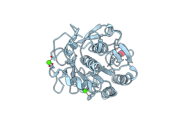 |
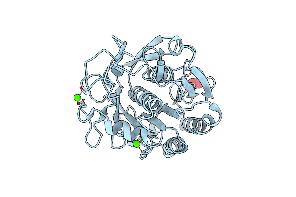
Serial Microseconds Crystallography At Id29 Using Fixed-Target (Small Foils): Proteinase K With 10 Um Spacing Between X-Ray Pulses
Organism: Parengyodontium album
Method: X-RAY DIFFRACTION Resolution:2.00 Å Release Date: 2025-01-22 Classification: HYDROLASE Ligands: CA, NO3 |
Organism: Parengyodontium album
Method: X-RAY DIFFRACTION
Release Date: 2025-01-22
Ligands: CA, NO3
 |
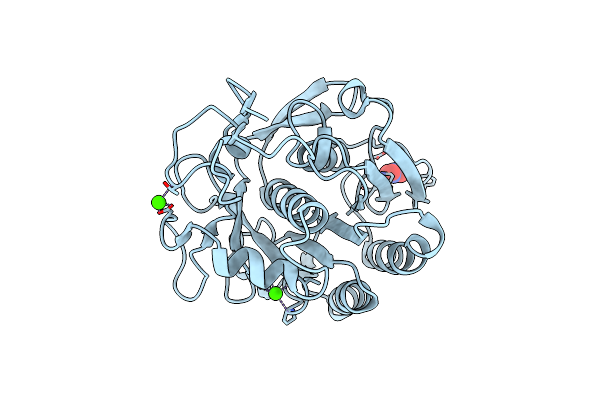
Serial Microseconds Crystallography At Id29 Using Fixed-Target (Small Foils): Proteinase K With 20 Um Spacing Between X-Ray Pulses
Organism: Parengyodontium album
Method: X-RAY DIFFRACTION Resolution:2.00 Å Release Date: 2025-01-22 Classification: HYDROLASE Ligands: CA, NO3 |
Organism: Parengyodontium album
Method: X-RAY DIFFRACTION
Release Date: 2025-01-22
Ligands: CA, NO3
 |
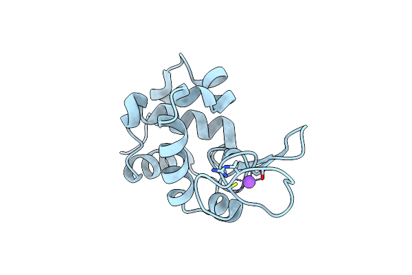
Serial Microseconds Crystallography At Id29 Using Fixed-Target (Large Foils): Proteinase K With 50 Um Spacing Between X-Ray Pulses
Organism: Parengyodontium album
Method: X-RAY DIFFRACTION Resolution:2.00 Å Release Date: 2025-01-22 Classification: HYDROLASE Ligands: CA, NO3 |
Organism: Parengyodontium album
Method: X-RAY DIFFRACTION
Release Date: 2025-01-22
Ligands: CA, NO3
 |
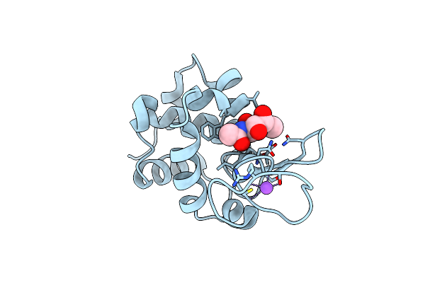
Serial Microseconds Crystallography At Id29 Using Fixed-Target (Si Chip): Lysozyme - Without Ligand Glcnac (Apo)
Organism: Gallus gallus
Method: X-RAY DIFFRACTION Resolution:2.05 Å Release Date: 2025-01-22 Classification: HYDROLASE |
Organism: Gallus gallus
Method: X-RAY DIFFRACTION
Release Date: 2025-01-22
 |
Serial Microseconds Crystallography At Id29 Using Fixed-Target (Si Chip): Lysozyme - With Ligand Glcnac
Organism: Gallus gallus
Method: X-RAY DIFFRACTION Resolution:2.05 Å Release Date: 2025-01-22 Classification: HYDROLASE |
Organism: Gallus gallus
Method: X-RAY DIFFRACTION
Release Date: 2025-01-22
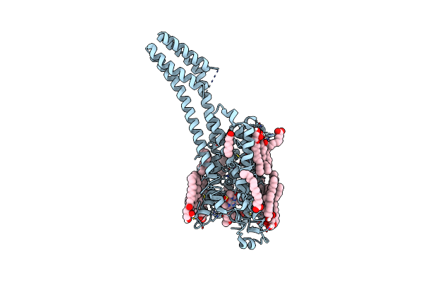 |
Serial Microseconds Crystallography At Id29 Using Fixed-Target (Small Foils): A2A Adenosine Receptor Co-Crystallised With Istradefylline
Organism: Homo sapiens
Method: X-RAY DIFFRACTION Resolution:2.50 Å Release Date: 2025-01-22 Classification: SIGNALING PROTEIN |
Organism: Homo sapiens
Method: X-RAY DIFFRACTION
Release Date: 2025-01-22
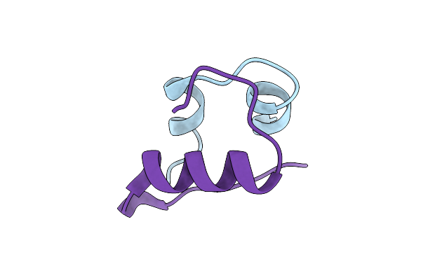 |
Cubic Insulin Sad Phasing At 14.2 Kev
Organism: Homo sapiens
Method: X-RAY DIFFRACTION Resolution:1.20 Å Release Date: 2022-06-01 Classification: HORMONE |
Organism: Homo sapiens
Method: X-RAY DIFFRACTION
Release Date: 2022-06-01
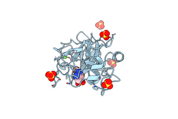 |
Trypsin Fast Data Collection
Organism: Bos taurus
Method: X-RAY DIFFRACTION Resolution:1.43 Å Release Date: 2020-05-20 Classification: HYDROLASE Ligands: CA, SO4, BEN |
Organism: Bos taurus
Method: X-RAY DIFFRACTION
Release Date: 2020-05-20
Ligands: CA, SO4, BEN
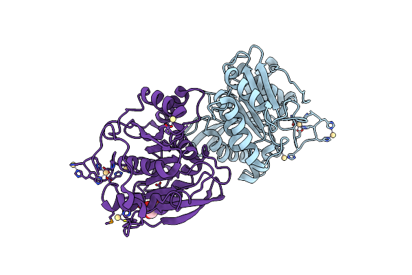 |
Selenomethionine Derivative Of Ferulic Acid Esterase (Fae)
Organism: Hungateiclostridium thermocellum
Method: X-RAY DIFFRACTION Resolution:1.80 Å Release Date: 2020-05-20 Classification: HYDROLASE Ligands: CD, GOL |
Organism: Hungateiclostridium thermocellum
Method: X-RAY DIFFRACTION
Release Date: 2020-05-20
Ligands: CD, GOL
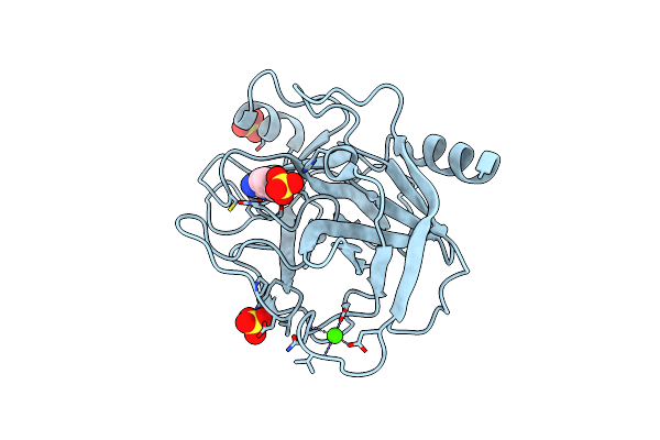 |
Bovine Trypsin Solved By S-Sad On Id30B
Organism: Bos taurus
Method: X-RAY DIFFRACTION Resolution:2.20 Å Release Date: 2018-02-07 Classification: HYDROLASE Ligands: CA, BEN, SO4 |
Organism: Bos taurus
Method: X-RAY DIFFRACTION
Release Date: 2018-02-07
Ligands: CA, BEN, SO4
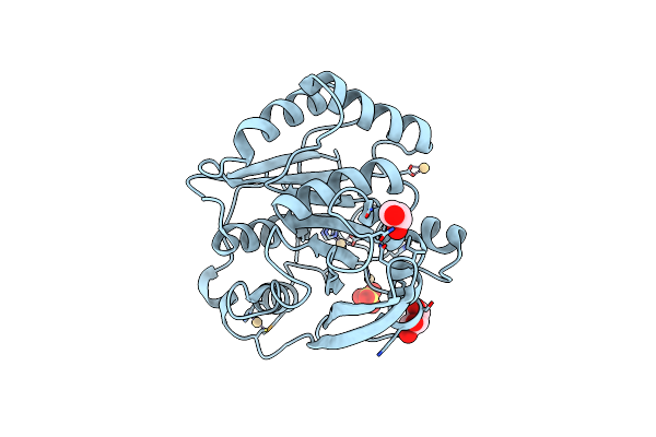 |
Structure Of Fae Solved By Sad From Data Collected At The Peak Of The Selenium Absorption Edge On Id30B
Organism: Clostridium thermocellum
Method: X-RAY DIFFRACTION Resolution:1.70 Å Release Date: 2018-02-07 Classification: HYDROLASE Ligands: CD, GOL, SO4 |
Organism: Clostridium thermocellum
Method: X-RAY DIFFRACTION
Release Date: 2018-02-07
Ligands: CD, GOL, SO4
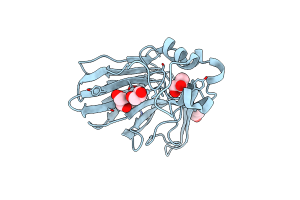 |
Structure Of Thaumatin Collected At 100K On Id30B
Organism: Thaumatococcus daniellii
Method: X-RAY DIFFRACTION Resolution:1.08 Å Release Date: 2018-02-07 Classification: PLANT PROTEIN Ligands: TLA, GOL |
Organism: Thaumatococcus daniellii
Method: X-RAY DIFFRACTION
Release Date: 2018-02-07
Ligands: TLA, GOL
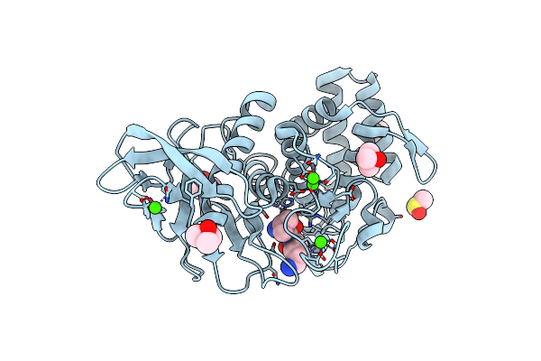 |
Structure Of Thermolysin Solved From Sad Data Collected At The Peak Of The Zn Absorption Edge On Id30B
Organism: Bacillus thermoproteolyticus
Method: X-RAY DIFFRACTION Resolution:1.43 Å Release Date: 2018-01-31 Classification: HYDROLASE Ligands: ZN, CA, DMS, TMO, VAL, LYS |
Organism: Bacillus thermoproteolyticus
Method: X-RAY DIFFRACTION
Release Date: 2018-01-31
Ligands: ZN, CA, DMS, TMO, VAL, LYS
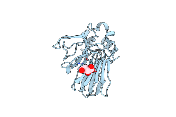 |
Structure Of Thaumatin Collected From An In Situ Crystal Collected On Id30B At 12.7 Kev.
Organism: Thaumatococcus daniellii
Method: X-RAY DIFFRACTION Resolution:1.50 Å Release Date: 2018-01-31 Classification: PLANT PROTEIN Ligands: TLA |
Organism: Thaumatococcus daniellii
Method: X-RAY DIFFRACTION
Release Date: 2018-01-31
Ligands: TLA
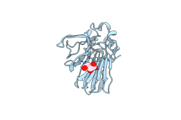 |
Structure Of Thaumatin Collected From An In Situ Crystal On Id30B At 17.5 Kev.
Organism: Thaumatococcus daniellii
Method: X-RAY DIFFRACTION Resolution:1.50 Å Release Date: 2018-01-31 Classification: PLANT PROTEIN Ligands: TLA |
Organism: Thaumatococcus daniellii
Method: X-RAY DIFFRACTION
Release Date: 2018-01-31
Ligands: TLA
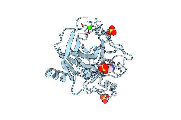 |
Structure Of Trypsin Solved By Mr From Data Collected By Direct Data Collection (Ddc) Using The Esrf Robodiff Goniometer
Organism: Bos taurus
Method: X-RAY DIFFRACTION Resolution:1.78 Å Release Date: 2016-03-16 Classification: HYDROLASE Ligands: CA, BEN, SO4 |
Organism: Bos taurus
Method: X-RAY DIFFRACTION
Release Date: 2016-03-16
Ligands: CA, BEN, SO4
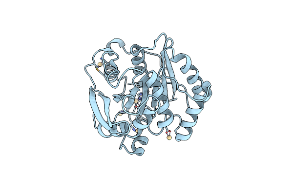 |
Structure Of Fae Solved By Sad From Data Collected By Direct Data Collection (Ddc) Using The Esrf Robodiff Goniometer
Organism: Ruminiclostridium thermocellum
Method: X-RAY DIFFRACTION Resolution:1.99 Å Release Date: 2016-03-16 Classification: HYDROLASE Ligands: CD |
Organism: Ruminiclostridium thermocellum
Method: X-RAY DIFFRACTION
Release Date: 2016-03-16
Ligands: CD