
Search Count: 54
All
Selected
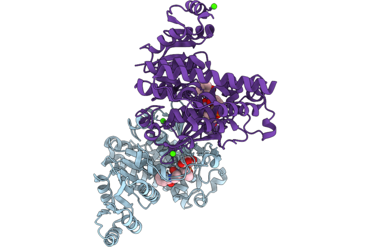 |
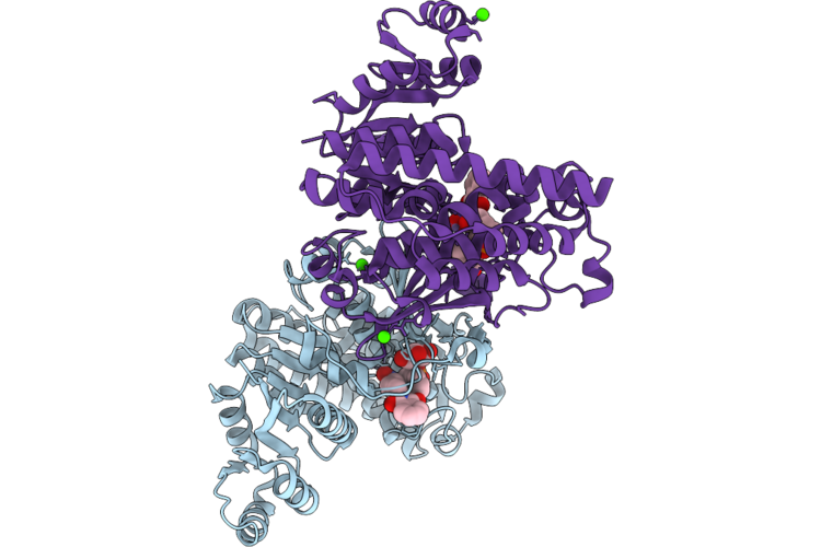
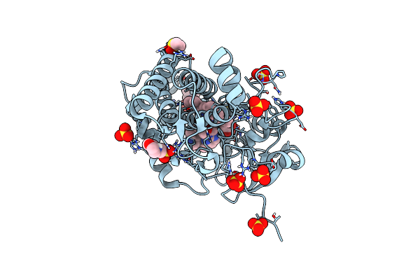
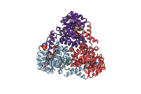
Organism: Plasmodium falciparum hb3
Method: X-RAY DIFFRACTION Resolution:1.53 Å Release Date: 2026-05-06 Classification: ISOMERASE Ligands: MN, A1L9Z, GOL, CA |
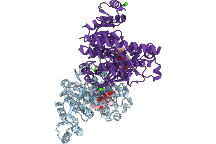 |
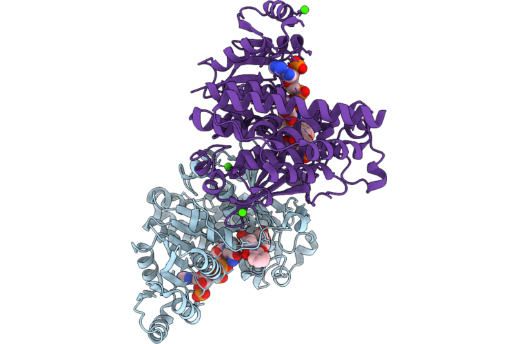
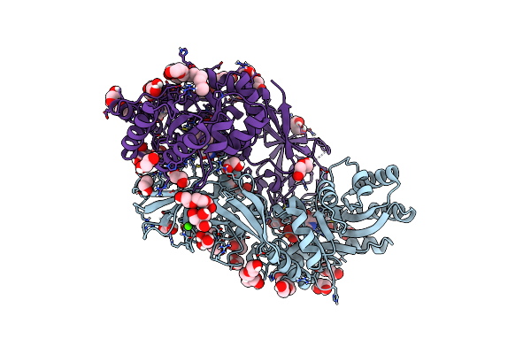
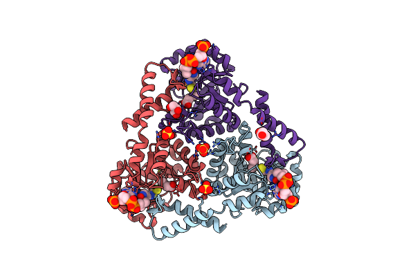
Organism: Plasmodium falciparum hb3
Method: X-RAY DIFFRACTION Resolution:1.59 Å Release Date: 2026-05-06 Classification: ISOMERASE Ligands: MN, A1L90, GOL, CA |
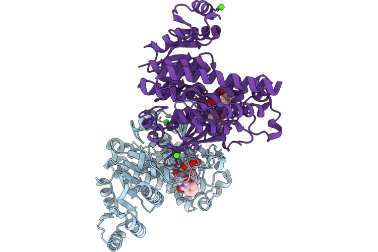 |
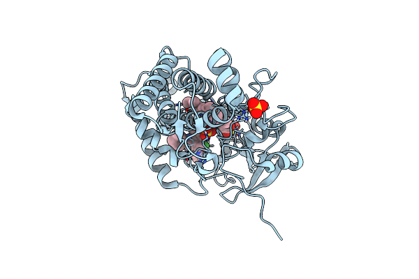
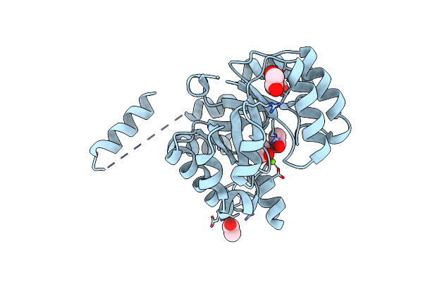
Organism: Plasmodium falciparum hb3
Method: X-RAY DIFFRACTION Resolution:1.67 Å Release Date: 2026-05-06 Classification: ISOMERASE Ligands: MN, A1L91, GOL, CA |
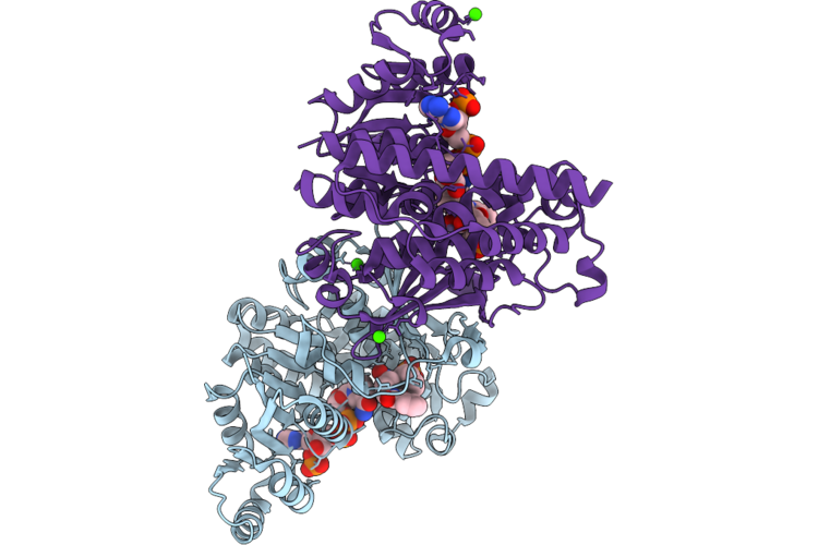 |
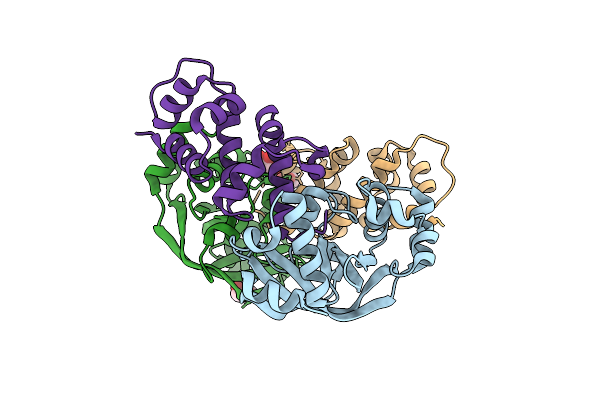
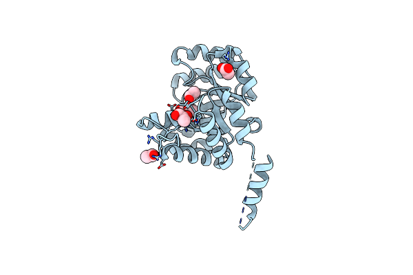
Organism: Plasmodium falciparum hb3
Method: X-RAY DIFFRACTION Resolution:1.69 Å Release Date: 2026-05-06 Classification: ISOMERASE Ligands: NDP, MN, A1L91, CA |
 |
Organism: Plasmodium falciparum hb3
Method: X-RAY DIFFRACTION Resolution:1.57 Å Release Date: 2026-05-06 Classification: ISOMERASE Ligands: MN, A1L92, GOL, CA |
 |
Organism: Plasmodium falciparum hb3
Method: X-RAY DIFFRACTION Resolution:1.58 Å Release Date: 2026-05-06 Classification: ISOMERASE Ligands: NDP, MN, A1L92, CA |
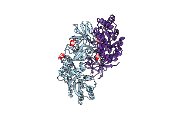 |
Structure Of Dcs-Resistant Variant D322N Of Alanine Racemase From Mycobacterium Tuberculosis
Organism: Mycobacterium tuberculosis h37rv
Method: X-RAY DIFFRACTION Resolution:1.58 Å Release Date: 2023-04-05 Classification: ISOMERASE Ligands: GOL, EDO |
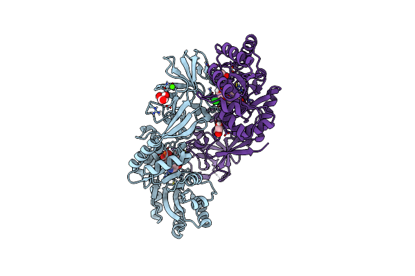 |
Structure Of Dcs-Resistant Variant D322N Of Alanine Racemase From M. Tuberculosis In Complex With Dcs
Organism: Mycobacterium tuberculosis h37rv
Method: X-RAY DIFFRACTION Resolution:1.78 Å Release Date: 2023-04-05 Classification: ISOMERASE Ligands: OJQ, L7N, CA, CL, GOL, EDO |
 |
Mycobacterium Tuberculosis Cytochrome P450 Cyp121 In Complex With Lead Compound 10
Organism: Mycobacterium tuberculosis (strain atcc 25618 / h37rv)
Method: X-RAY DIFFRACTION Resolution:1.60 Å Release Date: 2022-02-02 Classification: METAL BINDING PROTEIN Ligands: HEC, UMZ, MES, SO4, CL |
 |
Mycobacterium Tuberculosis Cytochrome P450 Cyp121 In Complex With Lead Compound 14
Organism: Mycobacterium tuberculosis (strain atcc 25618 / h37rv)
Method: X-RAY DIFFRACTION Resolution:1.60 Å Release Date: 2022-02-02 Classification: METAL BINDING PROTEIN Ligands: HEM, UMT, MES, SO4, CL |
 |
Mycobacterium Tuberculosis Cytochrome P450 Cyp121 In Complex With Lead Compound 21
Organism: Mycobacterium tuberculosis (strain atcc 25618 / h37rv)
Method: X-RAY DIFFRACTION Resolution:1.60 Å Release Date: 2022-02-02 Classification: METAL BINDING PROTEIN Ligands: HEC, ULZ, MES, DMS, SO4, CL |
 |
Organism: Mycobacterium tuberculosis h37rv
Method: X-RAY DIFFRACTION Resolution:1.57 Å Release Date: 2020-04-01 Classification: ISOMERASE Ligands: GOL, CA, CL, EDO, PEG, P4K, L7N, OJQ, TRS |
 |
Crystal Structure Of Mycobacterium Tuberculosis Cytochrome P450 Cyp121A1 In Complex With Triazole Pyrazole Inhibitor 10J
Organism: Mycobacterium tuberculosis (strain cdc 1551 / oshkosh)
Method: X-RAY DIFFRACTION Resolution:1.50 Å Release Date: 2019-08-07 Classification: OXIDOREDUCTASE Ligands: HEM, SO4, EW2 |
 |
Crystal Structure Of Mycobacterium Tuberculosis Cytochrome P450 Cyp121A1 In Complex With Triazole Pyrazole Inhibitor 14A
Organism: Mycobacterium tuberculosis (strain cdc 1551 / oshkosh)
Method: X-RAY DIFFRACTION Resolution:1.60 Å Release Date: 2019-08-07 Classification: OXIDOREDUCTASE Ligands: HEM, EW5, SO4 |
 |
Organism: Mycobacterium tuberculosis
Method: X-RAY DIFFRACTION Resolution:1.80 Å Release Date: 2019-02-27 Classification: TOXIN Ligands: GOL |
 |
Crystal Structure Of Protein Cite From Mycobacterium Tuberculosis In Complex With Magnesium, Pyruvate And Citramalyl-Coa
Organism: Mycobacterium tuberculosis h37rv
Method: X-RAY DIFFRACTION Resolution:1.83 Å Release Date: 2018-08-01 Classification: LYASE Ligands: MG, CQM, PO4, GOL, CL, PYR |
 |
Crystal Structure Of Protein Cite From Mycobacterium Tuberculosis In Complex With Magnesium, Pyruvate And Coenzyme A
Organism: Mycobacterium tuberculosis h37rv
Method: X-RAY DIFFRACTION Resolution:1.72 Å Release Date: 2018-08-01 Classification: LYASE Ligands: MG, PYR, COA, PO4, GOL |
 |
Crystal Structure Of Protein Cite From Mycobacterium Tuberculosis In Complex With Magnesium, Acetoacetate And Coenzyme A
Organism: Mycobacterium tuberculosis h37rv
Method: X-RAY DIFFRACTION Resolution:2.04 Å Release Date: 2018-08-01 Classification: LYASE Ligands: MG, COA, AAE, SO4, GOL |
 |
Crystal Structure Of Protein Cite From Mycobacterium Tuberculosis In Complex With Magnesium And Acetate
Organism: Mycobacterium tuberculosis h37rv
Method: X-RAY DIFFRACTION Resolution:1.61 Å Release Date: 2018-08-01 Classification: LYASE Ligands: MG, ACT, EDO |
 |
Crystal Structure Of Protein Cite From Mycobacterium Tuberculosis In Complex With Magnesium And Pyruvate
Organism: Mycobacterium tuberculosis h37rv
Method: X-RAY DIFFRACTION Resolution:1.73 Å Release Date: 2018-08-01 Classification: LYASE Ligands: MG, ACT, PYR |