

Search Count: 19
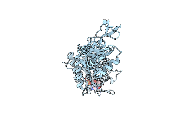 |
Crystal Structure Of Pseudomonas Aeruginosa Penicillin Binding Protein 3 (Pae-Pbp3) Bound To Etx0462
Organism: Pseudomonas aeruginosa
Method: X-RAY DIFFRACTION Resolution:2.20 Å Release Date: 2021-05-26 Classification: ANTIBIOTIC Ligands: CL, VMM |
Organism: Pseudomonas aeruginosa
Method: X-RAY DIFFRACTION
Release Date: 2021-05-26
Ligands: CL, VMM
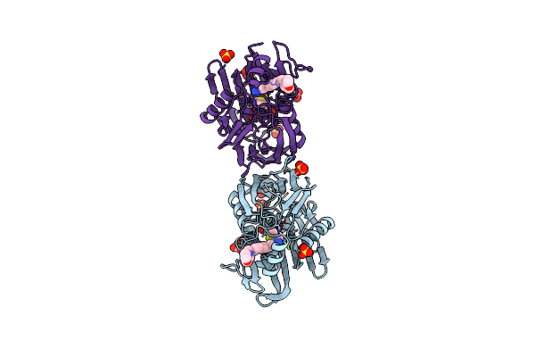 |
Crystal Structure Of P. Aeruginosa Lpxc With N-Hydroxyformamide Inhibitor 19
Organism: Pseudomonas aeruginosa (strain atcc 15692 / dsm 22644 / cip 104116 / jcm 14847 / lmg 12228 / 1c / prs 101 / pao1)
Method: X-RAY DIFFRACTION Resolution:1.90 Å Release Date: 2020-11-25 Classification: HYDROLASE Ligands: GOL, W4M, SO4, ZN |
Organism: Pseudomonas aeruginosa (strain atcc 15692 / dsm 22644 / cip 104116 / jcm 14847 / lmg 12228 / 1c / prs 101 / pao1)
Method: X-RAY DIFFRACTION
Release Date: 2020-11-25
Ligands: GOL, W4M, SO4, ZN
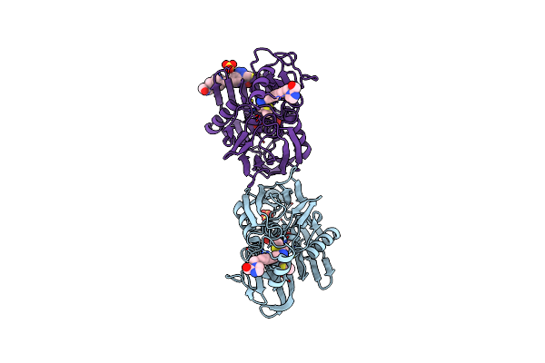 |
Crystal Structure Of P. Aeruginosa Lpxc With N-Hydroxyformamide Inhibitor
Organism: Pseudomonas aeruginosa (strain atcc 15692 / dsm 22644 / cip 104116 / jcm 14847 / lmg 12228 / 1c / prs 101 / pao1)
Method: X-RAY DIFFRACTION Resolution:2.00 Å Release Date: 2020-11-25 Classification: HYDROLASE Ligands: W4P, SO4, GOL, DMS, ZN, W8P |
Organism: Pseudomonas aeruginosa (strain atcc 15692 / dsm 22644 / cip 104116 / jcm 14847 / lmg 12228 / 1c / prs 101 / pao1)
Method: X-RAY DIFFRACTION
Release Date: 2020-11-25
Ligands: W4P, SO4, GOL, DMS, ZN, W8P
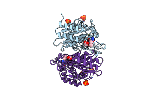 |
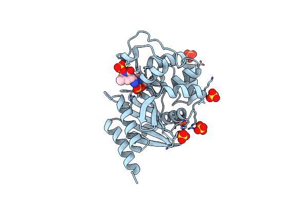
Crystal Structure Of Ctx-M-14 In Complex With Beta-Lactamase Inhibitor Etx1317
Organism: Escherichia coli
Method: X-RAY DIFFRACTION Resolution:1.28 Å Release Date: 2020-08-26 Classification: HYDROLASE Ligands: X57, PO4 |
Organism: Escherichia coli
Method: X-RAY DIFFRACTION
Release Date: 2020-08-26
Ligands: X57, PO4
 |
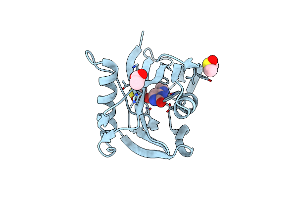
Diazabicyclooctenone Etx2514 Bound To Class D Beta Lactamase Oxa-24 From A. Baumannii
Organism: Acinetobacter baumannii
Method: X-RAY DIFFRACTION Resolution:1.93 Å Release Date: 2017-06-07 Classification: HYDROLASE/HYDROLASE INHIBITOR Ligands: SO4, 9CP, 9CM |
Organism: Acinetobacter baumannii
Method: X-RAY DIFFRACTION
Release Date: 2017-06-07
Ligands: SO4, 9CP, 9CM
 |
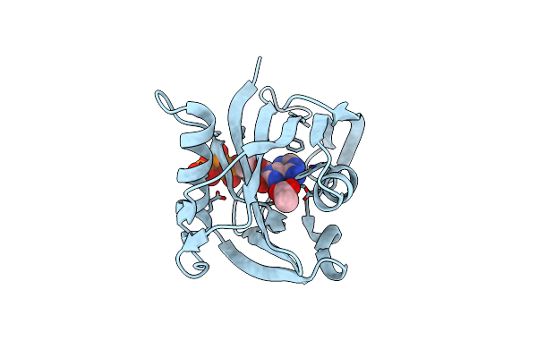
Mth1 Substrate Recognition: Complex With A Methylaminopyrimidinedione.
Organism: Homo sapiens
Method: X-RAY DIFFRACTION Resolution:1.67 Å Release Date: 2017-01-18 Classification: HYDROLASE Ligands: ACT, S76, DMS |
Organism: Homo sapiens
Method: X-RAY DIFFRACTION
Release Date: 2017-01-18
Ligands: ACT, S76, DMS
 |
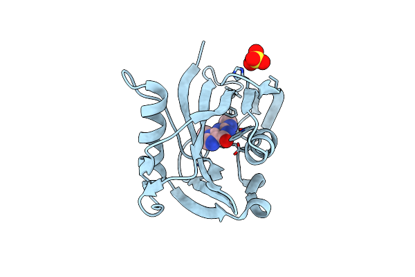
Mth1 Substrate Recognition: Complex With 8-Oxo-Dgtp.
Organism: Homo sapiens
Method: X-RAY DIFFRACTION Resolution:1.56 Å Release Date: 2016-07-20 Classification: HYDROLASE Ligands: H6Y, ACT |
Organism: Homo sapiens
Method: X-RAY DIFFRACTION
Release Date: 2016-07-20
Ligands: H6Y, ACT
 |
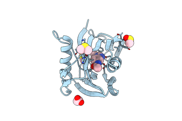
Mth1 Substrate Recognition: Complex With A Methylaminopurinone
Organism: Homo sapiens
Method: X-RAY DIFFRACTION Resolution:1.24 Å Release Date: 2016-07-20 Classification: HYDROLASE Ligands: UAN, SO4 |
Organism: Homo sapiens
Method: X-RAY DIFFRACTION
Release Date: 2016-07-20
Ligands: UAN, SO4
 |
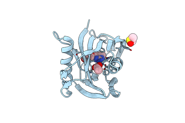
Mth1 Substrate Recognition: Complex With A Methylbenzimidazolyl Acetamide.
Organism: Homo sapiens
Method: X-RAY DIFFRACTION Resolution:1.67 Å Release Date: 2016-07-20 Classification: HYDROLASE Ligands: ACT, N91, DMS |
Organism: Homo sapiens
Method: X-RAY DIFFRACTION
Release Date: 2016-07-20
Ligands: ACT, N91, DMS
 |
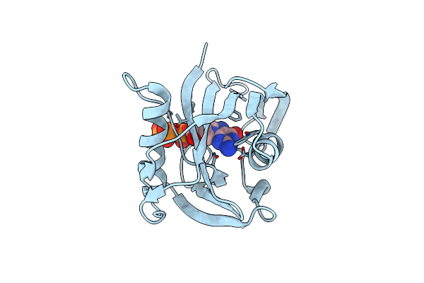
Mth1 Substrate Recognition: Complex With A Aminomethylpyrimidinyl Oxypropanol.
Organism: Homo sapiens
Method: X-RAY DIFFRACTION Resolution:1.69 Å Release Date: 2016-07-20 Classification: HYDROLASE Ligands: 6Q3, ACT, DMS |
Organism: Homo sapiens
Method: X-RAY DIFFRACTION
Release Date: 2016-07-20
Ligands: 6Q3, ACT, DMS
 |
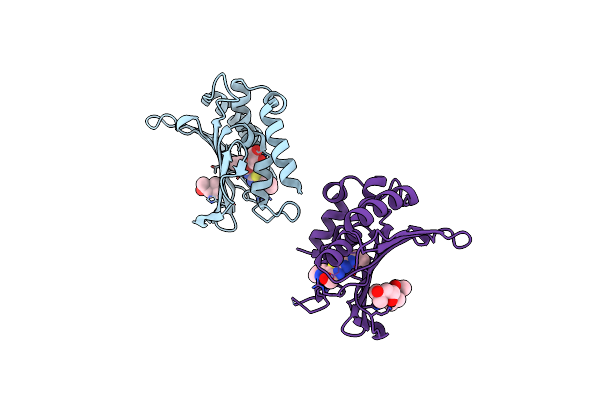
Mth1 Substrate Recognition: Complex With 8-Oxo-Dgtp.
Organism: Homo sapiens
Method: X-RAY DIFFRACTION Resolution:1.63 Å Release Date: 2016-07-11 Classification: HYDROLASE Ligands: 8DG |
Organism: Homo sapiens
Method: X-RAY DIFFRACTION
Release Date: 2016-07-11
Ligands: 8DG
 |
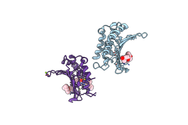
Crystal Structure Of The Atp Binding Domain Of S. Aureus Gyrb Complexed With A Ligand
Organism: Staphylococcus aureus
Method: X-RAY DIFFRACTION Resolution:2.05 Å Release Date: 2015-11-25 Classification: ISOMERASE/ISOMERASE INHIBITOR Ligands: 57U, MPD, GOL |
Organism: Staphylococcus aureus
Method: X-RAY DIFFRACTION
Release Date: 2015-11-25
Ligands: 57U, MPD, GOL
 |
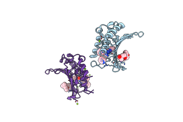
Crystal Structure Of The Atp Binding Domain Of S. Aureus Gyrb Complexed With A Ligand
Organism: Staphylococcus aureus
Method: X-RAY DIFFRACTION Resolution:1.50 Å Release Date: 2015-11-25 Classification: ISOMERASE/ISOMERASE INHIBITOR Ligands: MPD, 57V, MG |
Organism: Staphylococcus aureus
Method: X-RAY DIFFRACTION
Release Date: 2015-11-25
Ligands: MPD, 57V, MG
 |
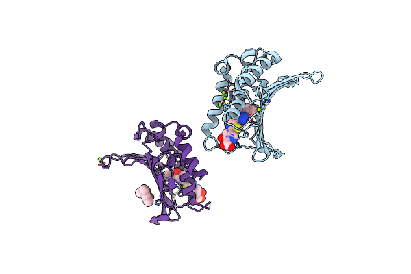
Crystal Structure Of The Atp Binding Domain Of S. Aureus Gyrb Complexed With A Ligand
Organism: Staphylococcus aureus
Method: X-RAY DIFFRACTION Resolution:1.55 Å Release Date: 2015-11-25 Classification: ISOMERASE/ISOMERASE INHIBITOR Ligands: MPD, 57W, MG |
Organism: Staphylococcus aureus
Method: X-RAY DIFFRACTION
Release Date: 2015-11-25
Ligands: MPD, 57W, MG
 |
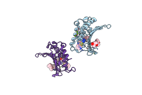
Crystal Structure Of The Atp Binding Domain Of S. Aureus Gyrb Complexed With A Ligand
Organism: Staphylococcus aureus
Method: X-RAY DIFFRACTION Resolution:1.55 Å Release Date: 2015-11-18 Classification: ISOMERASE/ISOMERASE INHIBITOR Ligands: MG, CL, 57Y, MPD |
Organism: Staphylococcus aureus
Method: X-RAY DIFFRACTION
Release Date: 2015-11-18
Ligands: MG, CL, 57Y, MPD
 |
Crystal Structure Of The Atp Binding Domain Of S. Aureus Gyrb Complexed With A Ligand
Organism: Staphylococcus aureus
Method: X-RAY DIFFRACTION Resolution:1.60 Å Release Date: 2015-11-11 Classification: ISOMERASE/ISOMERASE INHIBITOR Ligands: MPD, CL, MG, 57X |
Organism: Staphylococcus aureus
Method: X-RAY DIFFRACTION
Release Date: 2015-11-11
Ligands: MPD, CL, MG, 57X
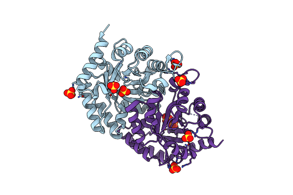 |
Structure Of Leishmania Donovani Omp Decarboxylase
Organism: Leishmania infantum
Method: X-RAY DIFFRACTION Resolution:1.70 Å Release Date: 2011-04-20 Classification: TRANSFERASE, LYASE Ligands: SO4 |
Organism: Leishmania infantum
Method: X-RAY DIFFRACTION
Release Date: 2011-04-20
Ligands: SO4
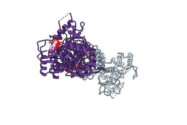 |
Structure Of Leishmania Donovani Ump Synthase
Organism: Leishmania donovani
Method: X-RAY DIFFRACTION Resolution:3.00 Å Release Date: 2011-04-20 Classification: TRANSFERASE, LYASE Ligands: U5P, CL |
Organism: Leishmania donovani
Method: X-RAY DIFFRACTION
Release Date: 2011-04-20
Ligands: U5P, CL
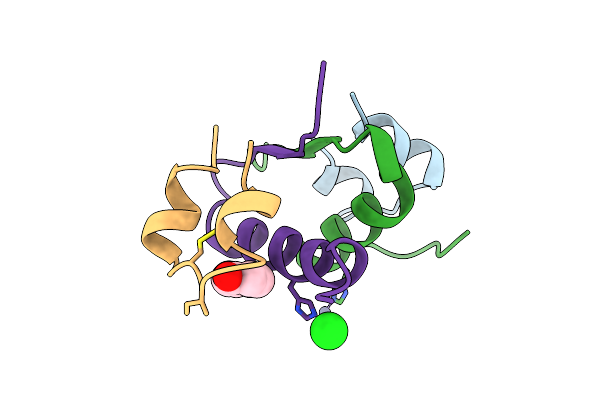 |
Lys(B28)Pro(B29)-Human Insulin
Organism: Homo sapiens
Method: X-RAY DIFFRACTION Resolution:2.30 Å Release Date: 1996-06-20 Classification: HORMONE Ligands: ZN, IPH, CL |
Organism: Homo sapiens
Method: X-RAY DIFFRACTION
Release Date: 1996-06-20
Ligands: ZN, IPH, CL