

Search Count: 41
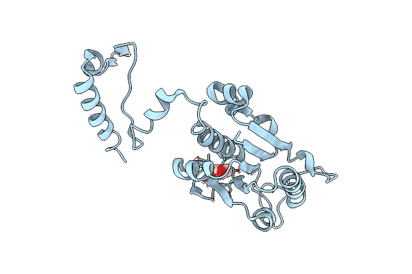 |
Crystal Structure Of The Trna (M1G37) Methyltransferase Apoenzyme From Anaplasma Phagocytophilum
Organism: Anaplasma phagocytophilum
Method: X-RAY DIFFRACTION Resolution:2.90 Å Release Date: 2024-04-17 Classification: TRANSFERASE Ligands: GOL |
Organism: Anaplasma phagocytophilum
Method: X-RAY DIFFRACTION
Release Date: 2024-04-17
Ligands: GOL
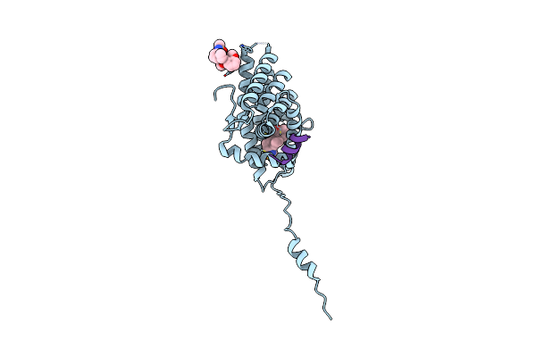 |
Crystal Structure Of The Glucocorticoid Receptor Ligand Binding Domain Bound To A Dibenzoxapine Sulfonamide
Organism: Homo sapiens
Method: X-RAY DIFFRACTION Resolution:2.35 Å Release Date: 2014-01-29 Classification: TRANSCRIPTION Ligands: LSJ |
Organism: Homo sapiens
Method: X-RAY DIFFRACTION
Release Date: 2014-01-29
Ligands: LSJ
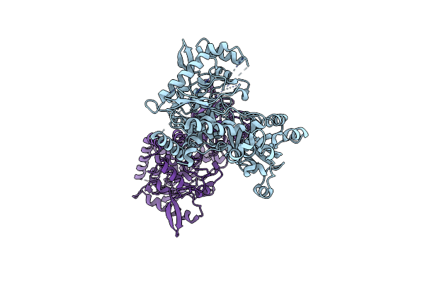 |
Structure Of [Fes]Cluster-Free Apo Form Of 6-Phosphogluconate Dehydratase From Shewanella Oneidensis
Organism: Shewanella oneidensis
Method: X-RAY DIFFRACTION Resolution:2.49 Å Release Date: 2006-05-09 Classification: LYASE |
Organism: Shewanella oneidensis
Method: X-RAY DIFFRACTION
Release Date: 2006-05-09
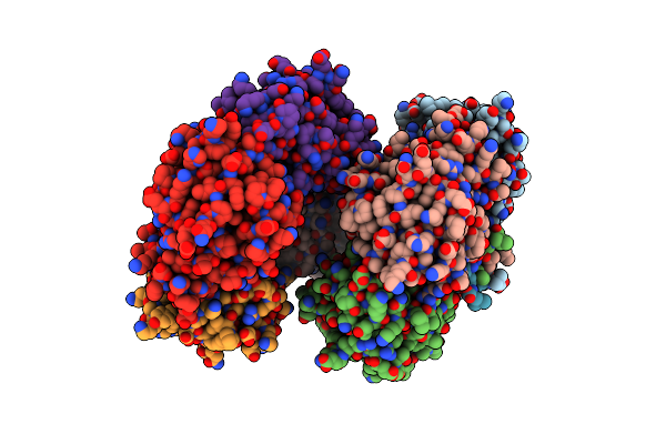 |
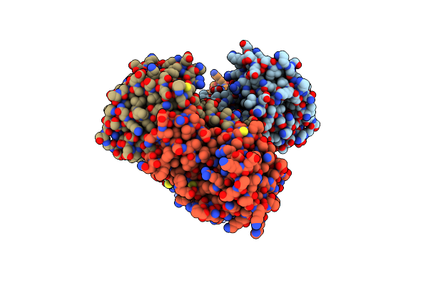
Structure Of Mar1 Ribonuclease From Caenorhabditis Elegans
Organism: Caenorhabditis elegans
Method: X-RAY DIFFRACTION Resolution:2.14 Å Release Date: 2005-09-27 Classification: HYDROLASE |
Organism: Caenorhabditis elegans
Method: X-RAY DIFFRACTION
Release Date: 2005-09-27
 |
Crystal Structure Of The Putative Carbonyl Reductase Sniffer Of Caenorhabditis Elegans
Organism: Caenorhabditis elegans
Method: X-RAY DIFFRACTION Resolution:2.60 Å Release Date: 2005-02-15 Classification: OXIDOREDUCTASE |
Organism: Caenorhabditis elegans
Method: X-RAY DIFFRACTION
Release Date: 2005-02-15
 |
Structural Genomics Of Caenorhabditis Elegans: Glutathione S-Transferase
Organism: Caenorhabditis elegans
Method: X-RAY DIFFRACTION Resolution:3.00 Å Release Date: 2005-02-15 Classification: TRANSFERASE |
Organism: Caenorhabditis elegans
Method: X-RAY DIFFRACTION
Release Date: 2005-02-15
 |
Crystal Structure Of Short-Chain Dehydrogenase/Reductase Of Unknown Function From Caenorhabditis Elegans With Cofactor
Organism: Caenorhabditis elegans
Method: X-RAY DIFFRACTION Resolution:2.10 Å Release Date: 2004-10-12 Classification: UNKNOWN FUNCTION Ligands: NDP |
Organism: Caenorhabditis elegans
Method: X-RAY DIFFRACTION
Release Date: 2004-10-12
Ligands: NDP
 |
Crystal Structure Of Putative Tropinone Reductase-Ii From Caenorhabditis Elegans With Cofactor And Substrate
Organism: Caenorhabditis elegans
Method: X-RAY DIFFRACTION Resolution:2.40 Å Release Date: 2004-09-28 Classification: OXIDOREDUCTASE Ligands: NDP, TNE |
Organism: Caenorhabditis elegans
Method: X-RAY DIFFRACTION
Release Date: 2004-09-28
Ligands: NDP, TNE
 |
Structural Genomics Of Caenorhabditis Elegans: Cap-Gly Domain Of F53F4.3
Organism: Caenorhabditis elegans
Method: X-RAY DIFFRACTION Resolution:1.77 Å Release Date: 2004-07-27 Classification: STRUCTURAL GENOMICS, UNKNOWN FUNCTION Ligands: SO4 |
Organism: Caenorhabditis elegans
Method: X-RAY DIFFRACTION
Release Date: 2004-07-27
Ligands: SO4
 |
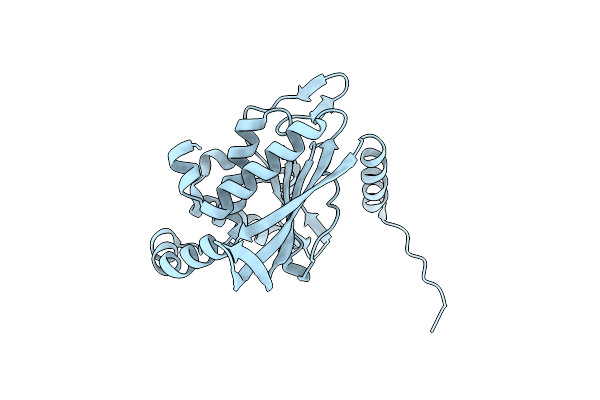
Crystal Structure Of Glucose Dehydrogenase Of Caenorhabditis Elegans In The Apo-Form
Organism: Caenorhabditis elegans
Method: X-RAY DIFFRACTION Resolution:2.10 Å Release Date: 2004-03-23 Classification: OXIDOREDUCTASE |
Organism: Caenorhabditis elegans
Method: X-RAY DIFFRACTION
Release Date: 2004-03-23
 |
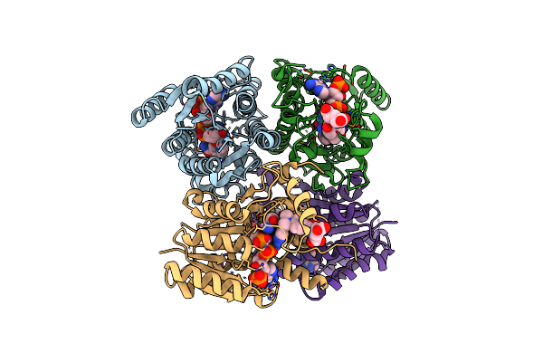
Improved Structural Model For The Catalytic Domain Of E.Coli Dihydrolipoamide Succinyltransferase
Organism: Escherichia coli
Method: X-RAY DIFFRACTION Resolution:2.20 Å Release Date: 2004-03-02 Classification: TRANSFERASE |
Organism: Escherichia coli
Method: X-RAY DIFFRACTION
Release Date: 2004-03-02
 |
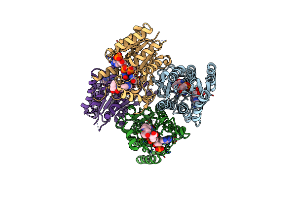
Crystal Structure Of Inhibitor Complex Of Putative Pteridine Reductase 2 (Ptr2) From Trypanosoma Cruzi
Organism: Trypanosoma cruzi
Method: X-RAY DIFFRACTION Resolution:2.30 Å Release Date: 2003-10-14 Classification: OXIDOREDUCTASE Ligands: NDP, MTX |
Organism: Trypanosoma cruzi
Method: X-RAY DIFFRACTION
Release Date: 2003-10-14
Ligands: NDP, MTX
 |
Crystal Structure Of Substrate Complex Of Putative Pteridine Reductase 2 (Ptr2) From Trypanosoma Cruzi
Organism: Trypanosoma cruzi
Method: X-RAY DIFFRACTION Resolution:2.20 Å Release Date: 2003-10-14 Classification: OXIDOREDUCTASE Ligands: NAP, DHF |
Organism: Trypanosoma cruzi
Method: X-RAY DIFFRACTION
Release Date: 2003-10-14
Ligands: NAP, DHF
 |
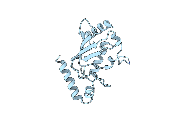
Crystal Structures Of Two Ubc (E2) Enzymes Of The Ubiquitin-Conjugating System In Caenorhabditis Elegans
Organism: Caenorhabditis elegans
Method: X-RAY DIFFRACTION Resolution:2.90 Å Release Date: 2003-08-05 Classification: LIGASE |
Organism: Caenorhabditis elegans
Method: X-RAY DIFFRACTION
Release Date: 2003-08-05
 |
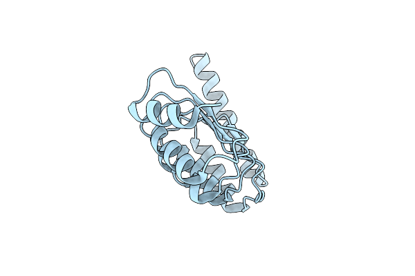
Crystal Structures Of Two Ubc (E2) Enzymes Of The Ubiquitin-Conjugating System In Caenorhabditis Elegans
Organism: Caenorhabditis elegans
Method: X-RAY DIFFRACTION Resolution:2.52 Å Release Date: 2003-07-22 Classification: LIGASE |
Organism: Caenorhabditis elegans
Method: X-RAY DIFFRACTION
Release Date: 2003-07-22
 |
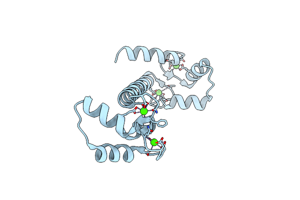
Structural Genomics Of Caenorhabditis Elegans: Tropomodulin C-Terminal Domain
Organism: Caenorhabditis elegans
Method: X-RAY DIFFRACTION Resolution:1.80 Å Release Date: 2003-06-10 Classification: PROTEIN BINDING |
Organism: Caenorhabditis elegans
Method: X-RAY DIFFRACTION
Release Date: 2003-06-10
 |
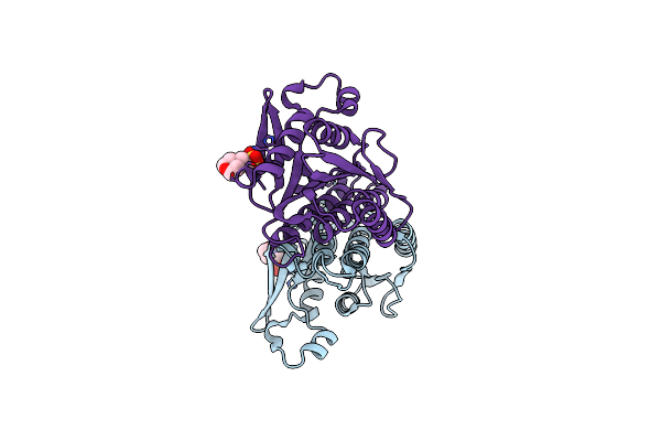
Structural Genomics Of Caenorhabditis Elegans : Calmodulin
Organism: Caenorhabditis elegans
Method: X-RAY DIFFRACTION Resolution:2.11 Å Release Date: 2003-03-25 Classification: METAL BINDING PROTEIN Ligands: CA |
Organism: Caenorhabditis elegans
Method: X-RAY DIFFRACTION
Release Date: 2003-03-25
Ligands: CA
 |
Structural Genomics Of Caenorhabditis Elegans : Dihydropteridine Reductase
Organism: Caenorhabditis elegans
Method: X-RAY DIFFRACTION Resolution:1.65 Å Release Date: 2003-03-18 Classification: OXIDOREDUCTASE Ligands: MES |
Organism: Caenorhabditis elegans
Method: X-RAY DIFFRACTION
Release Date: 2003-03-18
Ligands: MES
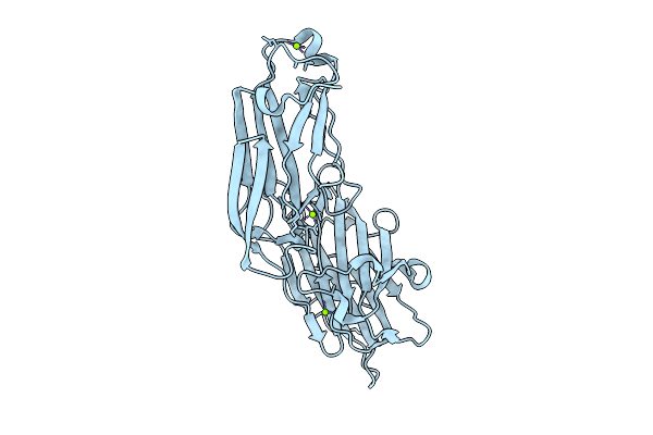 |
Clumping Factor A From Staphylococcus Aureus
Organism: Staphylococcus aureus
Method: X-RAY DIFFRACTION Resolution:1.90 Å Release Date: 2003-03-04 Classification: CELL ADHESION Ligands: MG |
Organism: Staphylococcus aureus
Method: X-RAY DIFFRACTION
Release Date: 2003-03-04
Ligands: MG
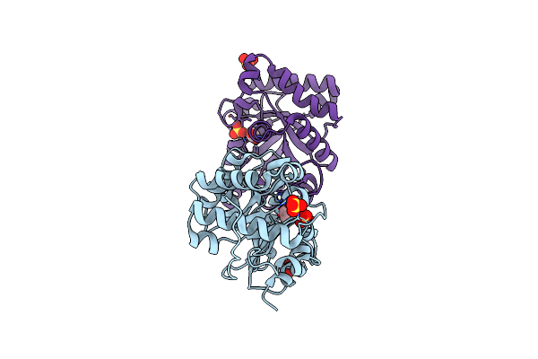 |
Structural Genomics Of Caenorhabditis Elegans: Triose Phosphate Isomerase
Organism: Caenorhabditis elegans
Method: X-RAY DIFFRACTION Resolution:1.70 Å Release Date: 2002-09-13 Classification: ISOMERASE Ligands: ACT, SO4 |
Organism: Caenorhabditis elegans
Method: X-RAY DIFFRACTION
Release Date: 2002-09-13
Ligands: ACT, SO4