

Search Count: 80
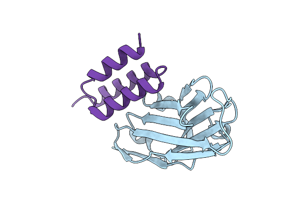 |
Crystal Structure Of S. Aureus Protein A Bound To A Camelid Single-Domain Antibody
Organism: Camelidae, Staphylococcus aureus subsp. aureus nctc 8325
Method: X-RAY DIFFRACTION Resolution:2.00 Å Release Date: 2025-11-12 Classification: IMMUNE SYSTEM |
Organism: Camelidae, Staphylococcus aureus subsp. aureus nctc 8325
Method: X-RAY DIFFRACTION
Release Date: 2025-11-12
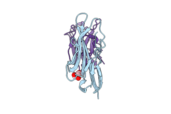 |
Structure Of Single-Domain Antibody Vhh_H4 In Complex With Human Vsig4
Organism: Homo sapiens, Camelidae
Method: X-RAY DIFFRACTION Resolution:1.94 Å Release Date: 2025-10-29 Classification: IMMUNE SYSTEM Ligands: GOL |
Organism: Homo sapiens, Camelidae
Method: X-RAY DIFFRACTION
Release Date: 2025-10-29
Ligands: GOL
 |
Structure Of Single-Domain Antibody Vhh_H5 In Complex With Human Vsig4
Organism: Homo sapiens, Camelidae
Method: X-RAY DIFFRACTION Resolution:1.97 Å Release Date: 2025-10-29 Classification: IMMUNE SYSTEM |
Organism: Homo sapiens, Camelidae
Method: X-RAY DIFFRACTION
Release Date: 2025-10-29
 |
Structure Of The Single-Domain Antibody Vhh_H1 In Complex With Human Vsig4
Organism: Homo sapiens, Camelidae
Method: X-RAY DIFFRACTION Resolution:1.05 Å Release Date: 2025-10-29 Classification: IMMUNE SYSTEM Ligands: SO4 |
Organism: Homo sapiens, Camelidae
Method: X-RAY DIFFRACTION
Release Date: 2025-10-29
Ligands: SO4
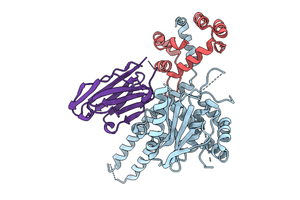 |
Crystal Structure Of The Uch37 Rpn13 Deubad Complex Bound To An Inhibitory Nanobody In The Canonical Ubiquitin Binding Site
Organism: Homo sapiens, Camelidae mixed library
Method: X-RAY DIFFRACTION Resolution:2.41 Å Release Date: 2025-09-03 Classification: HYDROLASE Ligands: CL |
Organism: Homo sapiens, Camelidae mixed library
Method: X-RAY DIFFRACTION
Release Date: 2025-09-03
Ligands: CL
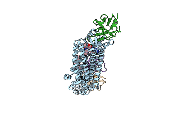 |
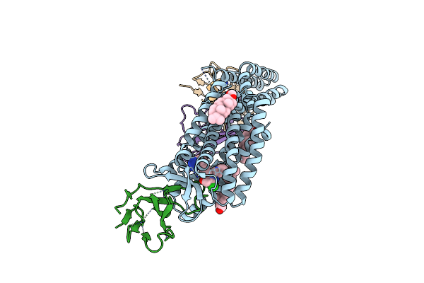
Structure Of Nanobody At209 In Complex With The Olmesartan-Bound Angiotensin Ii Type I Receptor (At1R)
Organism: Camelidae, Homo sapiens, Escherichia coli, Synthetic construct
Method: ELECTRON MICROSCOPY Release Date: 2025-03-05 Classification: MEMBRANE PROTEIN Ligands: OLM, CLR |
Organism: Camelidae, Homo sapiens, Escherichia coli, Synthetic construct
Method: ELECTRON MICROSCOPY
Release Date: 2025-03-05
Ligands: OLM, CLR
 |
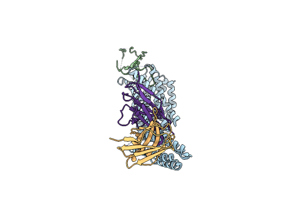
Structure Of Nanobody At206 In Complex With The Losartan-Bound Angiotensin Ii Type I Receptor (At1R)
Organism: Camelidae, Homo sapiens, Escherichia coli, Synthetic construct
Method: ELECTRON MICROSCOPY Release Date: 2025-03-05 Classification: MEMBRANE PROTEIN Ligands: LSN, Y01, CLR |
Organism: Camelidae, Homo sapiens, Escherichia coli, Synthetic construct
Method: ELECTRON MICROSCOPY
Release Date: 2025-03-05
Ligands: LSN, Y01, CLR
 |
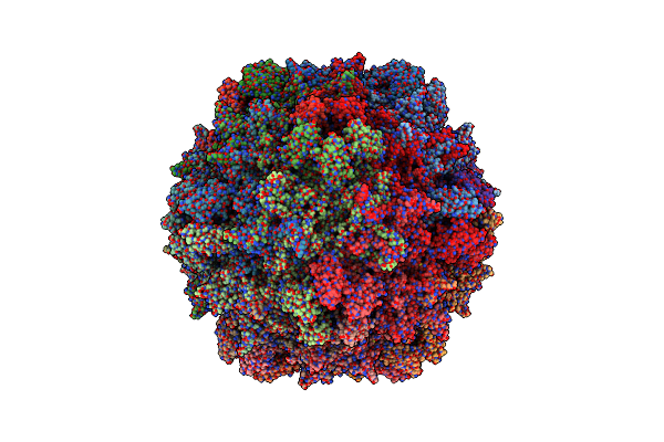
Structure Of Nanobody At206 In Complex With The Angiotensin Ii Type I Receptor (At1R)
Organism: Camelidae, Homo sapiens, Escherichia coli, Synthetic construct
Method: ELECTRON MICROSCOPY Release Date: 2025-03-05 Classification: MEMBRANE PROTEIN |
Organism: Camelidae, Homo sapiens, Escherichia coli, Synthetic construct
Method: ELECTRON MICROSCOPY
Release Date: 2025-03-05
 |
Aav8 In Complex With The Aavx Affinity Ligand
Organism: Adeno-associated virus 8, Camelidae
Method: ELECTRON MICROSCOPY Release Date: 2024-11-27 Classification: VIRUS |
Organism: Adeno-associated virus 8, Camelidae
Method: ELECTRON MICROSCOPY
Release Date: 2024-11-27
 |
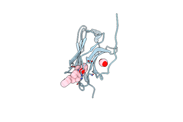
X-Ray Crystal Structure Of Jgfn4 Complexed With Fentanyl
Organism: Camelidae
Method: X-RAY DIFFRACTION Resolution:1.68 Å Release Date: 2024-08-07 Classification: ANTITOXIN Ligands: 7V7, EDO |
Organism: Camelidae
Method: X-RAY DIFFRACTION
Release Date: 2024-08-07
Ligands: 7V7, EDO
 |
X-Ray Crystal Structure Of Jgfn4 Complex With Fentanyl
Organism: Camelidae
Method: X-RAY DIFFRACTION Resolution:1.55 Å Release Date: 2024-08-07 Classification: ANTITOXIN Ligands: 7V7, EDO |
Organism: Camelidae
Method: X-RAY DIFFRACTION
Release Date: 2024-08-07
Ligands: 7V7, EDO
 |
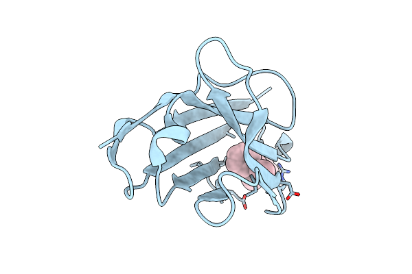
X-Ray Crystal Structure Of Nanobody Jgfn4
Organism: Camelidae
Method: X-RAY DIFFRACTION Resolution:1.76 Å Release Date: 2024-08-07 Classification: ANTITOXIN |
Organism: Camelidae
Method: X-RAY DIFFRACTION
Release Date: 2024-08-07
 |
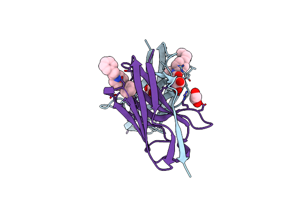
X-Ray Crystal Structure Of Jgfn4 N76D Complexed With Fentanyl In Monomer Form
Organism: Camelidae
Method: X-RAY DIFFRACTION Resolution:1.60 Å Release Date: 2024-08-07 Classification: ANTITOXIN Ligands: 7V7 |
Organism: Camelidae
Method: X-RAY DIFFRACTION
Release Date: 2024-08-07
Ligands: 7V7
 |
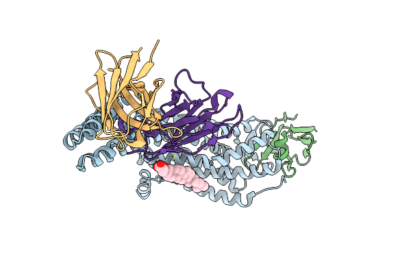
X-Ray Crystal Structure Of Jgfn4 N76D Complexed With Fentanyl In Dimer Form
Organism: Camelidae
Method: X-RAY DIFFRACTION Resolution:1.85 Å Release Date: 2024-08-07 Classification: ANTITOXIN Ligands: 7V7, PGE, EDO |
Organism: Camelidae
Method: X-RAY DIFFRACTION
Release Date: 2024-08-07
Ligands: 7V7, PGE, EDO
 |
Structure Of At118-H Nanobody Antagonist In Complex With The Angiotensin Ii Type I Receptor
Organism: Camelidae, Homo sapiens, Escherichia coli, Synthetic construct
Method: ELECTRON MICROSCOPY Release Date: 2024-05-22 Classification: SIGNALING PROTEIN/IMMUNE SYSTEM Ligands: Y01 |
Organism: Camelidae, Homo sapiens, Escherichia coli, Synthetic construct
Method: ELECTRON MICROSCOPY
Release Date: 2024-05-22
Ligands: Y01
 |
Human Igd Fab In Complex With An Orthosteric Inhibitor Of Phl P 7
Organism: Homo sapiens, Camelidae mixed library
Method: X-RAY DIFFRACTION Resolution:2.10 Å Release Date: 2023-12-06 Classification: IMMUNE SYSTEM Ligands: EDO, ACT, NA |
Organism: Homo sapiens, Camelidae mixed library
Method: X-RAY DIFFRACTION
Release Date: 2023-12-06
Ligands: EDO, ACT, NA
 |
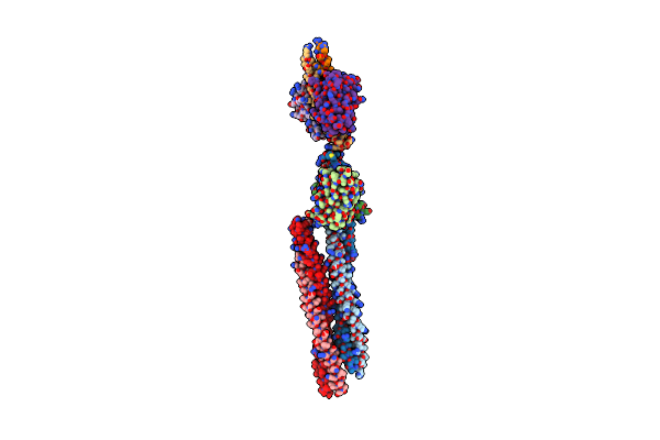
Crystal Structure Of A Nanobody Bound To The V5 Peptide.
Organism: Camelidae, Simian virus 12
Method: X-RAY DIFFRACTION Resolution:2.01 Å Release Date: 2023-11-22 Classification: BIOSYNTHETIC PROTEIN |
Organism: Camelidae, Simian virus 12
Method: X-RAY DIFFRACTION
Release Date: 2023-11-22
 |
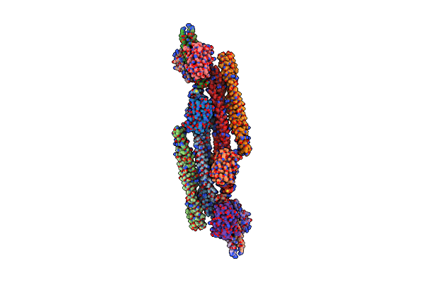
Cryo-Em Structure Of Crescentin Filaments (Stutter Mutant, C1 Symmetry And Small Box)
Organism: Caulobacter vibrioides, Camelidae
Method: ELECTRON MICROSCOPY Release Date: 2023-08-16 Classification: STRUCTURAL PROTEIN |
Organism: Caulobacter vibrioides, Camelidae
Method: ELECTRON MICROSCOPY
Release Date: 2023-08-16
 |
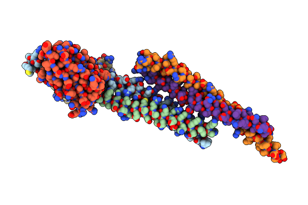
Cryo-Em Structure Of Crescentin Filaments (Stutter Mutant, C2, Symmetry And Small Box)
Organism: Caulobacter vibrioides, Camelidae
Method: ELECTRON MICROSCOPY Release Date: 2023-08-16 Classification: STRUCTURAL PROTEIN |
Organism: Caulobacter vibrioides, Camelidae
Method: ELECTRON MICROSCOPY
Release Date: 2023-08-16
 |
Cryo-Em Structure Of Crescentin Filaments (Wildtype, C1 Symmetry And Small Box)
Organism: Caulobacter vibrioides, Camelidae
Method: ELECTRON MICROSCOPY Release Date: 2023-08-16 Classification: STRUCTURAL PROTEIN |
Organism: Caulobacter vibrioides, Camelidae
Method: ELECTRON MICROSCOPY
Release Date: 2023-08-16