

Search Count: 1,184
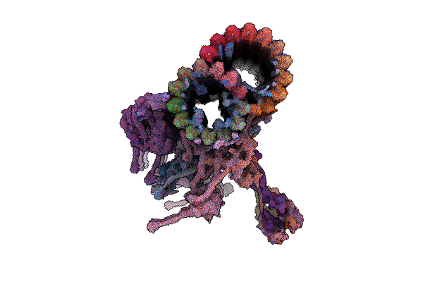 |
96Nm Repeat Of Human Respiratory Doublet Microtubule And Associated Axonemal Complexes
Organism: Homo sapiens
Method: ELECTRON MICROSCOPY Release Date: 2023-07-26 Classification: STRUCTURAL PROTEIN Ligands: GTP, MG, GDP |
Organism: Homo sapiens
Method: ELECTRON MICROSCOPY
Release Date: 2023-07-26
Ligands: GTP, MG, GDP
 |
96-Nm Repeat Of Axonemal Doublet Microtubules From Bovine Sperm
Organism: Bos taurus
Method: ELECTRON MICROSCOPY Release Date: 2025-03-26 Classification: STRUCTURAL PROTEIN Ligands: GTP, MG, GDP |
Organism: Bos taurus
Method: ELECTRON MICROSCOPY
Release Date: 2025-03-26
Ligands: GTP, MG, GDP
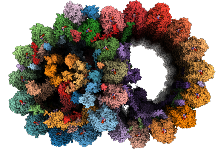 |
Cryo-Em Structure Of The 48-Nm Repeat Doublet Microtubule From Mouse Sperm
Organism: Mus musculus
Method: ELECTRON MICROSCOPY Release Date: 2026-05-27 Classification: STRUCTURAL PROTEIN Ligands: GTP, MG, GDP, ATP |
Organism: Mus musculus
Method: ELECTRON MICROSCOPY
Release Date: 2026-05-27
Ligands: GTP, MG, GDP, ATP
 |
Cryo-Em Structure Of The 48-Nm Repeat Doublet Microtubule From Mouse Sperm
Organism: Mus musculus
Method: ELECTRON MICROSCOPY Release Date: 2023-07-05 Classification: STRUCTURAL PROTEIN Ligands: GTP, MG, GDP |
Organism: Mus musculus
Method: ELECTRON MICROSCOPY
Release Date: 2023-07-05
Ligands: GTP, MG, GDP
 |
In Situ Structure Of Axonemal Doublet Microtubules In Mouse Sperm With 48-Nm Repeat
Organism: Mus musculus
Method: ELECTRON MICROSCOPY Release Date: 2023-10-11 Classification: STRUCTURAL PROTEIN Ligands: GTP |
Organism: Mus musculus
Method: ELECTRON MICROSCOPY
Release Date: 2023-10-11
Ligands: GTP
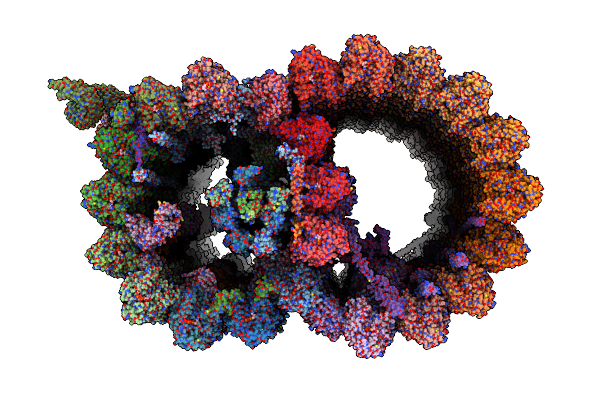 |
Structure Of The 48-Nm Repeat Doublet Microtubule From Bovine Tracheal Cilia
Organism: Bos taurus
Method: ELECTRON MICROSCOPY Release Date: 2021-10-27 Classification: STRUCTURAL PROTEIN Ligands: GTP, MG, GDP |
Organism: Bos taurus
Method: ELECTRON MICROSCOPY
Release Date: 2021-10-27
Ligands: GTP, MG, GDP
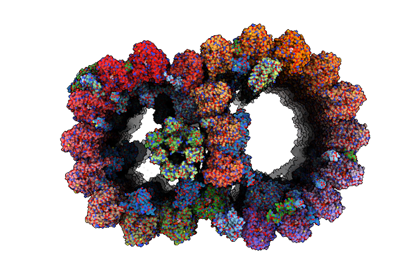 |
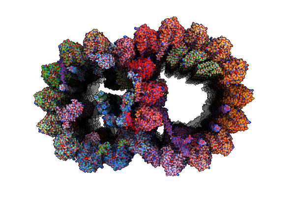
Atomic Model Of Bovine Fallopian Tube Cilia Doublet Microtubule (48-Nm Periodicity)
Organism: Bos taurus
Method: ELECTRON MICROSCOPY Release Date: 2024-10-30 Classification: STRUCTURAL PROTEIN Ligands: GDP, MG, GTP |
Organism: Bos taurus
Method: ELECTRON MICROSCOPY
Release Date: 2024-10-30
Ligands: GDP, MG, GTP
 |
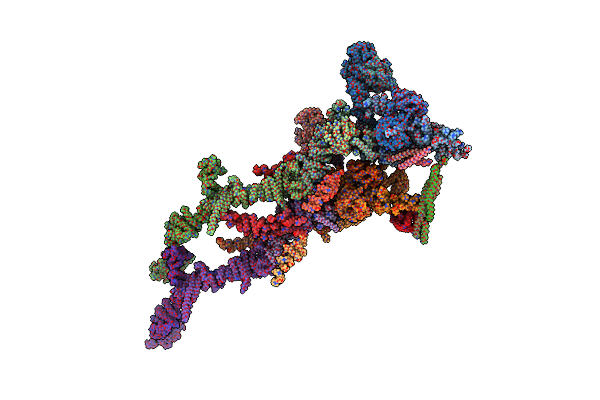
48-Nm Repeat Of The Human Respiratory Doublet Microtubule
Organism: Homo sapiens
Method: ELECTRON MICROSCOPY Release Date: 2022-10-05 Classification: STRUCTURAL PROTEIN Ligands: GTP, MG, GDP |
Organism: Homo sapiens
Method: ELECTRON MICROSCOPY
Release Date: 2022-10-05
Ligands: GTP, MG, GDP
 |
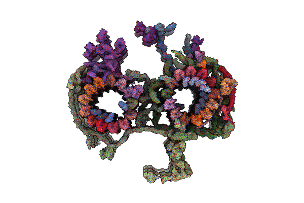
Structure Of Xenopus Laevis Cytoplasmic Ring Subunit.
Organism: Xenopus laevis
Method: ELECTRON MICROSCOPY Release Date: 2021-07-21 Classification: STRUCTURAL PROTEIN |
Organism: Xenopus laevis
Method: ELECTRON MICROSCOPY
Release Date: 2021-07-21
 |
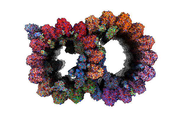
In Situ Structure Of The Central Apparatus In Mouse Sperm Axoneme.
Organism: Mus musculus
Method: ELECTRON MICROSCOPY Release Date: 2025-06-04 Classification: PROTEIN TRANSPORT |
Organism: Mus musculus
Method: ELECTRON MICROSCOPY
Release Date: 2025-06-04
 |
Atomic Model Of Porcine Brain Ventricles Cilia Doublet Microtubule (48-Nm Periodicity)
Organism: Sus scrofa
Method: ELECTRON MICROSCOPY Release Date: 2024-10-23 Classification: STRUCTURAL PROTEIN Ligands: GTP, GDP |
Organism: Sus scrofa
Method: ELECTRON MICROSCOPY
Release Date: 2024-10-23
Ligands: GTP, GDP
 |
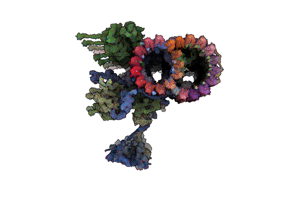
96-Nm Repeat Of The Leishmania Tarentolae Doublet Microtubule
Organism: Leishmania tarentolae, Leishmania mexicana
Method: ELECTRON MICROSCOPY Release Date: 2025-12-17 Classification: STRUCTURAL PROTEIN Ligands: GDP, MG, GTP, ZN, CA, ATP |
Organism: Leishmania tarentolae, Leishmania mexicana
Method: ELECTRON MICROSCOPY
Release Date: 2025-12-17
Ligands: GDP, MG, GTP, ZN, CA, ATP
 |
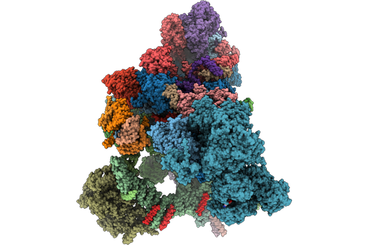
Cryo-Em Structure Of 96 Nm Repeat Of Microtubule Doublet From T. Brucei Flagellum
Organism: Trypanosoma brucei brucei treu927
Method: ELECTRON MICROSCOPY Release Date: 2025-03-12 Classification: MOTOR PROTEIN Ligands: ZN, ATP, ADP, GDP, MG, GTP |
Organism: Trypanosoma brucei brucei treu927
Method: ELECTRON MICROSCOPY
Release Date: 2025-03-12
Ligands: ZN, ATP, ADP, GDP, MG, GTP
 |
Human Minor Spliceosome Branching-Completed C Complex (After Step-I)
Method: ELECTRON MICROSCOPY
Release Date: 2026-08-12 Classification: SPLICING Ligands: MG, K, IHP, GTP, ADP, ZN, ATP |
Method: ELECTRON MICROSCOPY
Release Date: 2026-08-12
Ligands: MG, K, IHP, GTP, ADP, ZN, ATP
 |
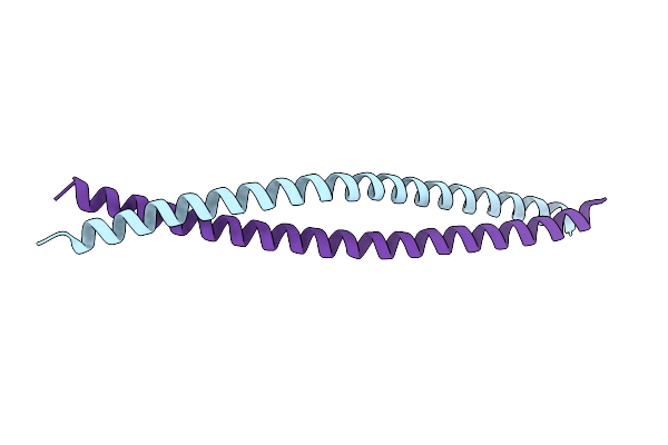
High-Resolution Solution Nmr Structure Of The Trimeric Coiled-Coil Domain Of Chicken Cartilage Matrix Protein, 20 Structures
|
Organism: Gallus gallus
Method: SOLUTION NMR
Release Date: 1998-02-11
 |
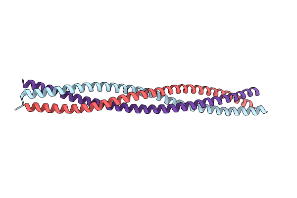
Coiled-Coil Structure Of The Rhoa-Binding Domain In Rho-Kinase
Organism: Bos taurus
Method: X-RAY DIFFRACTION Resolution:1.80 Å Release Date: 2003-10-21 Classification: TRANSFERASE |
Organism: Bos taurus
Method: X-RAY DIFFRACTION
Release Date: 2003-10-21
 |
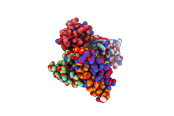
Crystal Structure Of The Zebrafish Cavin4A Hr1 Domain
Organism: Danio rerio
Method: X-RAY DIFFRACTION Resolution:1.70 Å Release Date: 2015-03-18 Classification: SIGNALING PROTEIN Ligands: CL |
Organism: Danio rerio
Method: X-RAY DIFFRACTION
Release Date: 2015-03-18
Ligands: CL
 |
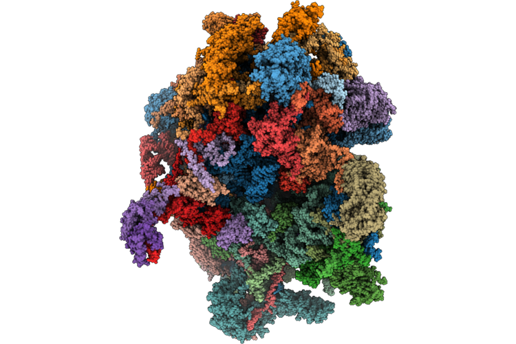
Crystal Structure Of The P5P6 Coiled-Coil In Complex With Nanobody Nb34.
Organism: Lama glama, Synthetic construct
Method: X-RAY DIFFRACTION Resolution:2.16 Å Release Date: 2021-05-05 Classification: DE NOVO PROTEIN Ligands: SO4, EDO |
Organism: Lama glama, Synthetic construct
Method: X-RAY DIFFRACTION
Release Date: 2021-05-05
Ligands: SO4, EDO
 |
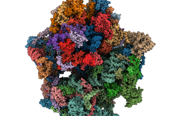
Human Mitoribosome Bound To Taco1 (Translational Activator Of Cox1), Mrna, A/A-, P/P- And (Partial) E/E-Trnas
Organism: Homo sapiens
Method: ELECTRON MICROSCOPY Resolution:2.64 Å Release Date: 2026-07-15 Classification: RIBOSOME Ligands: NAD, MG, K, 5F0, ZN, FES, ATP, GDP, SPD, VOL |
Organism: Homo sapiens
Method: ELECTRON MICROSCOPY
Release Date: 2026-07-15
Ligands: NAD, MG, K, 5F0, ZN, FES, ATP, GDP, SPD, VOL
 |
In Situ Structure Of The Human Mitoribosome In The P State
Organism: Homo sapiens
Method: ELECTRON MICROSCOPY Release Date: 2026-02-18 Classification: RIBOSOME Ligands: ZN, K, SPD, PUT, MG, VAL, FES, NAD, SPM, ATP, GDP |
Organism: Homo sapiens
Method: ELECTRON MICROSCOPY
Release Date: 2026-02-18
Ligands: ZN, K, SPD, PUT, MG, VAL, FES, NAD, SPM, ATP, GDP