
Search Count: 2,736
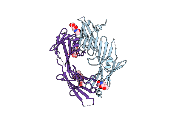 |
Crystal Structure Of The Zp-N1 And Zp-N2 Domains Of Human Zp2 (Hzp2-N1N2)
Organism: Homo sapiens
Method: X-RAY DIFFRACTION Resolution:3.20 Å Release Date: 2024-03-13 Classification: CELL ADHESION Ligands: NAG |
Organism: Homo sapiens
Method: X-RAY DIFFRACTION
Release Date: 2024-03-13
Ligands: NAG
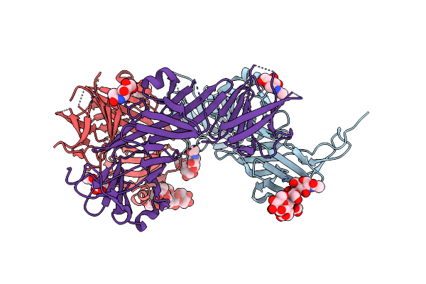 |
Crystal Structure Of The Complete N-Terminal Region Of Human Zp2 (Hzp2-N1N2N3)
Organism: Homo sapiens
Method: X-RAY DIFFRACTION Resolution:2.70 Å Release Date: 2024-03-13 Classification: CELL ADHESION Ligands: NAG |
Organism: Homo sapiens
Method: X-RAY DIFFRACTION
Release Date: 2024-03-13
Ligands: NAG
 |
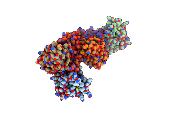
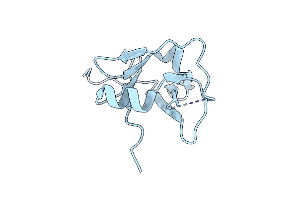
Solution Structure Of The Hyaluronan Binding Domain Of Human Cd44
|
Organism: Homo sapiens
Method: SOLUTION NMR
Release Date: 2004-03-16
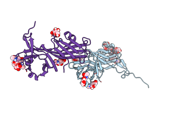 |
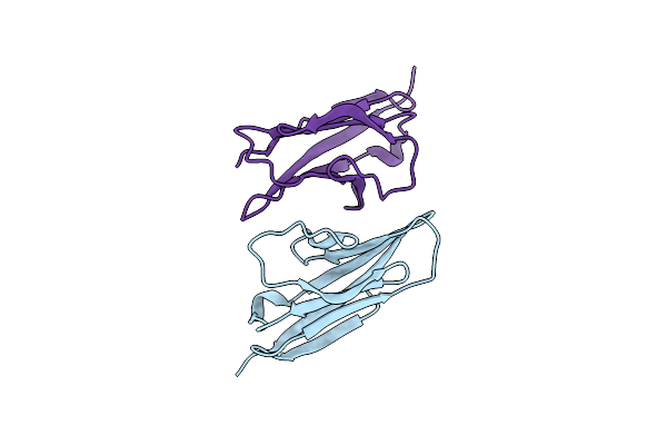
Crystal Structure Of The Zp-N2 And Zp-N3 Domains Of Mouse Zp2 (Mzp2-N2N3)
Organism: Mus musculus
Method: X-RAY DIFFRACTION Resolution:1.90 Å Release Date: 2024-03-13 Classification: CELL ADHESION Ligands: NAG |
Organism: Mus musculus
Method: X-RAY DIFFRACTION
Release Date: 2024-03-13
Ligands: NAG
 |
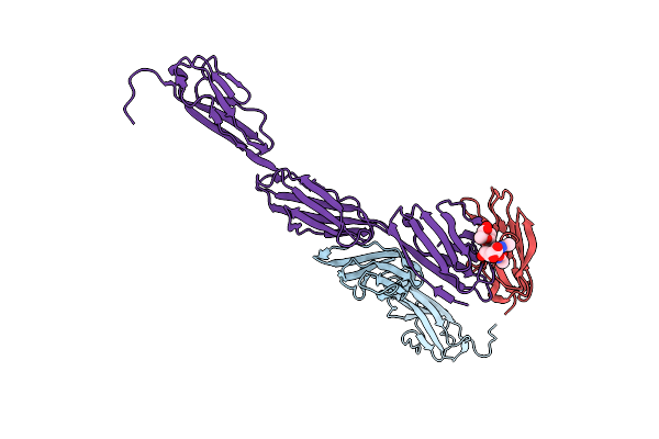
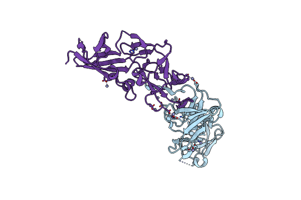
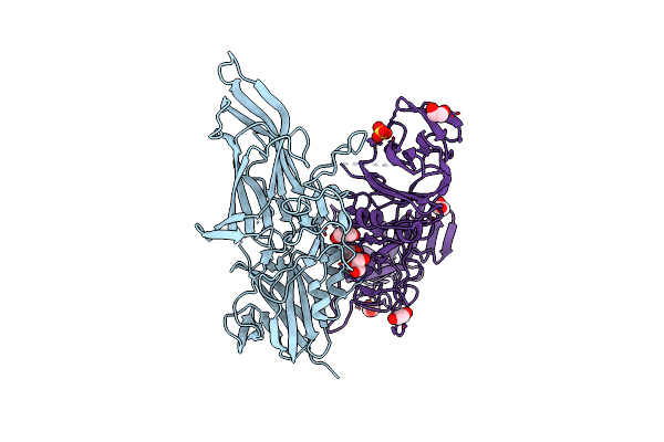
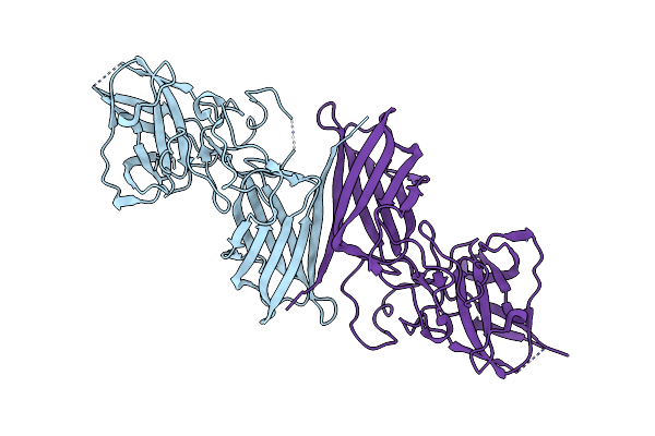
Structure Of Fertilization-Blocking Monoclonal Antibody Ie-3 Vhvl Bound To The Zp-N1 Domain Of Mouse Zp2 (Crystal Form Ii)
Organism: Mus musculus, Rattus norvegicus
Method: X-RAY DIFFRACTION Resolution:2.02 Å Release Date: 2025-03-12 Classification: CELL ADHESION |
Organism: Mus musculus, Rattus norvegicus
Method: X-RAY DIFFRACTION
Release Date: 2025-03-12
 |
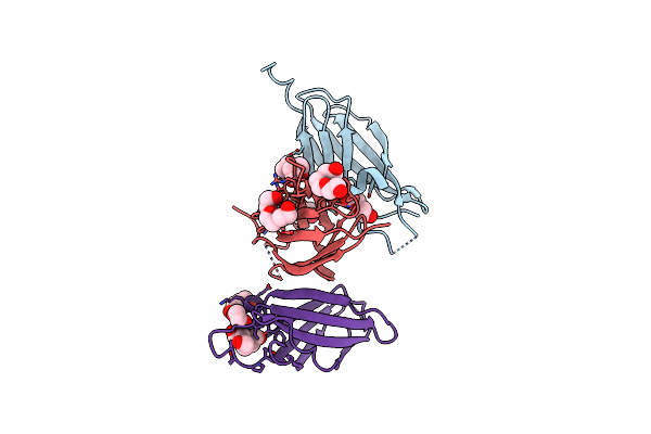
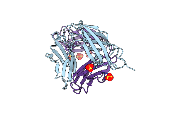
Structure Of Fertilization-Blocking Monoclonal Antibody Ie-3 Vhvl Bound To The Zp-N1 Domain Of Mouse Zp2 (Crystal Form I)
Organism: Mus musculus, Rattus norvegicus
Method: X-RAY DIFFRACTION Resolution:1.53 Å Release Date: 2025-03-12 Classification: CELL ADHESION |
Organism: Mus musculus, Rattus norvegicus
Method: X-RAY DIFFRACTION
Release Date: 2025-03-12
 |
Crystal Structure Of Mouse Nectin-Like Molecule 4 (Mnecl-4) Full Ectodomain In Complex With Mouse Nectin-Like Molecule 1 (Mnecl-1) Ig1 Domain, 3.3A
Organism: Mus musculus
Method: X-RAY DIFFRACTION Resolution:3.29 Å Release Date: 2019-01-30 Classification: CELL ADHESION |
Organism: Mus musculus
Method: X-RAY DIFFRACTION
Release Date: 2019-01-30
 |
Crystal Structure Of Human Opioid Binding Protein/Cell Adhesion Molecule Like (Opcml)
Organism: Homo sapiens
Method: X-RAY DIFFRACTION Resolution:2.65 Å Release Date: 2018-03-21 Classification: CELL ADHESION Ligands: NAG |
Organism: Homo sapiens
Method: X-RAY DIFFRACTION
Release Date: 2018-03-21
Ligands: NAG
 |
Complex Structure Of Necl-2 And Crtam
Organism: Homo sapiens
Method: X-RAY DIFFRACTION Resolution:1.70 Å Release Date: 2013-08-07 Classification: CELL ADHESION |
Organism: Homo sapiens
Method: X-RAY DIFFRACTION
Release Date: 2013-08-07
 |
Crystal Structure Of The First Fibronectin Type Iii Domain Of Neural Cell Adhesion Molecule Splicing Isoform From Human Muscle Culture Lambda-4.4
Organism: Homo sapiens
Method: X-RAY DIFFRACTION Resolution:1.95 Å Release Date: 2007-06-05 Classification: CELL ADHESION Ligands: PEG, PGE, BTB, EDO |
Organism: Homo sapiens
Method: X-RAY DIFFRACTION
Release Date: 2007-06-05
Ligands: PEG, PGE, BTB, EDO
 |
Crystal Structure Of The Zp-N1 Domain Of Mouse Sperm Receptor Zp2 At 0.95 A Resolution
Organism: Mus musculus
Method: X-RAY DIFFRACTION Resolution:0.95 Å Release Date: 2017-06-14 Classification: CELL ADHESION |
Organism: Mus musculus
Method: X-RAY DIFFRACTION
Release Date: 2017-06-14
 |
Crystal Structure Of A Putative Cell Adhesion Protein (Bt0320) From Bacteroides Thetaiotaomicron Vpi-5482 At 2.37 A Resolution
Organism: Bacteroides thetaiotaomicron
Method: X-RAY DIFFRACTION Resolution:2.37 Å Release Date: 2012-02-15 Classification: CELL ADHESION Ligands: ZN, CL |
Organism: Bacteroides thetaiotaomicron
Method: X-RAY DIFFRACTION
Release Date: 2012-02-15
Ligands: ZN, CL
 |
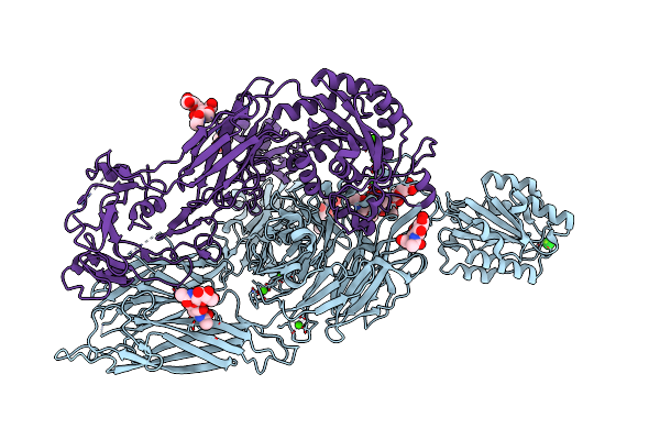
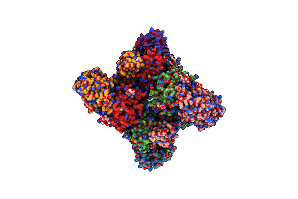
Structure Of Integrin Alphaxbeta2 Ectodomain
Organism: Homo sapiens
Method: X-RAY DIFFRACTION Resolution:3.50 Å Release Date: 2010-01-12 Classification: CELL ADHESION Ligands: MAN, NAG, CA, MG |
Organism: Homo sapiens
Method: X-RAY DIFFRACTION
Release Date: 2010-01-12
Ligands: MAN, NAG, CA, MG
 |
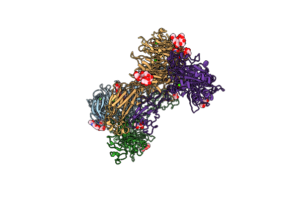
An Internal Ligand-Bound, Metastable State Of A Leukocyte Integrin, Axb2
Organism: Homo sapiens
Method: X-RAY DIFFRACTION Resolution:2.90 Å Release Date: 2014-01-15 Classification: CELL ADHESION Ligands: CA, MG, CL |
Organism: Homo sapiens
Method: X-RAY DIFFRACTION
Release Date: 2014-01-15
Ligands: CA, MG, CL
 |
An Internal Ligand-Bound, Metastable State Of A Leukocyte Integrin, Axb2
Organism: Homo sapiens
Method: X-RAY DIFFRACTION Resolution:2.75 Å Release Date: 2014-01-15 Classification: CELL ADHESION Ligands: CA, MG, NAG, CL, NA |
Organism: Homo sapiens
Method: X-RAY DIFFRACTION
Release Date: 2014-01-15
Ligands: CA, MG, NAG, CL, NA
 |
Structure Of Integrin Alphax Beta2
Organism: Homo sapiens
Method: X-RAY DIFFRACTION Resolution:3.70 Å Release Date: 2010-01-12 Classification: CELL ADHESION Ligands: NAG, CA, MAN |
Organism: Homo sapiens
Method: X-RAY DIFFRACTION
Release Date: 2010-01-12
Ligands: NAG, CA, MAN
 |
Structure Of Integrin Alphax Beta2 Ectodomain
Organism: Homo sapiens
Method: X-RAY DIFFRACTION Resolution:3.95 Å Release Date: 2010-01-12 Classification: CELL ADHESION Ligands: NAG, CA, MG |
Organism: Homo sapiens
Method: X-RAY DIFFRACTION
Release Date: 2010-01-12
Ligands: NAG, CA, MG
 |
Crystal Structure Of Putative Cell Adhesion Protein (Yp_001304840.1) From Parabacteroides Distasonis Atcc 8503 At 2.05 A Resolution
Organism: Parabacteroides distasonis
Method: X-RAY DIFFRACTION Resolution:2.05 Å Release Date: 2010-02-09 Classification: CELL ADHESION Ligands: CL, GOL, SO4, EDO |
Organism: Parabacteroides distasonis
Method: X-RAY DIFFRACTION
Release Date: 2010-02-09
Ligands: CL, GOL, SO4, EDO
 |
Human Ncam, Fn3 Domains 1 And 2
Organism: Homo sapiens
Method: X-RAY DIFFRACTION Resolution:2.30 Å Release Date: 2008-02-26 Classification: CELL ADHESION Ligands: SO4 |
Organism: Homo sapiens
Method: X-RAY DIFFRACTION
Release Date: 2008-02-26
Ligands: SO4
 |
Crystal Structure Of A Putative Cell Adhesion Protein (Bf2867) From Bacteroides Fragilis Nctc 9343 At 2.57 A Resolution
Organism: Bacteroides fragilis
Method: X-RAY DIFFRACTION Resolution:2.57 Å Release Date: 2012-10-03 Classification: CELL ADHESION Ligands: CL |
Organism: Bacteroides fragilis
Method: X-RAY DIFFRACTION
Release Date: 2012-10-03
Ligands: CL