

Search Count: 21
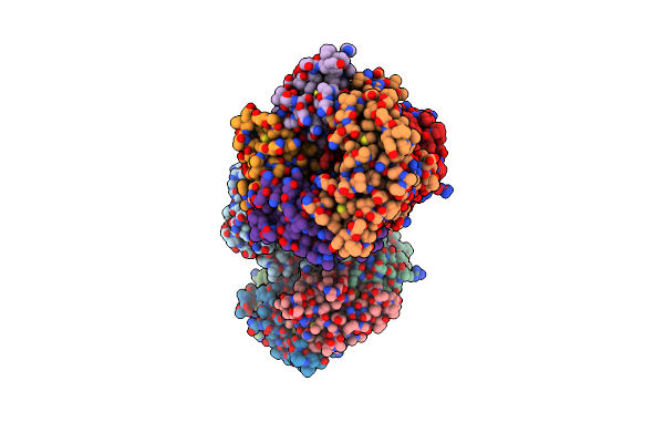 |
Structural Basis Of Subtilase Cytotoxin Subab Assembly
Organism: Escherichia coli
Method: X-RAY DIFFRACTION Resolution:2.60 Å Release Date: 2013-08-14 Classification: TOXIN Ligands: GOL, SO4 |
Organism: Escherichia coli
Method: X-RAY DIFFRACTION
Release Date: 2013-08-14
Ligands: GOL, SO4
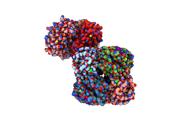 |
Green-Fluorescent Variant Of The Non-Fluorescent Chromoprotein Rtms5
Organism: Montipora efflorescens
Method: X-RAY DIFFRACTION Resolution:2.20 Å Release Date: 2012-06-06 Classification: FLUORESCENT PROTEIN Ligands: IOD, CL |
Organism: Montipora efflorescens
Method: X-RAY DIFFRACTION
Release Date: 2012-06-06
Ligands: IOD, CL
 |
Green-Fluorescent Variant Of The Non-Fluorescent Chromoprotein Rtms5
Organism: Montipora efflorescens
Method: X-RAY DIFFRACTION Resolution:2.20 Å Release Date: 2012-06-06 Classification: FLUORESCENT PROTEIN Ligands: IOD, CL |
Organism: Montipora efflorescens
Method: X-RAY DIFFRACTION
Release Date: 2012-06-06
Ligands: IOD, CL
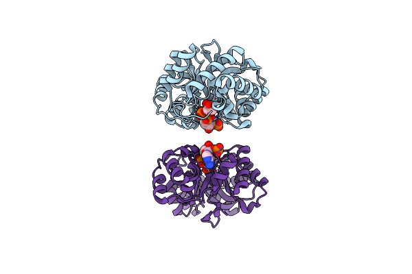 |
Crystal Structure Of A Mycobacterium Aldo-Keto Reductase In Its Apo And Liganded Form
Organism: Mycobacterium smegmatis
Method: X-RAY DIFFRACTION Resolution:1.64 Å Release Date: 2010-02-16 Classification: OXIDOREDUCTASE Ligands: NA7 |
Organism: Mycobacterium smegmatis
Method: X-RAY DIFFRACTION
Release Date: 2010-02-16
Ligands: NA7
 |
Crystal Structure Of A Mycobacterium Aldo-Keto Reductase In Its Apo And Liganded Form
Organism: Mycobacterium smegmatis
Method: X-RAY DIFFRACTION Resolution:1.90 Å Release Date: 2010-02-16 Classification: OXIDOREDUCTASE |
Organism: Mycobacterium smegmatis
Method: X-RAY DIFFRACTION
Release Date: 2010-02-16
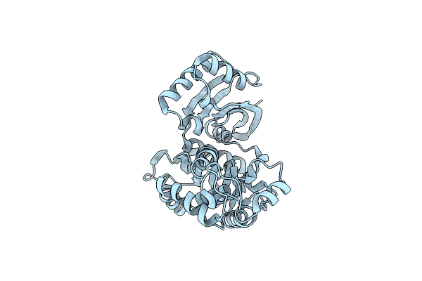 |
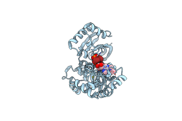
Crystal Structure Of Lp1Ntpdase From Legionella Pneumophila
Organism: Legionella pneumophila
Method: X-RAY DIFFRACTION Resolution:1.60 Å Release Date: 2010-02-09 Classification: HYDROLASE |
Organism: Legionella pneumophila
Method: X-RAY DIFFRACTION
Release Date: 2010-02-09
 |
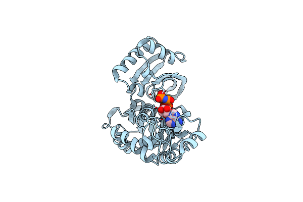
Crystal Structure Of Lp1Ntpdase From Legionella Pneumophila In Complex With The Inhibitor Arl 67156
Organism: Legionella pneumophila
Method: X-RAY DIFFRACTION Resolution:2.00 Å Release Date: 2010-02-09 Classification: HYDROLASE Ligands: ARU |
Organism: Legionella pneumophila
Method: X-RAY DIFFRACTION
Release Date: 2010-02-09
Ligands: ARU
 |
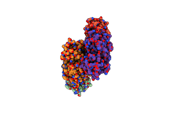
Crystal Structure Of Lp1Ntpdase From Legionella Pneumophila In Complex With Amppnp
Organism: Legionella pneumophila
Method: X-RAY DIFFRACTION Resolution:1.65 Å Release Date: 2010-02-09 Classification: HYDROLASE Ligands: ANP |
Organism: Legionella pneumophila
Method: X-RAY DIFFRACTION
Release Date: 2010-02-09
Ligands: ANP
 |
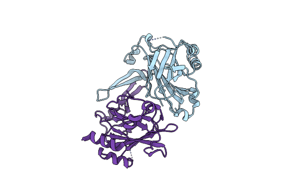
The Oxidoreductase Nmdsba1 From N. Meningitidis
Organism: Neisseria meningitidis serogroup b
Method: X-RAY DIFFRACTION Resolution:2.10 Å Release Date: 2009-10-20 Classification: OXIDOREDUCTASE |
Organism: Neisseria meningitidis serogroup b
Method: X-RAY DIFFRACTION
Release Date: 2009-10-20
 |
Crystal Structure Of Lipl32, The Most Abundant Surface Protein Of Pathogenic Leptospira Spp
Organism: Leptospira interrogans
Method: X-RAY DIFFRACTION Resolution:2.01 Å Release Date: 2009-02-24 Classification: UNKNOWN FUNCTION |
Organism: Leptospira interrogans
Method: X-RAY DIFFRACTION
Release Date: 2009-02-24
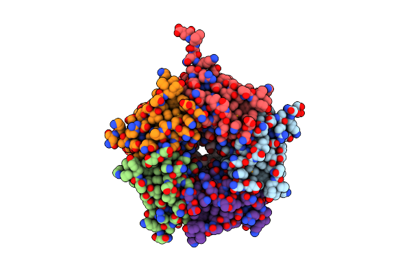 |
Crystal Structure Of The B-Subunit Of The Ab5 Toxin From E. Coli
Organism: Escherichia coli
Method: X-RAY DIFFRACTION Resolution:2.08 Å Release Date: 2008-11-04 Classification: TOXIN Ligands: 1PE |
Organism: Escherichia coli
Method: X-RAY DIFFRACTION
Release Date: 2008-11-04
Ligands: 1PE
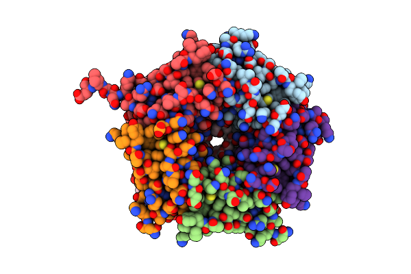 |
Crystal Structure Of The B-Subunit Of The Ab5 Toxin From E. Coli With Neu5Gc
Organism: Escherichia coli
Method: X-RAY DIFFRACTION Resolution:2.20 Å Release Date: 2008-11-04 Classification: TOXIN Ligands: NGC, 1PE |
Organism: Escherichia coli
Method: X-RAY DIFFRACTION
Release Date: 2008-11-04
Ligands: NGC, 1PE
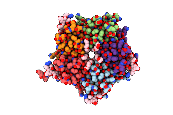 |
Crystal Structure Of The A-Subunit Of The Ab5 Toxin From E. Coli With Neu5Gc-2,3Gal-1,3Glcnac
Organism: Escherichia coli
Method: X-RAY DIFFRACTION Resolution:2.10 Å Release Date: 2008-11-04 Classification: TOXIN Ligands: POL, AZI, 1PE |
Organism: Escherichia coli
Method: X-RAY DIFFRACTION
Release Date: 2008-11-04
Ligands: POL, AZI, 1PE
 |
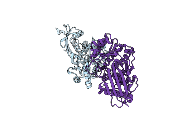
Oxidoreductase Nmdsba3 From Neisseria Meningitidis
Organism: Neisseria meningitidis serogroup b
Method: X-RAY DIFFRACTION Resolution:2.30 Å Release Date: 2008-08-19 Classification: OXIDOREDUCTASE |
Organism: Neisseria meningitidis serogroup b
Method: X-RAY DIFFRACTION
Release Date: 2008-08-19
 |
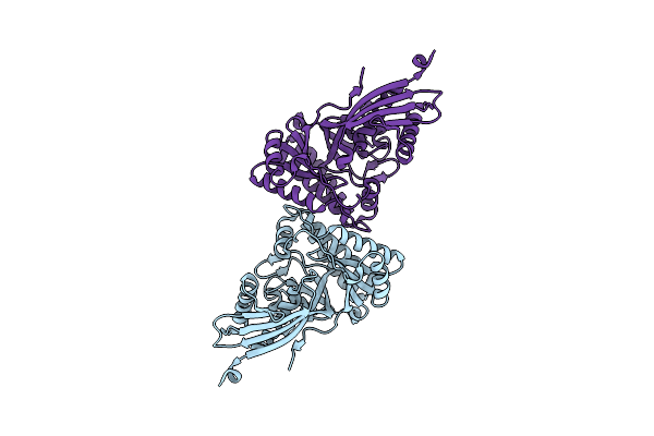
Crystal Structure Of Mevalonate Diphosphate Decarboxylase From Staphylococcus Aureus (Monoclinic Form)
Organism: Staphylococcus aureus
Method: X-RAY DIFFRACTION Resolution:2.30 Å Release Date: 2007-06-12 Classification: LYASE |
Organism: Staphylococcus aureus
Method: X-RAY DIFFRACTION
Release Date: 2007-06-12
 |
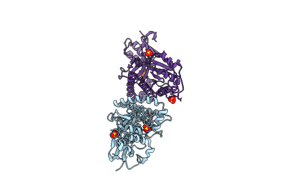
Crystal Structure Of Mevalonate Diphosphate Decarboxylase From Staphylococcus Aureus (Orthorhombic Form)
Organism: Staphylococcus aureus
Method: X-RAY DIFFRACTION Resolution:2.30 Å Release Date: 2007-06-12 Classification: LYASE |
Organism: Staphylococcus aureus
Method: X-RAY DIFFRACTION
Release Date: 2007-06-12
 |
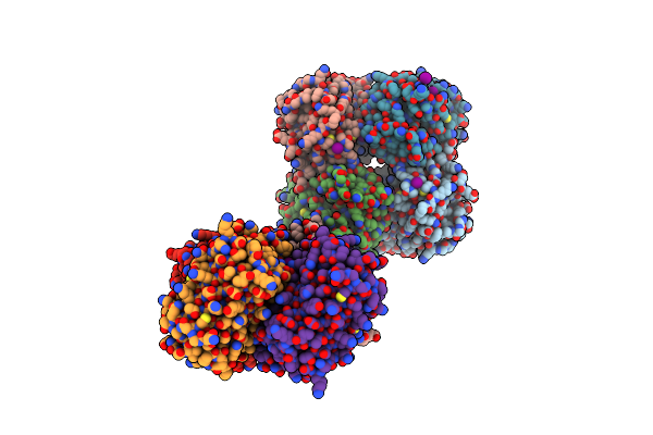
Mevalonate Diphosphate Decarboxylase From Trypanosoma Brucei
Organism: Trypanosoma brucei
Method: X-RAY DIFFRACTION Resolution:1.80 Å Release Date: 2007-06-12 Classification: LYASE Ligands: SO4 |
Organism: Trypanosoma brucei
Method: X-RAY DIFFRACTION
Release Date: 2007-06-12
Ligands: SO4
 |
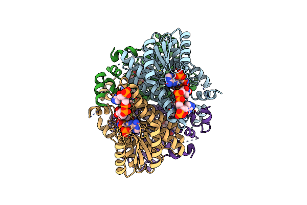
High Ph Structure Of Rtms5 H146S Variant
Organism: Montipora efflorescens
Method: X-RAY DIFFRACTION Resolution:1.80 Å Release Date: 2007-04-03 Classification: LUMINESCENT PROTEIN Ligands: IOD |
Organism: Montipora efflorescens
Method: X-RAY DIFFRACTION
Release Date: 2007-04-03
Ligands: IOD
 |
Structure Of Human Decr Ternary Complex
Organism: Homo sapiens
Method: X-RAY DIFFRACTION Resolution:1.75 Å Release Date: 2004-10-28 Classification: OXIDOREDUCTASE Ligands: NAP, HXC |
Organism: Homo sapiens
Method: X-RAY DIFFRACTION
Release Date: 2004-10-28
Ligands: NAP, HXC
 |
Binary Structure Of Human Decr Solved By Semet Sad.
Organism: Homo sapiens
Method: X-RAY DIFFRACTION Resolution:2.10 Å Release Date: 2004-10-28 Classification: OXIDOREDUCTASE Ligands: GOL, NAP |
Organism: Homo sapiens
Method: X-RAY DIFFRACTION
Release Date: 2004-10-28
Ligands: GOL, NAP