

Search Count: 30
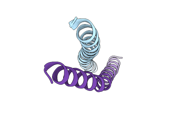 |
Crystal Structure Of Human Striatin-3 Coiled Coil Domain
Organism: Homo sapiens
Method: X-RAY DIFFRACTION Resolution:2.00 Å Release Date: 2014-02-26 Classification: SIGNALING PROTEIN |
Organism: Homo sapiens
Method: X-RAY DIFFRACTION
Release Date: 2014-02-26
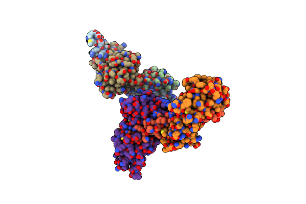 |
Crystal Structure Of A Light Chain Domain Of Scallop Smooth Muscle Myosin
Organism: Placopecten magellanicus
Method: X-RAY DIFFRACTION Resolution:2.39 Å Release Date: 2011-11-23 Classification: STRUCTURAL PROTEIN Ligands: MG, CA |
Organism: Placopecten magellanicus
Method: X-RAY DIFFRACTION
Release Date: 2011-11-23
Ligands: MG, CA
 |
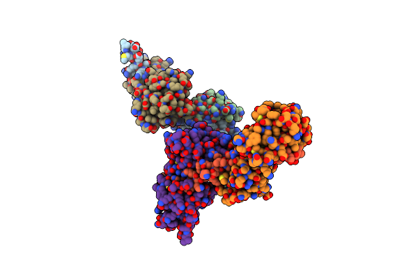
Phosphorylated Light Chain Domain Of Scallop Smooth Muscle Myosin
Organism: Placopecten magellanicus
Method: X-RAY DIFFRACTION Resolution:2.50 Å Release Date: 2011-11-23 Classification: STRUCTURAL PROTEIN Ligands: MG, CA |
Organism: Placopecten magellanicus
Method: X-RAY DIFFRACTION
Release Date: 2011-11-23
Ligands: MG, CA
 |
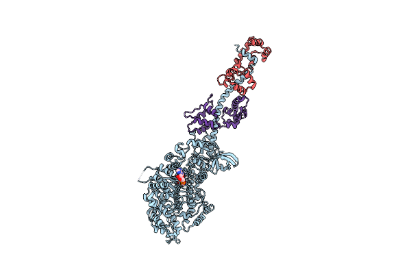
Visualizing New Hinges And A Potential Major Source Of Compliance In The Lever Arm Of Myosin
Organism: Placopecten magellanicus
Method: X-RAY DIFFRACTION Resolution:2.25 Å Release Date: 2011-01-05 Classification: PROTEIN BINDING Ligands: MG, CA |
Organism: Placopecten magellanicus
Method: X-RAY DIFFRACTION
Release Date: 2011-01-05
Ligands: MG, CA
 |
Crystal Structure Of Squid Mg.Adp Myosin S1
Organism: Loligo pealei, Todarodes pacificus
Method: X-RAY DIFFRACTION Resolution:3.10 Å Release Date: 2009-08-04 Classification: CONTRACTILE PROTEIN Ligands: MG, ADP |
Organism: Loligo pealei, Todarodes pacificus
Method: X-RAY DIFFRACTION
Release Date: 2009-08-04
Ligands: MG, ADP
 |
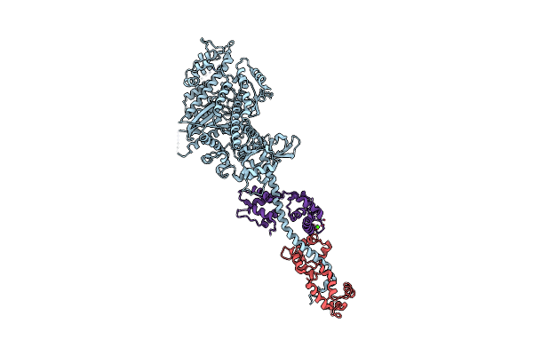
Crystal Structure Of Rigor-Like Squid Myosin S1
Organism: Loligo pealei, Todarodes pacificus
Method: X-RAY DIFFRACTION Resolution:2.60 Å Release Date: 2009-07-28 Classification: CONTRACTILE PROTEIN Ligands: MLI, CA |
Organism: Loligo pealei, Todarodes pacificus
Method: X-RAY DIFFRACTION
Release Date: 2009-07-28
Ligands: MLI, CA
 |
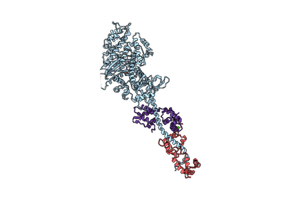
The Crystal Structure Of Rigor Like Squid Myosin S1 In The Absence Of Nucleotide
Organism: Loligo pealei, Todarodes pacificus
Method: X-RAY DIFFRACTION Resolution:3.40 Å Release Date: 2009-07-28 Classification: CONTRACTILE PROTEIN Ligands: CA |
Organism: Loligo pealei, Todarodes pacificus
Method: X-RAY DIFFRACTION
Release Date: 2009-07-28
Ligands: CA
 |
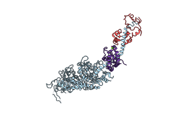
The Crystal Structure Of Squid Myosin S1 In The Presence Of So4 2-
Organism: Loligo pealei, Todarodes pacificus
Method: X-RAY DIFFRACTION Resolution:3.30 Å Release Date: 2009-07-28 Classification: CONTRACTILE PROTEIN Ligands: SO4, CA |
Organism: Loligo pealei, Todarodes pacificus
Method: X-RAY DIFFRACTION
Release Date: 2009-07-28
Ligands: SO4, CA
 |
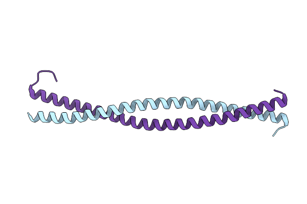
Placopecten Striated Muscle Myosin Ii
Organism: Placopecten magellanicus
Method: X-RAY DIFFRACTION Resolution:3.25 Å Release Date: 2008-02-26 Classification: CONTRACTILE PROTEIN Ligands: CA |
Organism: Placopecten magellanicus
Method: X-RAY DIFFRACTION
Release Date: 2008-02-26
Ligands: CA
 |
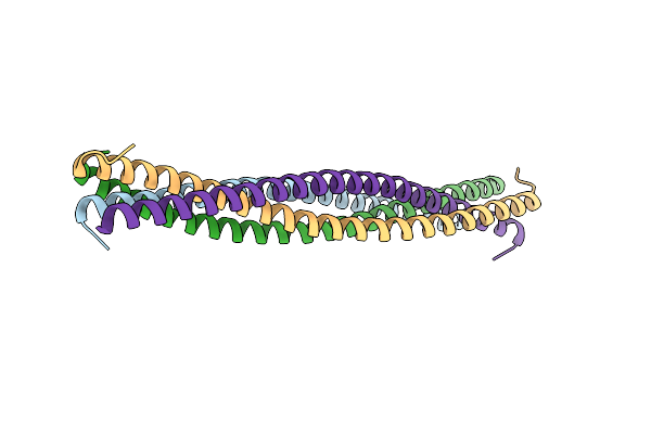
Crystal Structure Of The N-Terminal Region Of The Scallop Myosin Rod, Monoclinic (C2) Form
Organism: Argopecten irradians, Saccharomyces cerevisiae
Method: X-RAY DIFFRACTION Resolution:2.30 Å Release Date: 2008-01-08 Classification: CONTRACTILE PROTEIN Ligands: IOD |
Organism: Argopecten irradians, Saccharomyces cerevisiae
Method: X-RAY DIFFRACTION
Release Date: 2008-01-08
Ligands: IOD
 |
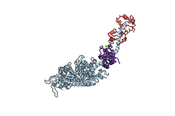
Crystal Structure Of The N-Terminal Region Of The Scallop Myosin Rod, Monoclinic (P21) Form
Organism: Argopecten irradians, Saccharomyces cerevisiae
Method: X-RAY DIFFRACTION Resolution:2.30 Å Release Date: 2008-01-08 Classification: CONTRACTILE PROTEIN |
Organism: Argopecten irradians, Saccharomyces cerevisiae
Method: X-RAY DIFFRACTION
Release Date: 2008-01-08
 |
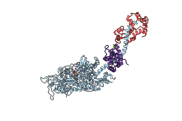
Rigor-Like Structures Of Muscle Myosins Reveal Key Mechanical Elements In The Transduction Pathways Of This Allosteric Motor
Organism: Placopecten magellanicus
Method: X-RAY DIFFRACTION Resolution:3.27 Å Release Date: 2007-05-29 Classification: CONTRACTILE PROTEIN Ligands: MG, CA |
Organism: Placopecten magellanicus
Method: X-RAY DIFFRACTION
Release Date: 2007-05-29
Ligands: MG, CA
 |
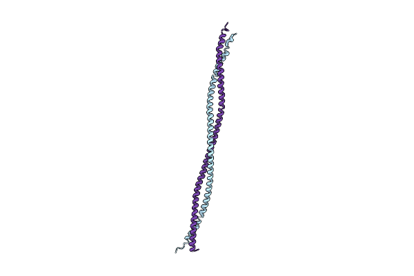
Rigor-Like Structures Of Muscle Myosins Reveal Key Mechanical Elements In The Transduction Pathways Of This Allosteric Motor
Organism: Placopecten magellanicus
Method: X-RAY DIFFRACTION Resolution:3.12 Å Release Date: 2007-05-29 Classification: CONTRACTILE PROTEIN Ligands: MG, ADP, CA |
Organism: Placopecten magellanicus
Method: X-RAY DIFFRACTION
Release Date: 2007-05-29
Ligands: MG, ADP, CA
 |
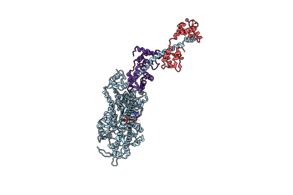
Structure Of Tropomyosin'S Mid-Region: Bending And Binding Sites For Actin
Organism: Rattus norvegicus
Method: X-RAY DIFFRACTION Resolution:2.30 Å Release Date: 2006-01-03 Classification: CONTRACTILE PROTEIN |
Organism: Rattus norvegicus
Method: X-RAY DIFFRACTION
Release Date: 2006-01-03
 |
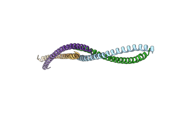
Crystal Structure Of Scallop Myosin S1 In The Pre-Power Stroke State To 2.6 Angstrom Resolution: Flexibility And Function In The Head
Organism: Argopecten irradians
Method: X-RAY DIFFRACTION Resolution:2.54 Å Release Date: 2003-12-16 Classification: CONTRACTILE PROTEIN Ligands: MG, VO4, ADP, CA |
Organism: Argopecten irradians
Method: X-RAY DIFFRACTION
Release Date: 2003-12-16
Ligands: MG, VO4, ADP, CA
 |
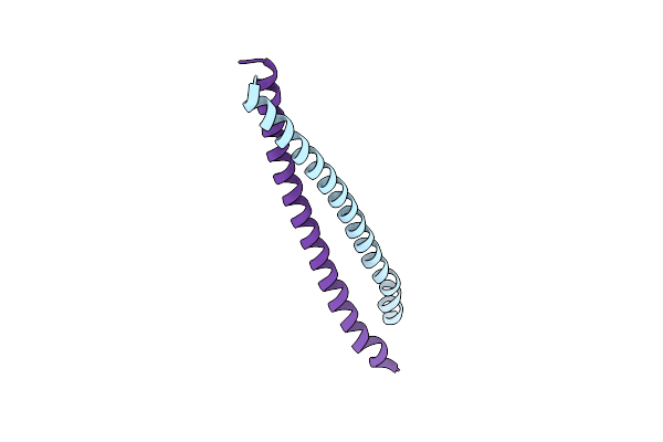
Visualizing An Unstable Coiled Coil: The Crystal Structure Of An N-Terminal Segment Of The Scallop Myosin Rod
Organism: Argopecten irradians, saccharomyces cerevisiae
Method: X-RAY DIFFRACTION Resolution:2.50 Å Release Date: 2003-07-22 Classification: CONTRACTILE PROTEIN |
Organism: Argopecten irradians, saccharomyces cerevisiae
Method: X-RAY DIFFRACTION
Release Date: 2003-07-22
 |
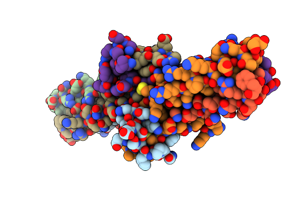
Crystal Structure Of The C-Terminal Region Of Striated Muscle Alpha-Tropomyosin At 2.7 Angstrom Resolution
Organism: Saccharomyces cerevisiae, Rattus norvegicus
Method: X-RAY DIFFRACTION Resolution:2.70 Å Release Date: 2002-05-29 Classification: CONTRACTILE PROTEIN |
Organism: Saccharomyces cerevisiae, Rattus norvegicus
Method: X-RAY DIFFRACTION
Release Date: 2002-05-29
 |
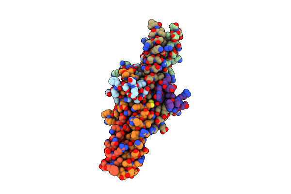
Crystal Structure Of The Central Region Of Bovine Fibrinogen (E5 Fragment) At 1.4 Angstroms Resolution
Organism: Bos taurus
Method: X-RAY DIFFRACTION Resolution:1.40 Å Release Date: 2001-10-17 Classification: BLOOD CLOTTING |
Organism: Bos taurus
Method: X-RAY DIFFRACTION
Release Date: 2001-10-17
 |
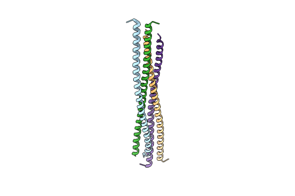
Crystal Structure Of The Central Region Of Bovine Fibrinogen (E5 Fragment) At 1.4 Angstroms Resolution
Organism: Bos taurus
Method: X-RAY DIFFRACTION Resolution:1.60 Å Release Date: 2001-10-17 Classification: BLOOD CLOTTING |
Organism: Bos taurus
Method: X-RAY DIFFRACTION
Release Date: 2001-10-17
 |
Deciphering The Design Of The Tropomyosin Molecule
Organism: Gallus gallus
Method: X-RAY DIFFRACTION Resolution:2.00 Å Release Date: 2001-07-25 Classification: CONTRACTILE PROTEIN |
Organism: Gallus gallus
Method: X-RAY DIFFRACTION
Release Date: 2001-07-25