

Search Count: 14
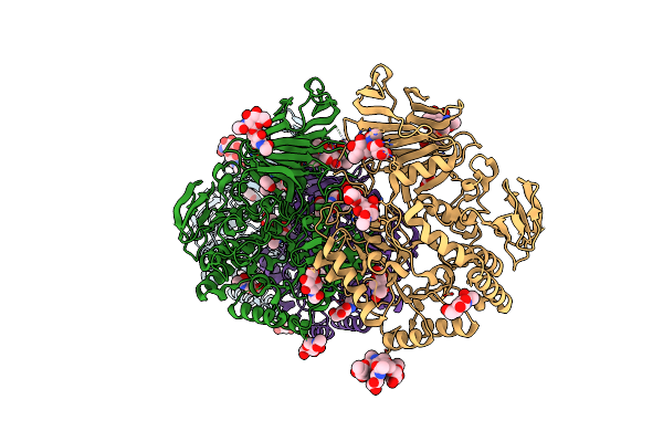 |
Crystal Structure Of Unliganded Myorg
Organism: Homo sapiens
Method: X-RAY DIFFRACTION Resolution:2.43 Å Release Date: 2022-08-31 Classification: HYDROLASE Ligands: NAG, ACT, MLI |
Organism: Homo sapiens
Method: X-RAY DIFFRACTION
Release Date: 2022-08-31
Ligands: NAG, ACT, MLI
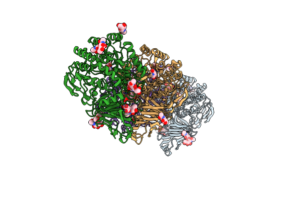 |
Crystal Structure Of Myorg Bound To 1-Deoxygalactonojirimycin
Organism: Homo sapiens
Method: X-RAY DIFFRACTION Resolution:2.43 Å Release Date: 2022-08-31 Classification: HYDROLASE Ligands: NAG, DGJ, MLI |
Organism: Homo sapiens
Method: X-RAY DIFFRACTION
Release Date: 2022-08-31
Ligands: NAG, DGJ, MLI
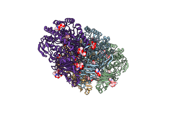 |
Crystal Structure Of Myorg (D520N) In Complex With Gal-A1,4-Glc
Organism: Homo sapiens
Method: X-RAY DIFFRACTION Resolution:2.25 Å Release Date: 2022-08-31 Classification: HYDROLASE Ligands: NAG, EDO, MLI |
Organism: Homo sapiens
Method: X-RAY DIFFRACTION
Release Date: 2022-08-31
Ligands: NAG, EDO, MLI
 |
Crystal Structure Of The Helical Cell Shape Determining Protein Pgp2 From Campylobacter Jejuni
Organism: Campylobacter jejuni subsp. jejuni
Method: X-RAY DIFFRACTION Resolution:1.50 Å Release Date: 2021-03-17 Classification: HYDROLASE |
Organism: Campylobacter jejuni subsp. jejuni
Method: X-RAY DIFFRACTION
Release Date: 2021-03-17
 |
Crystal Structure Of The Helical Cell Shape Determining Protein Pgp2 (K307A Mutant) From Campylobacter Jejuni
Organism: Campylobacter jejuni subsp. jejuni
Method: X-RAY DIFFRACTION Resolution:1.85 Å Release Date: 2021-03-17 Classification: HYDROLASE |
Organism: Campylobacter jejuni subsp. jejuni
Method: X-RAY DIFFRACTION
Release Date: 2021-03-17
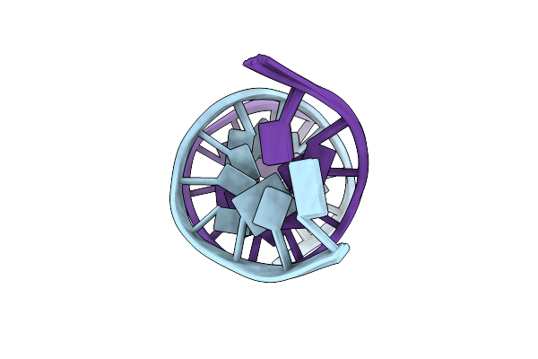 |
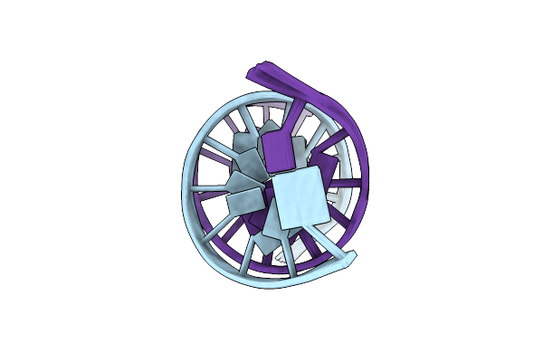
Solution Structure Of Dna Dodecamer With 8-Oxoguanine At 10Th Position
|
Organism: Synthetic construct
Method: SOLUTION NMR
Release Date: 2016-08-03
 |
Solution Structure Of Dna Dodecamer With 8-Oxoguanine At 4Th Position
|
Organism: Synthetic construct
Method: SOLUTION NMR
Release Date: 2016-07-27
 |
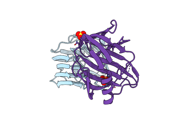
Crystal Structure Of The Bacillus Circulans Endo-Beta-(1,4)-Xylanase (Bcx) E172H Mutant
Organism: Bacillus circulans
Method: X-RAY DIFFRACTION Resolution:2.41 Å Release Date: 2013-05-08 Classification: HYDROLASE Ligands: SO4 |
Organism: Bacillus circulans
Method: X-RAY DIFFRACTION
Release Date: 2013-05-08
Ligands: SO4
 |
Crystal Structure Of The Bacillus Circulans Endo-Beta-(1,4)-Xylanase (Bcx) N35E Mutant
Organism: Bacillus circulans
Method: X-RAY DIFFRACTION Resolution:1.55 Å Release Date: 2013-05-08 Classification: HYDROLASE Ligands: SO4 |
Organism: Bacillus circulans
Method: X-RAY DIFFRACTION
Release Date: 2013-05-08
Ligands: SO4
 |
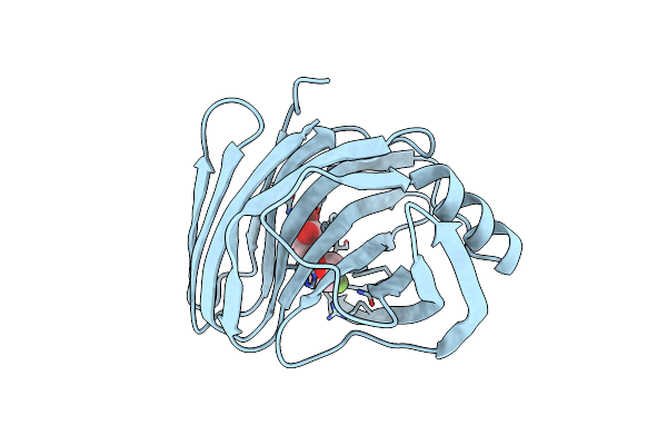
Crystal Structure Of The Bacillus Circulans Endo-Beta-(1,4)-Xylanase (Bcx) N35H Mutant
Organism: Bacillus circulans
Method: X-RAY DIFFRACTION Resolution:2.00 Å Release Date: 2013-05-08 Classification: HYDROLASE Ligands: SO4 |
Organism: Bacillus circulans
Method: X-RAY DIFFRACTION
Release Date: 2013-05-08
Ligands: SO4
 |
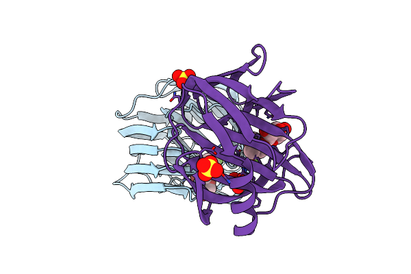
Crystal Structure Of The Bacillus Circulans Endo-Beta-(1,4)-Xylanase (Bcx) E172H Mutant With Glu78 Covalently Bonded To 2-Deoxy-2-Fluoro-Xylobiose
Organism: Bacillus circulans
Method: X-RAY DIFFRACTION Resolution:1.86 Å Release Date: 2013-05-08 Classification: HYDROLASE/HYDROLASE INHIBITOR |
Organism: Bacillus circulans
Method: X-RAY DIFFRACTION
Release Date: 2013-05-08
 |
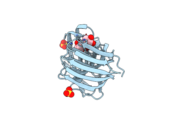
Crystal Structure Of The Bacillus Circulans Endo-Beta-(1,4)-Xylanase (Bcx) N35E Mutant With Glu78 Covalently Bonded To 2-Deoxy-2-Fluoro-Xylobiose
Organism: Bacillus circulans
Method: X-RAY DIFFRACTION Resolution:1.67 Å Release Date: 2013-05-08 Classification: HYDROLASE/HYDROLASE INHIBITOR Ligands: SO4 |
Organism: Bacillus circulans
Method: X-RAY DIFFRACTION
Release Date: 2013-05-08
Ligands: SO4
 |
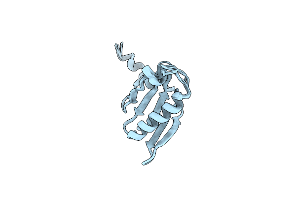
Crystal Structure Of The Bacillus Circulans Endo-Beta-(1,4)-Xylanase (Bcx) N35H Mutant With Glu78 Covalently Bonded To 2-Deoxy-2-Fluoro-Xylobiose
Organism: Bacillus circulans
Method: X-RAY DIFFRACTION Resolution:1.73 Å Release Date: 2013-05-08 Classification: HYDROLASE/HYDROLASE INHIBITOR Ligands: SO4 |
Organism: Bacillus circulans
Method: X-RAY DIFFRACTION
Release Date: 2013-05-08
Ligands: SO4
 |
Sixth Gelsolin-Like Domain Of Villin In 5 Mm Cacl2
Organism: Gallus gallus
Method: SOLUTION NMR Release Date: 2012-11-07 Classification: STRUCTURAL PROTEIN |
Organism: Gallus gallus
Method: SOLUTION NMR
Release Date: 2012-11-07