

Search Count: 24
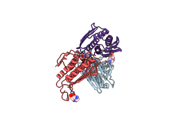 |
Crystal Structure Of Igpd From Pyrococcus Furiosus In Complex With (R)-C348
Organism: Pyrococcus furiosus
Method: X-RAY DIFFRACTION Resolution:1.80 Å Release Date: 2016-10-05 Classification: LYASE Ligands: MN, 5LD |
Organism: Pyrococcus furiosus
Method: X-RAY DIFFRACTION
Release Date: 2016-10-05
Ligands: MN, 5LD
 |
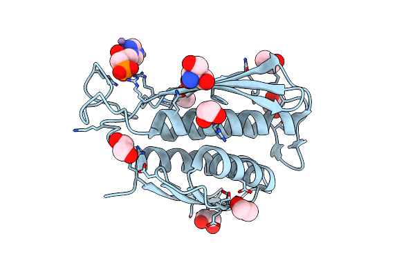
A. Thaliana Igpd2 In Complex With The Racemate Of The Triazole-Phosphonate Inhibitor, C348
Organism: Arabidopsis thaliana
Method: X-RAY DIFFRACTION Resolution:1.10 Å Release Date: 2016-10-05 Classification: LYASE Ligands: MN, 5DL, 5LD, EDO, TRS, CL |
Organism: Arabidopsis thaliana
Method: X-RAY DIFFRACTION
Release Date: 2016-10-05
Ligands: MN, 5DL, 5LD, EDO, TRS, CL
 |
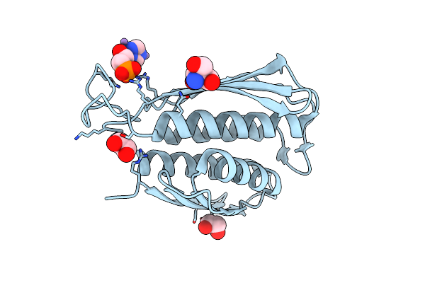
A. Thaliana Igpd2 In Complex With The Triazole-Phosphonate Inhibitor, (S)-C348, To 1.1A Resolution
Organism: Arabidopsis thaliana
Method: X-RAY DIFFRACTION Resolution:1.10 Å Release Date: 2016-10-05 Classification: LYASE Ligands: MN, 5DL, TRS, EDO |
Organism: Arabidopsis thaliana
Method: X-RAY DIFFRACTION
Release Date: 2016-10-05
Ligands: MN, 5DL, TRS, EDO
 |
A. Thaliana Igpd2 In Complex With The Triazole-Phosphonate Inhibitor, (R)-C348, To 1.36A Resolution
Organism: Arabidopsis thaliana
Method: X-RAY DIFFRACTION Resolution:1.40 Å Release Date: 2016-10-05 Classification: LYASE Ligands: MN, 5LD, EDO, TRS, CL |
Organism: Arabidopsis thaliana
Method: X-RAY DIFFRACTION
Release Date: 2016-10-05
Ligands: MN, 5LD, EDO, TRS, CL
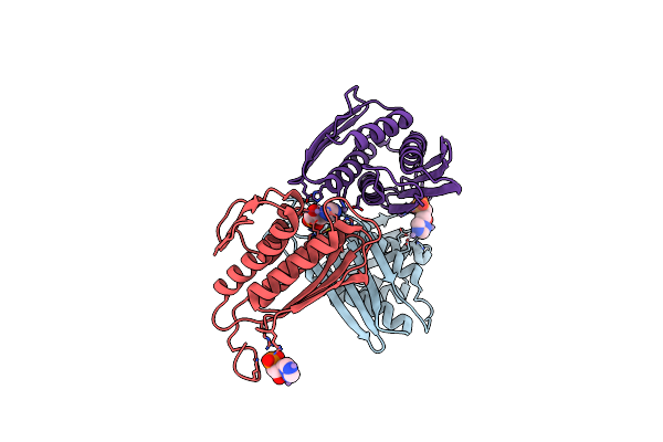 |
Crystal Structure Of Igpd From Pyrococcus Furiosus In Complex With (S)-C348
Organism: Pyrococcus furiosus
Method: X-RAY DIFFRACTION Resolution:1.53 Å Release Date: 2016-09-28 Classification: LYASE Ligands: MN, 5DL |
Organism: Pyrococcus furiosus
Method: X-RAY DIFFRACTION
Release Date: 2016-09-28
Ligands: MN, 5DL
 |
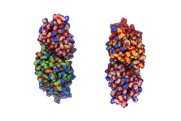
The Structure Of Wt A. Thaliana Igpd2 In Complex With Mn2+ And Formate At 1.3A Resolution
Organism: Arabidopsis thaliana
Method: X-RAY DIFFRACTION Resolution:1.30 Å Release Date: 2015-06-24 Classification: LYASE Ligands: MN, FMT, TRS, CL |
Organism: Arabidopsis thaliana
Method: X-RAY DIFFRACTION
Release Date: 2015-06-24
Ligands: MN, FMT, TRS, CL
 |
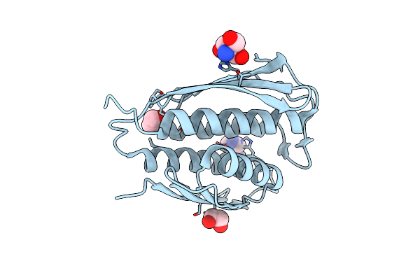
The Structure Of Wt A. Thaliana Igpd2 In Complex With Mn2+ And Phosphate
Organism: Arabidopsis thaliana
Method: X-RAY DIFFRACTION Resolution:1.75 Å Release Date: 2015-06-24 Classification: LYASE Ligands: MN, PO4, NA, EDO |
Organism: Arabidopsis thaliana
Method: X-RAY DIFFRACTION
Release Date: 2015-06-24
Ligands: MN, PO4, NA, EDO
 |
The Structure Of Wt A. Thaliana Igpd2 In Complex With Mn2+ And 1,2,4-Triazole At 1.3 A Resolution
Organism: Arabidopsis thaliana
Method: X-RAY DIFFRACTION Resolution:1.30 Å Release Date: 2014-09-24 Classification: LYASE Ligands: MN, TRI, TRS, CL, EDO |
Organism: Arabidopsis thaliana
Method: X-RAY DIFFRACTION
Release Date: 2014-09-24
Ligands: MN, TRI, TRS, CL, EDO
 |
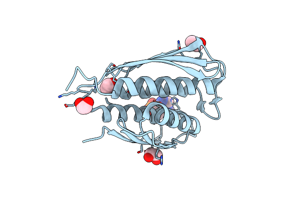
The Structure Of Wt A. Thaliana Igpd2 In Complex With Mn2+, Imidazole, And Sulfate At 1.5 A Resolution
Organism: Arabidopsis thaliana
Method: X-RAY DIFFRACTION Resolution:1.50 Å Release Date: 2014-09-24 Classification: LYASE Ligands: MN, IMD, SO4, EDO |
Organism: Arabidopsis thaliana
Method: X-RAY DIFFRACTION
Release Date: 2014-09-24
Ligands: MN, IMD, SO4, EDO
 |
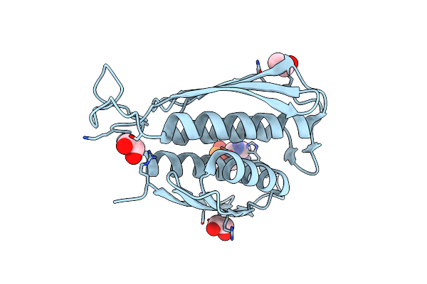
The Form A Structure Of An E21Q Catalytic Mutant Of A. Thaliana Igpd2 In Complex With Mn2+ And A Mixture Of Its Substrate, 2R3S-Igp, And An Inhibitor, 2S3S-Igp, To 1.12 A Resolution
Organism: Arabidopsis thaliana
Method: X-RAY DIFFRACTION Resolution:1.12 Å Release Date: 2014-09-24 Classification: LYASE/LYASE INHIBITOR Ligands: MN, IG2, IYP, EDO |
Organism: Arabidopsis thaliana
Method: X-RAY DIFFRACTION
Release Date: 2014-09-24
Ligands: MN, IG2, IYP, EDO
 |
The Form B Structure Of An E21Q Catalytic Mutant Of A. Thaliana Igpd2 In Complex With Mn2+ And Its Substrate, 2R3S-Igp, To 1.41 A Resolution
Organism: Arabidopsis thaliana
Method: X-RAY DIFFRACTION Resolution:1.41 Å Release Date: 2014-09-24 Classification: LYASE Ligands: MN, IYP, EDO |
Organism: Arabidopsis thaliana
Method: X-RAY DIFFRACTION
Release Date: 2014-09-24
Ligands: MN, IYP, EDO
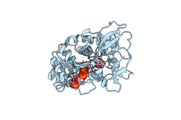 |
Haloferax Mediterranei Glucose Dehydrogenase In Complex With Nadp, Zn And Gluconolactone.
Organism: Haloferax mediterranei
Method: X-RAY DIFFRACTION Resolution:2.00 Å Release Date: 2009-01-13 Classification: OXIDOREDUCTASE Ligands: NAP, LGC, ZN |
Organism: Haloferax mediterranei
Method: X-RAY DIFFRACTION
Release Date: 2009-01-13
Ligands: NAP, LGC, ZN
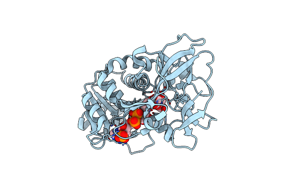 |
Haloferax Mediterranei Glucose Dehydrogenase In Complex With Nadp, Zn And Glucose.
Organism: Haloferax mediterranei
Method: X-RAY DIFFRACTION Resolution:2.03 Å Release Date: 2009-01-13 Classification: OXIDOREDUCTASE Ligands: NAP, BGC, ZN |
Organism: Haloferax mediterranei
Method: X-RAY DIFFRACTION
Release Date: 2009-01-13
Ligands: NAP, BGC, ZN
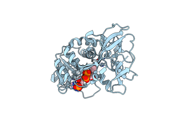 |
Haloferax Mediterranei Glucose Dehydrogenase In Complex With Nadph And Zn.
Organism: Haloferax mediterranei
Method: X-RAY DIFFRACTION Resolution:2.01 Å Release Date: 2009-01-13 Classification: OXIDOREDUCTASE Ligands: NDP, ZN |
Organism: Haloferax mediterranei
Method: X-RAY DIFFRACTION
Release Date: 2009-01-13
Ligands: NDP, ZN
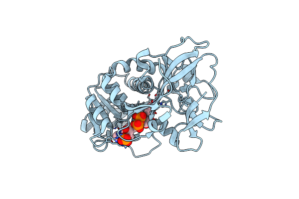 |
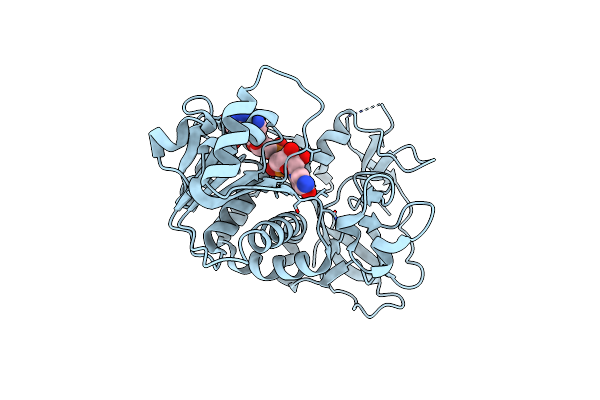
Haloferax Mediterranei Glucose Dehydrogenase In Complex With Nadp And Zn.
Organism: Haloferax mediterranei
Method: X-RAY DIFFRACTION Resolution:2.10 Å Release Date: 2009-01-13 Classification: OXIDOREDUCTASE Ligands: NAP, ZN |
Organism: Haloferax mediterranei
Method: X-RAY DIFFRACTION
Release Date: 2009-01-13
Ligands: NAP, ZN
 |
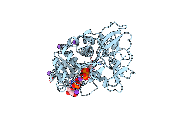
Crystal Structure Of Glucose Dehydrogenase From Haloferax Mediterranei
Organism: Haloferax mediterranei
Method: X-RAY DIFFRACTION Resolution:2.00 Å Release Date: 2006-04-04 Classification: OXIDOREDUCTASE Ligands: NAP |
Organism: Haloferax mediterranei
Method: X-RAY DIFFRACTION
Release Date: 2006-04-04
Ligands: NAP
 |
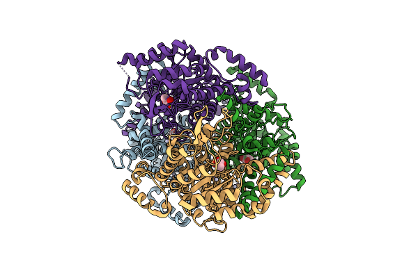
Crystal Structure Of D38C Glucose Dehydrogenase Mutant From Haloferax Mediterranei
Organism: Haloferax mediterranei
Method: X-RAY DIFFRACTION Resolution:1.60 Å Release Date: 2006-04-04 Classification: OXIDOREDUCTASE Ligands: ZN, FLC, K, NAP |
Organism: Haloferax mediterranei
Method: X-RAY DIFFRACTION
Release Date: 2006-04-04
Ligands: ZN, FLC, K, NAP
 |
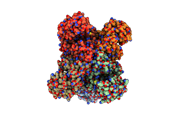
Crystal Structure Of The Isocitrate Lyase From The A219C Mutant Of Escherichia Coli
Organism: Escherichia coli
Method: X-RAY DIFFRACTION Resolution:2.10 Å Release Date: 2001-09-05 Classification: LYASE Ligands: HG, MG, PYR |
Organism: Escherichia coli
Method: X-RAY DIFFRACTION
Release Date: 2001-09-05
Ligands: HG, MG, PYR
 |
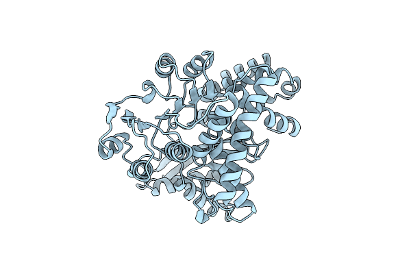
Glutamate Dehydrogenase From Thermococcus Litoralis
Organism: Thermococcus litoralis
Method: X-RAY DIFFRACTION Resolution:2.50 Å Release Date: 1999-09-18 Classification: OXIDOREDUCTASE |
Organism: Thermococcus litoralis
Method: X-RAY DIFFRACTION
Release Date: 1999-09-18
 |
K89L Mutant Of Glutamate Dehydrogenase
Organism: Clostridium symbiosum
Method: X-RAY DIFFRACTION Resolution:2.05 Å Release Date: 1999-01-27 Classification: OXIDOREDUCTASE |
Organism: Clostridium symbiosum
Method: X-RAY DIFFRACTION
Release Date: 1999-01-27