

Search Count: 24
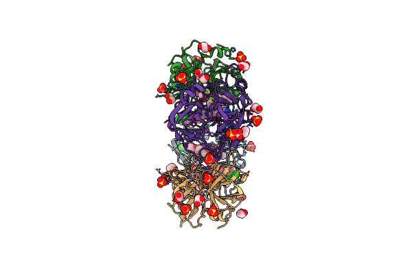 |
Human Tryptase Beta-2 (Htpsb2) Complexed With Covalent Inhibitor Compound #1
Organism: Homo sapiens
Method: X-RAY DIFFRACTION Resolution:1.98 Å Release Date: 2025-03-26 Classification: HYDROLASE Ligands: A1I52, SO4, PEG, EDO, ACT, GOL |
Organism: Homo sapiens
Method: X-RAY DIFFRACTION
Release Date: 2025-03-26
Ligands: A1I52, SO4, PEG, EDO, ACT, GOL
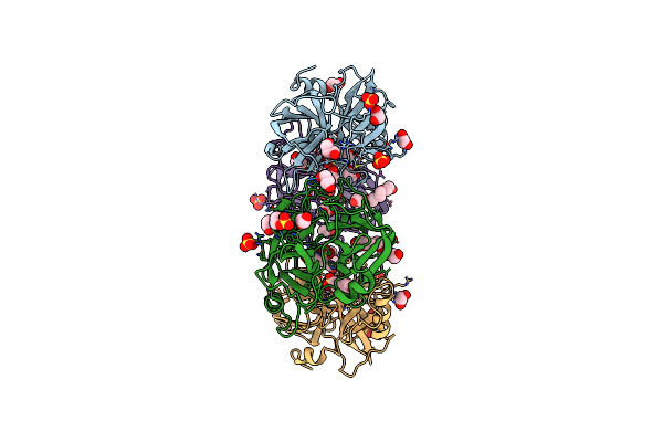 |
Human Tryptase Beta-2 (Htpsb2)
Organism: Homo sapiens
Method: X-RAY DIFFRACTION Resolution:2.06 Å Release Date: 2025-03-26 Classification: HYDROLASE Ligands: ACT, EDO, SO4, GOL, MES, 1PE |
Organism: Homo sapiens
Method: X-RAY DIFFRACTION
Release Date: 2025-03-26
Ligands: ACT, EDO, SO4, GOL, MES, 1PE
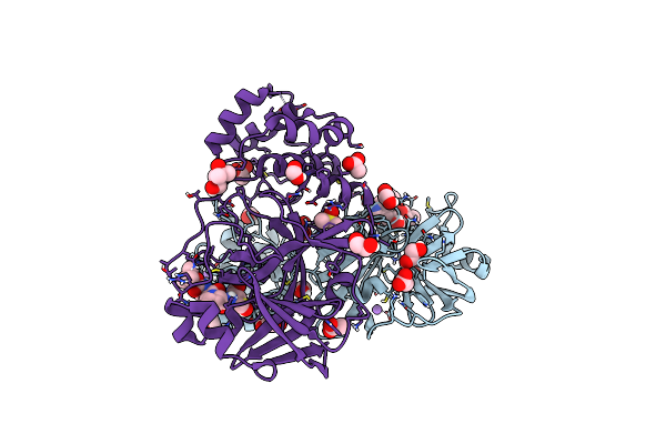 |
Crystal Structure Of F2F-2020184-00X Bound To The Main Protease (3Clpro/Mpro) Of Sars-Cov-2.
Organism: Severe acute respiratory syndrome coronavirus 2
Method: X-RAY DIFFRACTION Resolution:1.63 Å Release Date: 2023-05-03 Classification: VIRAL PROTEIN Ligands: 83F, ACT, EDO, FMT, NA, CL, DMS, MPD |
Organism: Severe acute respiratory syndrome coronavirus 2
Method: X-RAY DIFFRACTION
Release Date: 2023-05-03
Ligands: 83F, ACT, EDO, FMT, NA, CL, DMS, MPD
 |
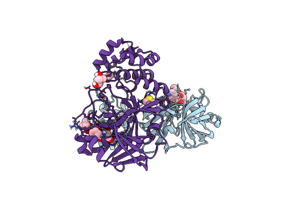
Crystal Structure Of F2F-2020185-01X Bound To The Main Protease (3Clpro/Mpro) Of Sars-Cov-2.
Organism: Severe acute respiratory syndrome coronavirus 2
Method: X-RAY DIFFRACTION Resolution:1.50 Å Release Date: 2023-05-03 Classification: VIRAL PROTEIN Ligands: 83N, EDO, FMT, NA, CL, MPD |
Organism: Severe acute respiratory syndrome coronavirus 2
Method: X-RAY DIFFRACTION
Release Date: 2023-05-03
Ligands: 83N, EDO, FMT, NA, CL, MPD
 |
Crystal Structure Of F2F-2020197-00X Bound To The Main Protease (3Clpro/Mpro) Of Sars-Cov-2.
Organism: Severe acute respiratory syndrome coronavirus 2
Method: X-RAY DIFFRACTION Resolution:1.66 Å Release Date: 2023-05-03 Classification: VIRAL PROTEIN Ligands: 84C, CL, BR, DMS, MPD, EDO, NA |
Organism: Severe acute respiratory syndrome coronavirus 2
Method: X-RAY DIFFRACTION
Release Date: 2023-05-03
Ligands: 84C, CL, BR, DMS, MPD, EDO, NA
 |
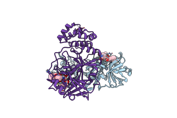
Crystal Structure Of F2F-2020198-00X Bound To The Main Protease (3Clpro/Mpro) Of Sars-Cov-2.
Organism: Severe acute respiratory syndrome coronavirus 2
Method: X-RAY DIFFRACTION Resolution:1.35 Å Release Date: 2023-05-03 Classification: VIRAL PROTEIN Ligands: 83W, CL, NA, MPD, EDO |
Organism: Severe acute respiratory syndrome coronavirus 2
Method: X-RAY DIFFRACTION
Release Date: 2023-05-03
Ligands: 83W, CL, NA, MPD, EDO
 |
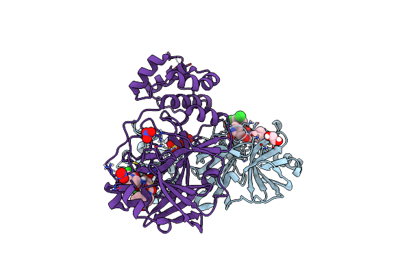
Crystal Structure Of F2F-2020209-00X Bound To The Main Protease (3Clpro/Mpro) Of Sars-Cov-2.
Organism: Severe acute respiratory syndrome coronavirus 2
Method: X-RAY DIFFRACTION Resolution:1.67 Å Release Date: 2022-11-23 Classification: VIRAL PROTEIN Ligands: 90I, CL, NA |
Organism: Severe acute respiratory syndrome coronavirus 2
Method: X-RAY DIFFRACTION
Release Date: 2022-11-23
Ligands: 90I, CL, NA
 |
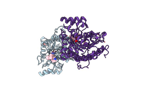
Crystal Structure Of F2F-2020216-01X Bound To The Main Protease (3Clpro/Mpro) Of Sars-Cov-2.
Organism: Severe acute respiratory syndrome coronavirus 2
Method: X-RAY DIFFRACTION Resolution:1.72 Å Release Date: 2022-11-23 Classification: VIRAL PROTEIN Ligands: 90X, SO4, EDO, CL, NO3, NA |
Organism: Severe acute respiratory syndrome coronavirus 2
Method: X-RAY DIFFRACTION
Release Date: 2022-11-23
Ligands: 90X, SO4, EDO, CL, NO3, NA
 |
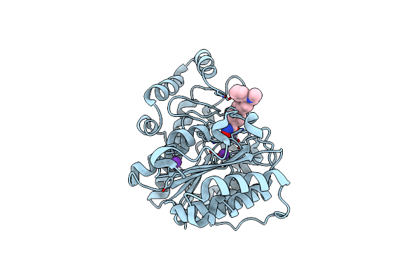
Crystal Structure Of Danio Rerio Histone Deacetylase 6 Catalytic Domain 2 (Cd2) Complexed With Nf2376
Organism: Danio rerio
Method: X-RAY DIFFRACTION Resolution:2.04 Å Release Date: 2020-12-02 Classification: HYDROLASE Ligands: ZN, K, QQD, EDO |
Organism: Danio rerio
Method: X-RAY DIFFRACTION
Release Date: 2020-12-02
Ligands: ZN, K, QQD, EDO
 |
Crystal Structure Of Danio Rerio Histone Deacetylase 6 Catalytic Domain 2 (Cd2) Complexed With Nf2657
Organism: Danio rerio
Method: X-RAY DIFFRACTION Resolution:2.09 Å Release Date: 2020-12-02 Classification: HYDROLASE Ligands: ZN, K, QQG |
Organism: Danio rerio
Method: X-RAY DIFFRACTION
Release Date: 2020-12-02
Ligands: ZN, K, QQG
 |
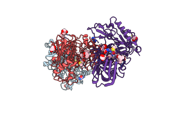
Tubulin-6J Complex
Organism: Rattus norvegicus, Gallus gallus, Bos taurus
Method: X-RAY DIFFRACTION Resolution:2.40 Å Release Date: 2018-12-05 Classification: CELL CYCLE Ligands: GTP, MG, CA, GDP, EZW, MES, ACP |
Organism: Rattus norvegicus, Gallus gallus, Bos taurus
Method: X-RAY DIFFRACTION
Release Date: 2018-12-05
Ligands: GTP, MG, CA, GDP, EZW, MES, ACP
 |
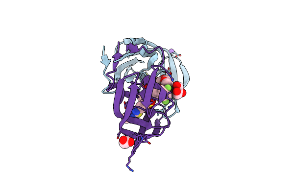
X-Ray Structure Of Bace1 In Complex With A Bicyclic Isoxazoline Carboxamide As The P3 Ligand
Organism: Homo sapiens
Method: X-RAY DIFFRACTION Resolution:2.85 Å Release Date: 2018-07-25 Classification: HYDROLASE Ligands: GHJ, GOL, URE, SO4 |
Organism: Homo sapiens
Method: X-RAY DIFFRACTION
Release Date: 2018-07-25
Ligands: GHJ, GOL, URE, SO4
 |
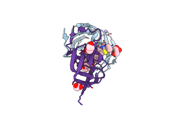
Hiv-1 Wild Type Protease With Grl-03314A, 6-5-5-Ring Fused Umbrella-Like Tetrahydropyranofuran As The P2-Ligand, A Cyclopropylaminobenzothiazole As The P2'-Ligand And 3,5-Difluorophenylmethyl As The P1-Ligand
Organism: Human immunodeficiency virus 1
Method: X-RAY DIFFRACTION Resolution:1.13 Å Release Date: 2018-05-30 Classification: HYDROLASE/HYDROLASE inhibitor Ligands: GR8, NA, CL, GOL, ACT |
Organism: Human immunodeficiency virus 1
Method: X-RAY DIFFRACTION
Release Date: 2018-05-30
Ligands: GR8, NA, CL, GOL, ACT
 |
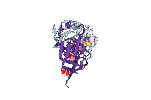
Hiv-1 Wild Type Protease With Grl-03214A, 6-5-5-Ring Fused Umbrella-Like Tetrahydropyranofuran As The P2-Ligand, A Cyclopropylaminobenzothiazole As The P2'-Ligand And 3,5-Difluorophenylmethyl As The P1-Ligand
Organism: Human immunodeficiency virus 1
Method: X-RAY DIFFRACTION Resolution:1.25 Å Release Date: 2018-05-30 Classification: HYDROLASE/HYDROLASE inhibitor |
Organism: Human immunodeficiency virus 1
Method: X-RAY DIFFRACTION
Release Date: 2018-05-30
 |
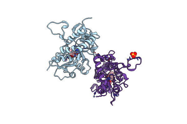
Crystal Structure Of Wild-Type Hiv-1 Protease With A Novel Hiv-1 Inhibitor Grl-14213A Of 6-5-5-Ring Fused Crown-Like Tetrahydropyranofuran As The P2-Ligand, A Cyclopropylaminobenzothiazole As The P2'-Ligand And 3,5-Difluorophenylmethyl As The P1-Ligand
Organism: Human immunodeficiency virus 1
Method: X-RAY DIFFRACTION Resolution:1.67 Å Release Date: 2018-02-28 Classification: HYDROLASE/HYDROLASE inhibitor |
Organism: Human immunodeficiency virus 1
Method: X-RAY DIFFRACTION
Release Date: 2018-02-28
 |
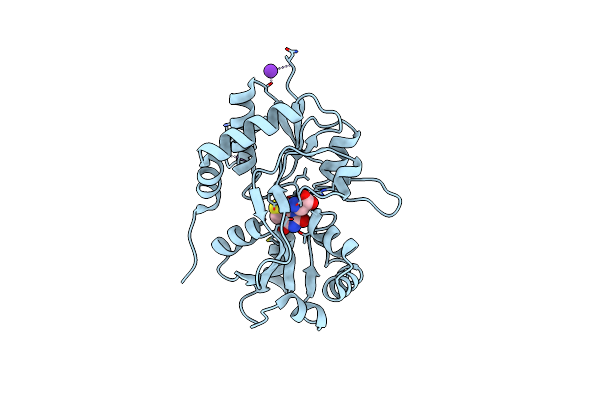
Crystal Structure Of The Kainate Receptor Gluk3 Ligand Binding Domain In Complex With (S)-1-[2'-Amino-2'-Carboxyethyl]-6-Methyl-5,7-Dihydropyrrolo[3,4-D]Pyrimidin-2,4(1H,3H)-Dione At Resolution 2.4A
Organism: Rattus norvegicus
Method: X-RAY DIFFRACTION Resolution:2.40 Å Release Date: 2018-02-28 Classification: MEMBRANE PROTEIN Ligands: ZN, CG8, CL, SO4 |
Organism: Rattus norvegicus
Method: X-RAY DIFFRACTION
Release Date: 2018-02-28
Ligands: ZN, CG8, CL, SO4
 |
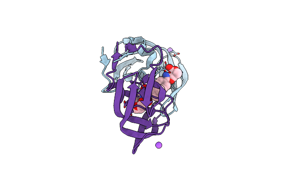
Crystal Structure Of The Kainate Receptor Gluk3 Ligand Binding Domain In Complex With (S)-1-[2-Amino-2-Carboxyethyl]-5,7-Dihydrothieno[3,4-D]Pyrimidin-2,4(1H,3H)-Dione At Resolution 2.6A
Organism: Rattus norvegicus
Method: X-RAY DIFFRACTION Resolution:2.60 Å Release Date: 2018-02-28 Classification: MEMBRANE PROTEIN Ligands: CGW, K, CL |
Organism: Rattus norvegicus
Method: X-RAY DIFFRACTION
Release Date: 2018-02-28
Ligands: CGW, K, CL
 |
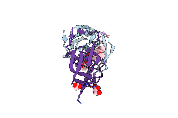
A Hydroxymethyl Functionality At The 4-Position Of The 2-Phenyloxazole Moiety Of Hiv-1 Protease Inhibitors Involving The P2' Ligands
Organism: Human immunodeficiency virus 1
Method: X-RAY DIFFRACTION Resolution:1.30 Å Release Date: 2017-11-22 Classification: HYDROLASE/HYDROLASE INHIBITOR Ligands: G53, NA, CL |
Organism: Human immunodeficiency virus 1
Method: X-RAY DIFFRACTION
Release Date: 2017-11-22
Ligands: G53, NA, CL
 |
A Novel 13-Ring Macrocyclic Hiv-1 Protease Inhibitors Involving The P1'-P2' Ligands
Organism: Human immunodeficiency virus 1
Method: X-RAY DIFFRACTION Resolution:1.27 Å Release Date: 2017-10-11 Classification: hydrolase/hydrolase inhibitor |
Organism: Human immunodeficiency virus 1
Method: X-RAY DIFFRACTION
Release Date: 2017-10-11
 |
Hiv-1 Wild Type Protease With Grl-1118A , An Isophthalamide-Derived P2-P3 Ligand With The Sulfonamide Isostere As The P2' Group
Organism: Human immunodeficiency virus 1
Method: X-RAY DIFFRACTION Resolution:1.33 Å Release Date: 2017-05-10 Classification: hydrolase/hydrolase inhibitor Ligands: 8FP, NA, CL, GOL, ACT |
Organism: Human immunodeficiency virus 1
Method: X-RAY DIFFRACTION
Release Date: 2017-05-10
Ligands: 8FP, NA, CL, GOL, ACT