

Search Count: 36
 |
Human Dnmt1(729-1600) Bound To Zebularine-Containing 12Mer Dsdna
Organism: Homo sapiens
Method: X-RAY DIFFRACTION Resolution:2.20 Å Release Date: 2021-07-07 Classification: TRANSFERASE Ligands: SAH, ZN, EDO, GOL |
Organism: Homo sapiens
Method: X-RAY DIFFRACTION
Release Date: 2021-07-07
Ligands: SAH, ZN, EDO, GOL
 |
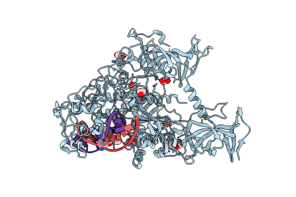
Human Dnmt1(729-1600) Bound To Zebularine-Containing 12Mer Dsdna And Inhibitor Gsk3830052
Organism: Homo sapiens
Method: X-RAY DIFFRACTION Resolution:1.79 Å Release Date: 2021-07-07 Classification: TRANSFERASE/TRANSFERASE INHIBITOR/DNA Ligands: ZN, EDO, X52 |
Organism: Homo sapiens
Method: X-RAY DIFFRACTION
Release Date: 2021-07-07
Ligands: ZN, EDO, X52
 |
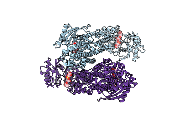
Human Dnmt1(729-1600) Bound To Zebularine-Containing 12Mer Dsdna And Inhibitor Gsk3685032A
Organism: Homo sapiens
Method: X-RAY DIFFRACTION Resolution:2.65 Å Release Date: 2021-07-07 Classification: TRANSFERASE/TRANSFERASE INHIBITOR/DNA Ligands: ZN, EDO, UXM |
Organism: Homo sapiens
Method: X-RAY DIFFRACTION
Release Date: 2021-07-07
Ligands: ZN, EDO, UXM
 |
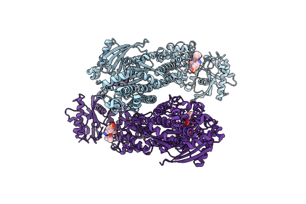
Crystal Structure Of Human Hexokinase 2 With Cmpd 30, A 2-Amino-6-Benzenesulfonamide Glucosamine
Organism: Homo sapiens
Method: X-RAY DIFFRACTION Resolution:2.73 Å Release Date: 2016-03-30 Classification: TRANSFERASE/TRANSFERASE INHIBITOR Ligands: 604 |
Organism: Homo sapiens
Method: X-RAY DIFFRACTION
Release Date: 2016-03-30
Ligands: 604
 |
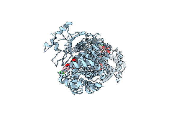
Crystal Structure Of Human Hexokinase 2 With Cmpd 27, A 2-Amido-6-Benzenesulfonamide Glucosamine
Organism: Homo sapiens
Method: X-RAY DIFFRACTION Resolution:2.92 Å Release Date: 2016-03-30 Classification: TRANSFERASE/TRANSFERASE INHIBITOR Ligands: 603 |
Organism: Homo sapiens
Method: X-RAY DIFFRACTION
Release Date: 2016-03-30
Ligands: 603
 |
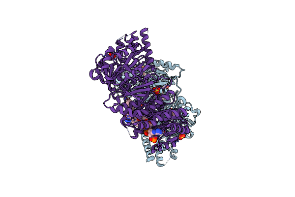
Crystal Structure Of Human Hexokinase 2 With Cmpd 1, A C-2-Substituted Glucosamine
Organism: Homo sapiens
Method: X-RAY DIFFRACTION Resolution:2.76 Å Release Date: 2016-03-30 Classification: TRANSFERASE/TRANSFERASE INHIBITOR Ligands: 62C, BG6, FLC |
Organism: Homo sapiens
Method: X-RAY DIFFRACTION
Release Date: 2016-03-30
Ligands: 62C, BG6, FLC
 |
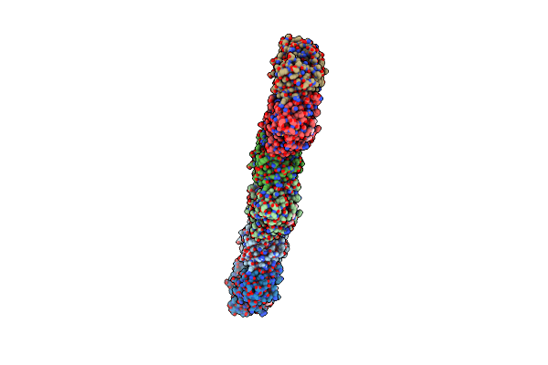
Human Fatty Acid Synthase Psi/Kr Tri-Domain With Nadph And Gsk2194069
Organism: Homo sapiens
Method: X-RAY DIFFRACTION Resolution:2.30 Å Release Date: 2014-07-23 Classification: TRANSFERASE/TRANSFERASE INHIBITOR Ligands: CAC, NDP, SO4, 2W4 |
Organism: Homo sapiens
Method: X-RAY DIFFRACTION
Release Date: 2014-07-23
Ligands: CAC, NDP, SO4, 2W4
 |
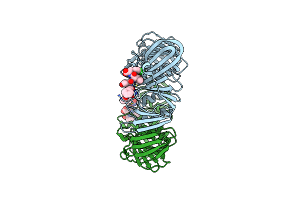
Structure Of A Small Peptide Ligand Bound To E.Coli Dna Sliding Clamp
Organism: Escherichia coli
Method: X-RAY DIFFRACTION Resolution:2.30 Å Release Date: 2011-12-28 Classification: transferase/transferase inhibitor |
Organism: Escherichia coli
Method: X-RAY DIFFRACTION
Release Date: 2011-12-28
 |
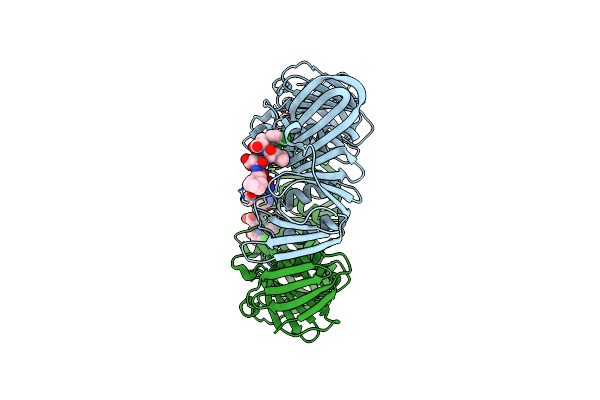
Structure Of A Small Peptide Ligand Bound To E.Coli Dna Sliding Clamp
Organism: Escherichia coli
Method: X-RAY DIFFRACTION Resolution:2.60 Å Release Date: 2011-12-28 Classification: transferase/transferase inhibitor |
Organism: Escherichia coli
Method: X-RAY DIFFRACTION
Release Date: 2011-12-28
 |
Structure Of A Small Peptide Ligand Bound To E.Coli Dna Sliding Clamp
Organism: Escherichia coli
Method: X-RAY DIFFRACTION Resolution:1.95 Å Release Date: 2011-12-28 Classification: transferase/transferase inhibitor |
Organism: Escherichia coli
Method: X-RAY DIFFRACTION
Release Date: 2011-12-28
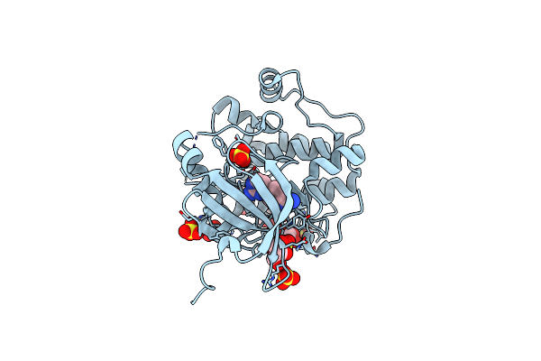 |
Phosphoinositide-Dependent Kinase-1 (Pdk1) With Lead Compound
Organism: Homo sapiens
Method: X-RAY DIFFRACTION Resolution:2.20 Å Release Date: 2011-06-22 Classification: TRANSFERASE/TRANSFERASE INHIBITOR Ligands: GOL, SO4, JMZ |
Organism: Homo sapiens
Method: X-RAY DIFFRACTION
Release Date: 2011-06-22
Ligands: GOL, SO4, JMZ
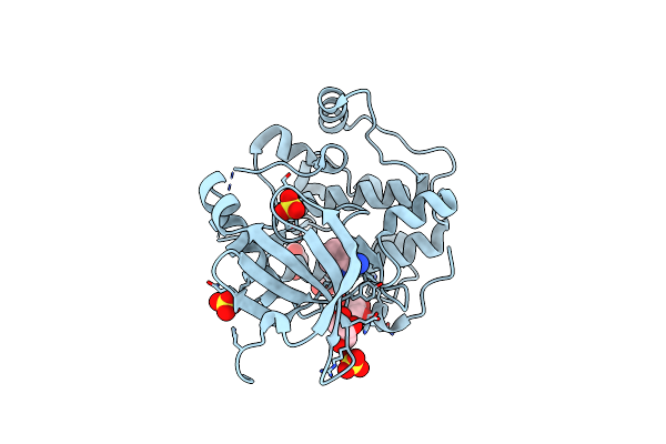 |
Phosphoinositide-Dependent Kinase-1 (Pdk1) With Fragment8
Organism: Homo sapiens
Method: X-RAY DIFFRACTION Resolution:2.75 Å Release Date: 2011-05-25 Classification: TRANSFERASE Ligands: GOL, JNZ, SO4 |
Organism: Homo sapiens
Method: X-RAY DIFFRACTION
Release Date: 2011-05-25
Ligands: GOL, JNZ, SO4
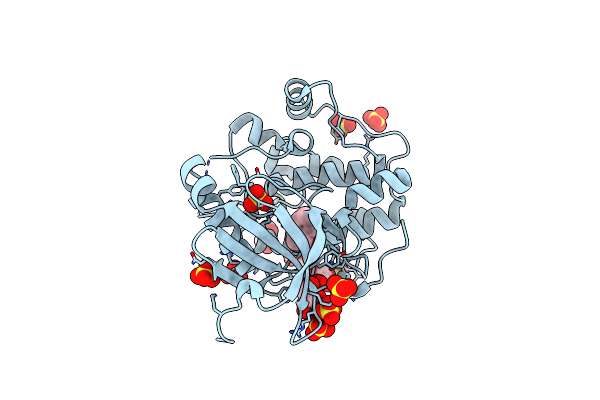 |
Phosphoinositide-Dependent Kinase-1 (Pdk1) With Fragment11
Organism: Homo sapiens
Method: X-RAY DIFFRACTION Resolution:1.98 Å Release Date: 2011-05-25 Classification: TRANSFERASE Ligands: GOL, SO4, JOZ |
Organism: Homo sapiens
Method: X-RAY DIFFRACTION
Release Date: 2011-05-25
Ligands: GOL, SO4, JOZ
 |
Phosphoinositide-Dependent Kinase-1 (Pdk1) With Fragment17
Organism: Homo sapiens
Method: X-RAY DIFFRACTION Resolution:2.10 Å Release Date: 2011-05-25 Classification: TRANSFERASE Ligands: GOL, SO4, JPZ |
Organism: Homo sapiens
Method: X-RAY DIFFRACTION
Release Date: 2011-05-25
Ligands: GOL, SO4, JPZ
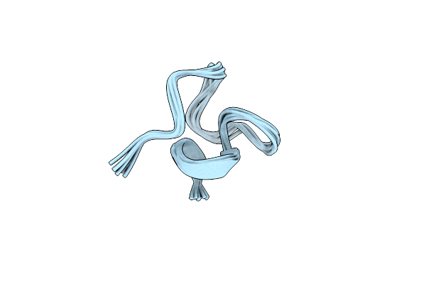 |
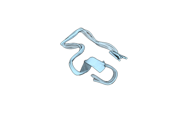
Structure Of The Spliceosomal Phosphopeptide P140 (Non-Phosphorylated Form)
|
Organism: Homo sapiens
Method: SOLUTION NMR
Release Date: 2010-12-01
 |
Structure Of The Spliceosomal Phosphopeptide P140 (Phosphorylated Form)
|
Organism: Homo sapiens
Method: SOLUTION NMR
Release Date: 2010-12-01
 |
Solution Structure Of Beta3-Analogue Peptide Corresponding To The Gp41 600-612 Loop Of Hiv.
|
Method: SOLUTION NMR
Release Date: 2003-07-01
 |
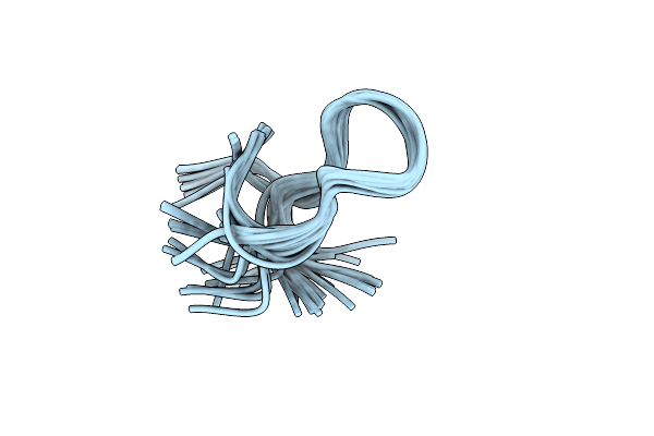
Solution Structure Of Beta3 Analogue Peptide (Hcys) Of Hiv Gp41 600-612 Loop.
|
Method: SOLUTION NMR
Release Date: 2003-07-01
 |
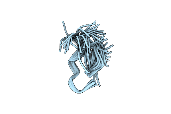
Solution Structure Of A Lactam Analogue (Dabd) Of Hiv Gp41 600-612 Loop.
|
Method: SOLUTION NMR
Release Date: 2003-07-01
 |
Solution Structure Of Lactam Analogue (Dape) Of Hiv Gp41 600-612 Loop.
|
Method: SOLUTION NMR
Release Date: 2003-07-01