

Search Count: 11
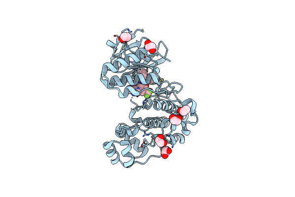 |
Dual Phosphorylated Human P38 Alpha Bound To Nilotinib
Organism: Homo sapiens
Method: X-RAY DIFFRACTION Resolution:1.80 Å Release Date: 2024-11-13 Classification: TRANSFERASE Ligands: EDO, NIL |
Organism: Homo sapiens
Method: X-RAY DIFFRACTION
Release Date: 2024-11-13
Ligands: EDO, NIL
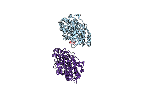 |
Dual Phosphorylated Human P38 Alpha
Organism: Homo sapiens
Method: X-RAY DIFFRACTION Resolution:2.83 Å Release Date: 2024-11-13 Classification: TRANSFERASE Ligands: PEG, EDO |
Organism: Homo sapiens
Method: X-RAY DIFFRACTION
Release Date: 2024-11-13
Ligands: PEG, EDO
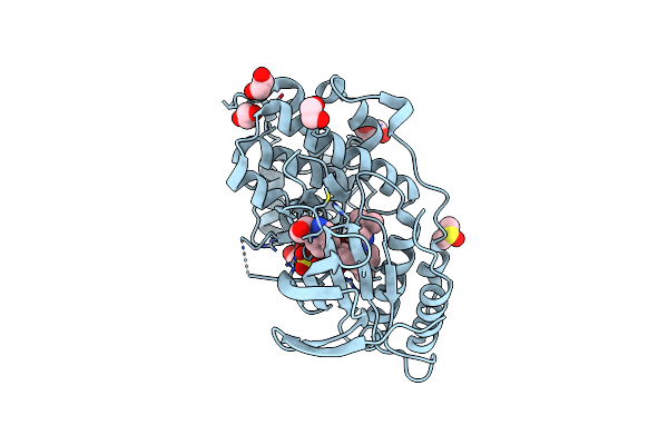 |
Dual Phosphorylated Human P38 Alpha Bound To Pexmetinib
Organism: Homo sapiens
Method: X-RAY DIFFRACTION Resolution:1.95 Å Release Date: 2024-11-13 Classification: TRANSFERASE Ligands: SO4, EDO, GOL, DMS, PDO, A1AWV |
Organism: Homo sapiens
Method: X-RAY DIFFRACTION
Release Date: 2024-11-13
Ligands: SO4, EDO, GOL, DMS, PDO, A1AWV
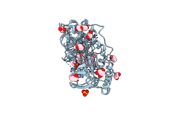 |
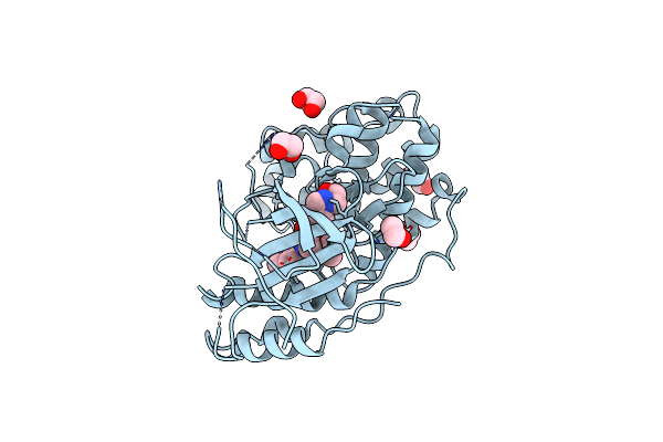
Dual Phosphorylated Human P38 Alpha Bound To Birb796
Organism: Homo sapiens
Method: X-RAY DIFFRACTION Resolution:1.80 Å Release Date: 2024-11-13 Classification: TRANSFERASE Ligands: B96, SO4, EDO, PEG, GOL |
Organism: Homo sapiens
Method: X-RAY DIFFRACTION
Release Date: 2024-11-13
Ligands: B96, SO4, EDO, PEG, GOL
 |
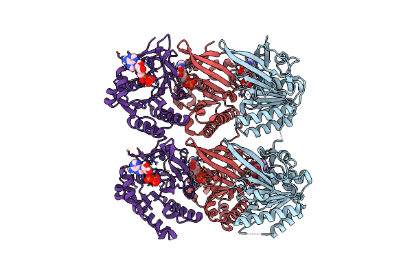
Unphosphorylated Human P38 Alpha Bound To Pexmetinib
Organism: Homo sapiens
Method: X-RAY DIFFRACTION Resolution:3.30 Å Release Date: 2024-11-13 Classification: TRANSFERASE Ligands: EDO, A1AWV, PDO |
Organism: Homo sapiens
Method: X-RAY DIFFRACTION
Release Date: 2024-11-13
Ligands: EDO, A1AWV, PDO
 |
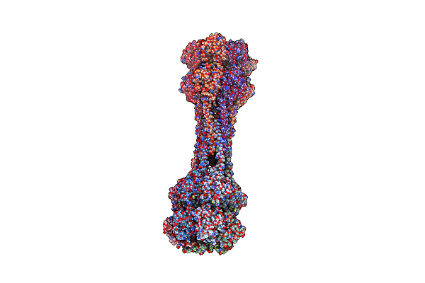
Crystal Structure Of Kaic With Truncated C-Terminal Coiled-Coil Domain
Organism: Cereibacter sphaeroides
Method: X-RAY DIFFRACTION Resolution:2.90 Å Release Date: 2023-03-22 Classification: CIRCADIAN CLOCK PROTEIN Ligands: ADP |
Organism: Cereibacter sphaeroides
Method: X-RAY DIFFRACTION
Release Date: 2023-03-22
Ligands: ADP
 |
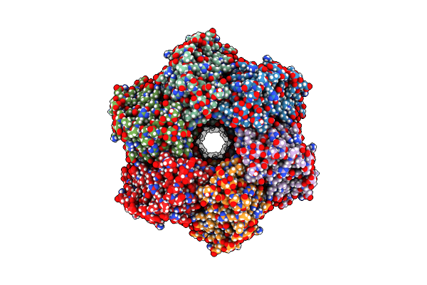
Crystal Structure Of Dodecameric Kaic
Organism: Cereibacter sphaeroides
Method: X-RAY DIFFRACTION Resolution:3.50 Å Release Date: 2023-03-22 Classification: CIRCADIAN CLOCK PROTEIN Ligands: ADP, MG |
Organism: Cereibacter sphaeroides
Method: X-RAY DIFFRACTION
Release Date: 2023-03-22
Ligands: ADP, MG
 |
Structure Of Dodecameric Kaic-Rs-S413E/S414E Solved By Cryo-Em
Organism: Cereibacter sphaeroides
Method: ELECTRON MICROSCOPY Release Date: 2023-03-22 Classification: CIRCADIAN CLOCK PROTEIN Ligands: ATP, ADP, MG |
Organism: Cereibacter sphaeroides
Method: ELECTRON MICROSCOPY
Release Date: 2023-03-22
Ligands: ATP, ADP, MG
 |
Structure Of Dodecameric Kaic-Rs-S413E/S414E Complexed With Kaib-Rs Solved By Cryo-Em
Organism: Cereibacter sphaeroides
Method: ELECTRON MICROSCOPY Release Date: 2023-03-22 Classification: CIRCADIAN CLOCK PROTEIN Ligands: ATP, ADP, MG |
Organism: Cereibacter sphaeroides
Method: ELECTRON MICROSCOPY
Release Date: 2023-03-22
Ligands: ATP, ADP, MG
 |
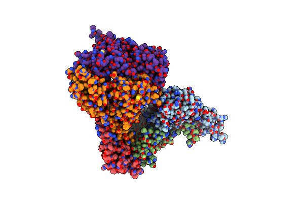
Structure Of The Phosphatase Domain Of The Cell Fate Determinant Spoiie From Bacillus Subtilis In A Crystal Form Without Domain Swapping
Organism: Bacillus subtilis
Method: X-RAY DIFFRACTION Resolution:2.45 Å Release Date: 2017-05-31 Classification: TRANSFERASE |
Organism: Bacillus subtilis
Method: X-RAY DIFFRACTION
Release Date: 2017-05-31
 |
Structure Of The Pp2C Phosphatase Domain And A Fragment Of The Regulatory Domain Of The Cell Fate Determinant Spoiie From Bacillus Subtilis
Organism: Bacillus subtilis (strain 168)
Method: X-RAY DIFFRACTION Resolution:3.91 Å Release Date: 2017-05-31 Classification: HYDROLASE |
Organism: Bacillus subtilis (strain 168)
Method: X-RAY DIFFRACTION
Release Date: 2017-05-31