

Search Count: 16
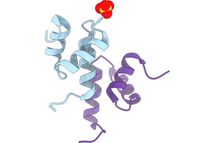 |
Stabilized Y188N Variant Of The Internal Uba Domain Of Hhr23A In The P1 21 1 Space Group
Organism: Homo sapiens
Method: X-RAY DIFFRACTION Resolution:1.15 Å Release Date: 2026-08-05 Classification: TRANSCRIPTION Ligands: SO4 |
Organism: Homo sapiens
Method: X-RAY DIFFRACTION
Release Date: 2026-08-05
Ligands: SO4
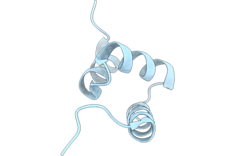 |
Stabilized Y188N Variant Of The Internal Uba Domain Of Hhr23A In The P43 Space Group
Organism: Homo sapiens
Method: X-RAY DIFFRACTION Resolution:1.20 Å Release Date: 2026-08-05 Classification: TRANSCRIPTION |
Organism: Homo sapiens
Method: X-RAY DIFFRACTION
Release Date: 2026-08-05
 |
E176T/Y188G Variant Of The Internal Uba Domain Of Hhr23A
Organism: Homo sapiens
Method: X-RAY DIFFRACTION Resolution:1.40 Å Release Date: 2023-04-12 Classification: DNA BINDING PROTEIN |
Organism: Homo sapiens
Method: X-RAY DIFFRACTION
Release Date: 2023-04-12
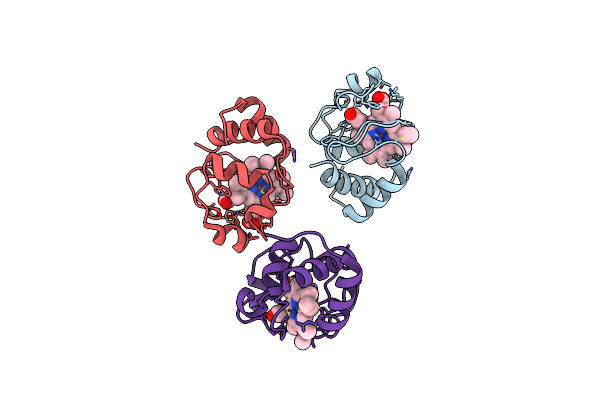 |
Crystal Structure Of N63T Yeast Iso-1-Cytochrome C
Organism: Saccharomyces cerevisiae (strain atcc 204508 / s288c)
Method: X-RAY DIFFRACTION Resolution:2.46 Å Release Date: 2022-05-11 Classification: ELECTRON TRANSPORT Ligands: HEC |
Organism: Saccharomyces cerevisiae (strain atcc 204508 / s288c)
Method: X-RAY DIFFRACTION
Release Date: 2022-05-11
Ligands: HEC
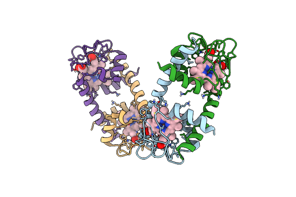 |
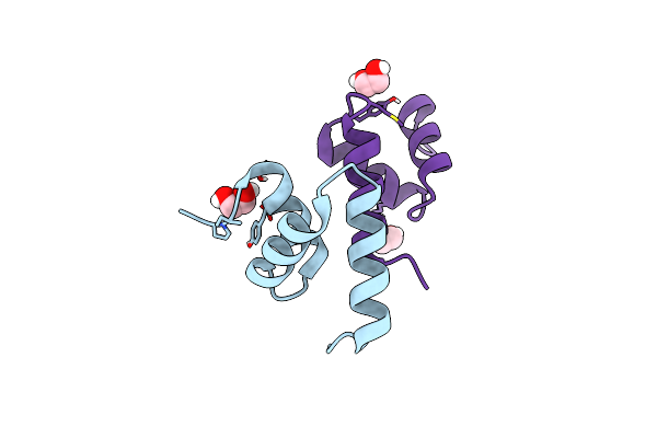
Crystal Structure Of Dimeric K72A Human Cytochrome C Alkaline Conformer
Organism: Homo sapiens
Method: X-RAY DIFFRACTION Resolution:2.08 Å Release Date: 2021-07-07 Classification: ELECTRON TRANSPORT Ligands: HEC |
Organism: Homo sapiens
Method: X-RAY DIFFRACTION
Release Date: 2021-07-07
Ligands: HEC
 |
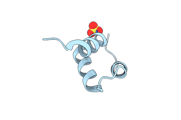
Crystal Structure Of Y188G Variant Of The Internal Uba Domain Of Hhr23A In Monoclinic Unit Cell
Organism: Homo sapiens
Method: X-RAY DIFFRACTION Resolution:1.10 Å Release Date: 2021-03-10 Classification: DNA BINDING PROTEIN Ligands: EDO |
Organism: Homo sapiens
Method: X-RAY DIFFRACTION
Release Date: 2021-03-10
Ligands: EDO
 |
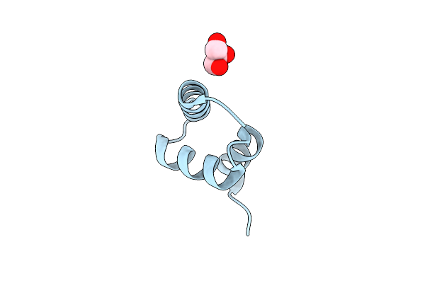
Crystal Structure Of The Internal Uba Domain Of Hhr23A
Organism: Homo sapiens
Method: X-RAY DIFFRACTION Resolution:1.60 Å Release Date: 2021-03-10 Classification: DNA BINDING PROTEIN Ligands: SO4 |
Organism: Homo sapiens
Method: X-RAY DIFFRACTION
Release Date: 2021-03-10
Ligands: SO4
 |
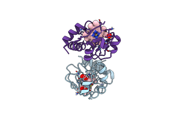
Crystal Structure Of Y188G Variant Of The Internal Uba Domain Of Hhr23A
Organism: Homo sapiens
Method: X-RAY DIFFRACTION Resolution:1.45 Å Release Date: 2021-03-10 Classification: DNA BINDING PROTEIN Ligands: GOL |
Organism: Homo sapiens
Method: X-RAY DIFFRACTION
Release Date: 2021-03-10
Ligands: GOL
 |
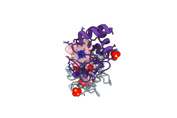
Crystal Structure Of A51V Variant Of Human Cytochrome C
Organism: Homo sapiens
Method: X-RAY DIFFRACTION Resolution:1.82 Å Release Date: 2019-06-26 Classification: APOPTOSIS Ligands: HEC |
Organism: Homo sapiens
Method: X-RAY DIFFRACTION
Release Date: 2019-06-26
Ligands: HEC
 |
Crystal Structure Of K72A Variant Of Human Cytochrome C
Organism: Homo sapiens
Method: X-RAY DIFFRACTION Resolution:1.25 Å Release Date: 2017-09-06 Classification: ELECTRON TRANSPORT Ligands: HEC, SO4 |
Organism: Homo sapiens
Method: X-RAY DIFFRACTION
Release Date: 2017-09-06
Ligands: HEC, SO4
 |
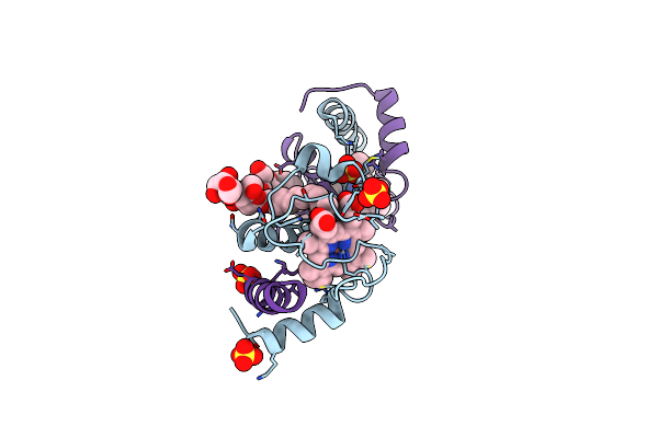
Crystal Structure Of A Domain-Swapped Dimer Of Yeast Iso-1-Cytochrome C With Cymal5
Organism: Saccharomyces cerevisiae (strain atcc 204508 / s288c)
Method: X-RAY DIFFRACTION Resolution:1.70 Å Release Date: 2017-03-22 Classification: ELECTRON TRANSPORT Ligands: HEC, SO4, CM5, CL, GOL |
Organism: Saccharomyces cerevisiae (strain atcc 204508 / s288c)
Method: X-RAY DIFFRACTION
Release Date: 2017-03-22
Ligands: HEC, SO4, CM5, CL, GOL
 |
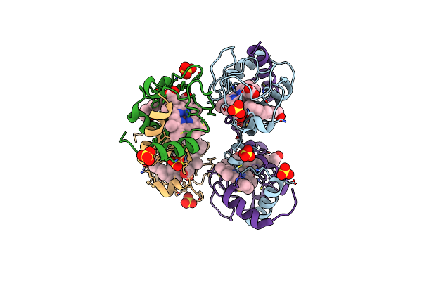
Crystal Structure Of A Domain-Swapped Dimer Of Yeast Iso-1-Cytochrome C With Omega-Undecylenyl-Beta-D-Maltopyranoside
Organism: Saccharomyces cerevisiae (strain atcc 204508 / s288c)
Method: X-RAY DIFFRACTION Resolution:1.99 Å Release Date: 2017-03-22 Classification: ELECTRON TRANSPORT Ligands: HEC, 6UZ, SO4 |
Organism: Saccharomyces cerevisiae (strain atcc 204508 / s288c)
Method: X-RAY DIFFRACTION
Release Date: 2017-03-22
Ligands: HEC, 6UZ, SO4
 |
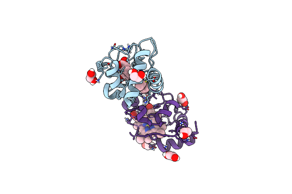
Crystal Structure Of Dimeric Yeast Iso-1-Cytochrome C With Cymal6
Organism: Saccharomyces cerevisiae (strain atcc 204508 / s288c)
Method: X-RAY DIFFRACTION Resolution:2.00 Å Release Date: 2017-03-22 Classification: ELECTRON TRANSPORT Ligands: SO4, ZE7, HEC |
Organism: Saccharomyces cerevisiae (strain atcc 204508 / s288c)
Method: X-RAY DIFFRACTION
Release Date: 2017-03-22
Ligands: SO4, ZE7, HEC
 |
Crystal Structure Of Spider Monkey Cytochrome C At 1.15 Angstrom
Organism: Ateles sp.
Method: X-RAY DIFFRACTION Resolution:1.15 Å Release Date: 2016-03-02 Classification: ELECTRON TRANSPORT Ligands: HEC, EDO, CL |
Organism: Ateles sp.
Method: X-RAY DIFFRACTION
Release Date: 2016-03-02
Ligands: HEC, EDO, CL
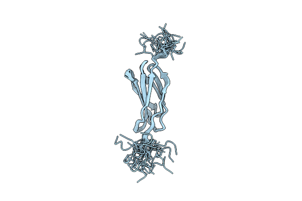 |
Structure Of The Third Type Iii Domain From Human Fibronectin
Organism: Homo sapiens
Method: SOLUTION NMR Release Date: 2015-11-11 Classification: STRUCTURAL PROTEIN |
Organism: Homo sapiens
Method: SOLUTION NMR
Release Date: 2015-11-11
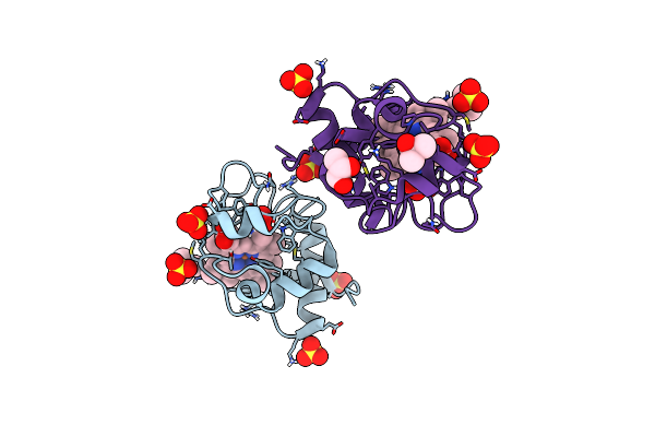 |
Crystal Structure Of An Oxidized Form Of Yeast Iso-1-Cytochrome C At Ph 8.8
Organism: Saccharomyces cerevisiae
Method: X-RAY DIFFRACTION Resolution:1.45 Å Release Date: 2014-06-04 Classification: ELECTRON TRANSPORT Ligands: HEC, SO4, TBU, GOL |
Organism: Saccharomyces cerevisiae
Method: X-RAY DIFFRACTION
Release Date: 2014-06-04
Ligands: HEC, SO4, TBU, GOL