

Search Count: 704
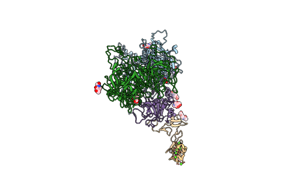 |
Cryo-Em Structure Of Factor Va Bound To Activated Protein C
Organism: Homo sapiens
Method: ELECTRON MICROSCOPY Release Date: 2025-05-21 Classification: BLOOD CLOTTING Ligands: NAG, CA |
Organism: Homo sapiens
Method: ELECTRON MICROSCOPY
Release Date: 2025-05-21
Ligands: NAG, CA
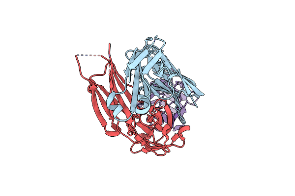 |
Human Factor Viii C2 Domain Complexed To Human Monoclonal Bo2C11 Fab.
Organism: Homo sapiens
Method: X-RAY DIFFRACTION Resolution:2.00 Å Release Date: 2001-08-15 Classification: IMMUNE SYSTEM/BLOOD CLOTTING |
Organism: Homo sapiens
Method: X-RAY DIFFRACTION
Release Date: 2001-08-15
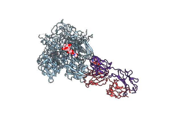 |
Structure Of Blood Coagulation Factor Viii In Complex With An Anti-C1 Domain Pathogenic Antibody Inhibitor
Organism: Sus scrofa, Homo sapiens, Mus musculus
Method: X-RAY DIFFRACTION Resolution:3.92 Å Release Date: 2020-10-14 Classification: BLOOD CLOTTING/Immune System Ligands: NAG, CU, CA, ZN |
Organism: Sus scrofa, Homo sapiens, Mus musculus
Method: X-RAY DIFFRACTION
Release Date: 2020-10-14
Ligands: NAG, CU, CA, ZN
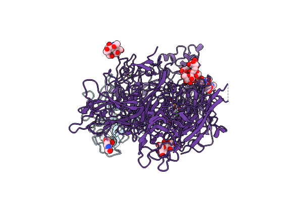 |
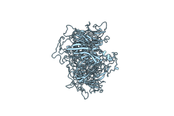
Crystal Structure Of The Prothrombinase Complex From The Venom Of Pseudonaja Textilis
Organism: Pseudonaja textilis
Method: X-RAY DIFFRACTION Resolution:3.32 Å Release Date: 2013-07-31 Classification: BLOOD CLOTTING Ligands: NAG, CA, CU |
Organism: Pseudonaja textilis
Method: X-RAY DIFFRACTION
Release Date: 2013-07-31
Ligands: NAG, CA, CU
 |
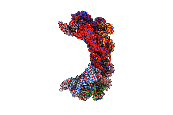
Cryo-Em Structure Of Coagulation Factor V Short
Organism: Homo sapiens
Method: ELECTRON MICROSCOPY Release Date: 2023-03-15 Classification: BLOOD CLOTTING |
Organism: Homo sapiens
Method: ELECTRON MICROSCOPY
Release Date: 2023-03-15
 |
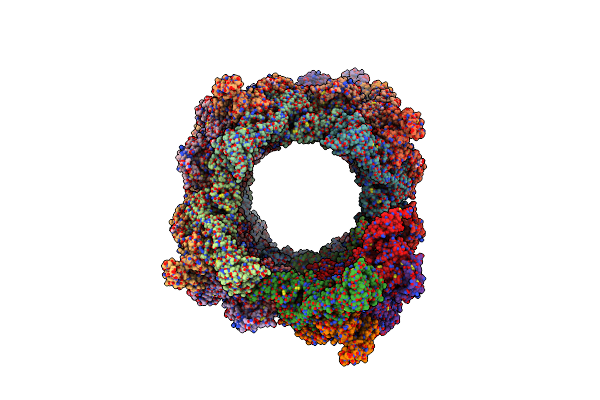
Cryo-Em Structure Of Vwf D'D3 Dimer Complexed With D1D2 At 3.27 Angstron Resolution (2 Units)
Organism: Homo sapiens
Method: ELECTRON MICROSCOPY Release Date: 2022-05-25 Classification: BLOOD CLOTTING Ligands: NAG, CA |
Organism: Homo sapiens
Method: ELECTRON MICROSCOPY
Release Date: 2022-05-25
Ligands: NAG, CA
 |
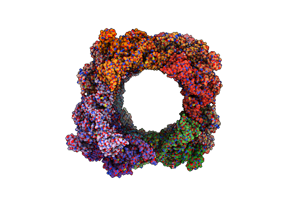
Vwf D'D3 Dimer Complexed With D1D2 At 4.39 Angstron Resolution(Vwf Tube)
Organism: Homo sapiens
Method: ELECTRON MICROSCOPY Release Date: 2022-05-25 Classification: BLOOD CLOTTING Ligands: NAG, CA |
Organism: Homo sapiens
Method: ELECTRON MICROSCOPY
Release Date: 2022-05-25
Ligands: NAG, CA
 |
Cryo-Em Structure Of Vwf D'D3 Dimer Complexed With D1D2 At 4.3 Angstron Resolution (7 Units)
Organism: Homo sapiens
Method: ELECTRON MICROSCOPY Release Date: 2022-05-25 Classification: BLOOD CLOTTING Ligands: NAG, CA |
Organism: Homo sapiens
Method: ELECTRON MICROSCOPY
Release Date: 2022-05-25
Ligands: NAG, CA
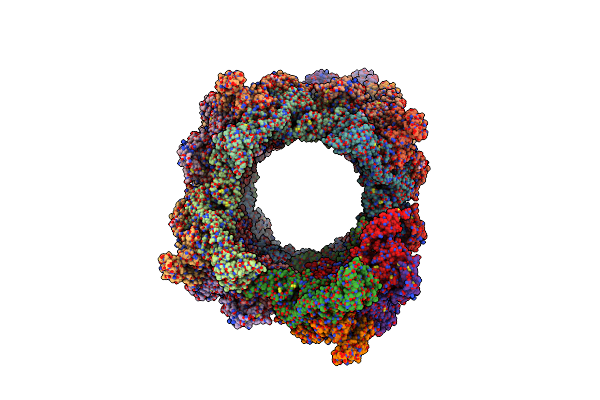 |
Cryo-Em Structure Of Vwf D'D3 Dimer Complexed With D1D2 At 4.3 Angstron Resolution (Vwf Tube)
Organism: Homo sapiens
Method: ELECTRON MICROSCOPY Release Date: 2022-05-25 Classification: BLOOD CLOTTING Ligands: NAG, CA |
Organism: Homo sapiens
Method: ELECTRON MICROSCOPY
Release Date: 2022-05-25
Ligands: NAG, CA
 |
Cryo-Em Structure Of Vwf D'D3 Dimer Complexed With D1D2 At 2.85 Angstron Resolution (1 Unit)
Organism: Homo sapiens
Method: ELECTRON MICROSCOPY Release Date: 2022-05-25 Classification: BLOOD CLOTTING Ligands: CA, NAG |
Organism: Homo sapiens
Method: ELECTRON MICROSCOPY
Release Date: 2022-05-25
Ligands: CA, NAG
 |
Cryo-Em Complex Of Meizothrombindesf1, Factor Xa, And Factor Va
Organism: Homo sapiens
Method: ELECTRON MICROSCOPY Release Date: 2026-05-20 Classification: BLOOD CLOTTING |
Organism: Homo sapiens
Method: ELECTRON MICROSCOPY
Release Date: 2026-05-20
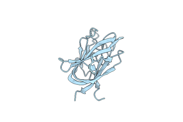 |
Crystal Structure Of Blood Coagulation Factor Viii C2 Domain Mutant L2251A/L2252A
Organism: Homo sapiens
Method: X-RAY DIFFRACTION Resolution:1.83 Å Release Date: 2024-09-04 Classification: BLOOD CLOTTING |
Organism: Homo sapiens
Method: X-RAY DIFFRACTION
Release Date: 2024-09-04
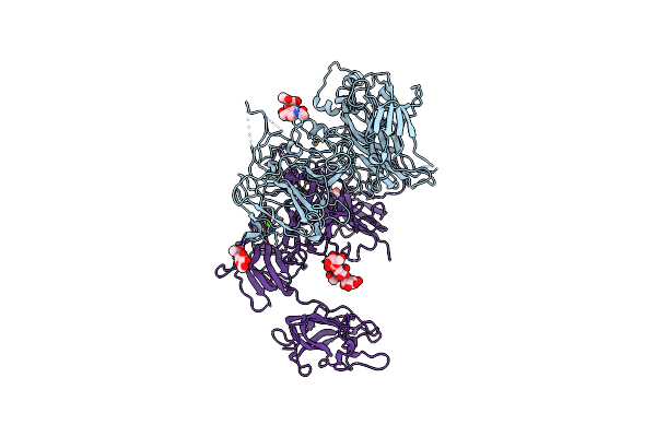 |
Model Of Membrane-Bound Factor Viii Organized In 2D Crystals
Organism: Homo sapiens
Method: ELECTRON CRYSTALLOGRAPHY Release Date: 2013-09-11 Classification: BLOOD CLOTTING Ligands: CU, CA, NAG |
Organism: Homo sapiens
Method: ELECTRON CRYSTALLOGRAPHY
Release Date: 2013-09-11
Ligands: CU, CA, NAG
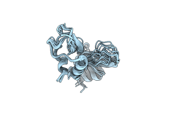 |
Nmr Structure Determination Of Ixolaris And Factor X Interaction Reveals A Noncanonical Mechanism Of Kunitz Inhibition
Organism: Ixodes scapularis
Method: SOLUTION NMR Release Date: 2019-06-12 Classification: BLOOD CLOTTING |
Organism: Ixodes scapularis
Method: SOLUTION NMR
Release Date: 2019-06-12
 |
Crystal Structure Of The Prothrombinase Complex From The Venom Of Pseudonaja Textilis
Organism: Pseudonaja textilis, Escherichia coli
Method: X-RAY DIFFRACTION Resolution:2.71 Å Release Date: 2013-07-31 Classification: BLOOD CLOTTING Ligands: 0GJ, NA, GOL |
Organism: Pseudonaja textilis, Escherichia coli
Method: X-RAY DIFFRACTION
Release Date: 2013-07-31
Ligands: 0GJ, NA, GOL
 |
Crystal Structure Of Bbetad432A Variant Fibrinogen Fragment D With The Peptide Ligand Gly-His-Arg-Pro-Amide
Organism: Homo sapiens, Synthetic construct
Method: X-RAY DIFFRACTION Resolution:2.30 Å Release Date: 2009-01-13 Classification: BLOOD CLOTTING Ligands: NAG, CA |
Organism: Homo sapiens, Synthetic construct
Method: X-RAY DIFFRACTION
Release Date: 2009-01-13
Ligands: NAG, CA
 |
Structure Of Engineered Coagulation Factor Viii With Enhanced Secretion And Coagulation Potential
Organism: Homo sapiens
Method: ELECTRON MICROSCOPY Release Date: 2025-09-10 Classification: BLOOD CLOTTING |
Organism: Homo sapiens
Method: ELECTRON MICROSCOPY
Release Date: 2025-09-10
 |
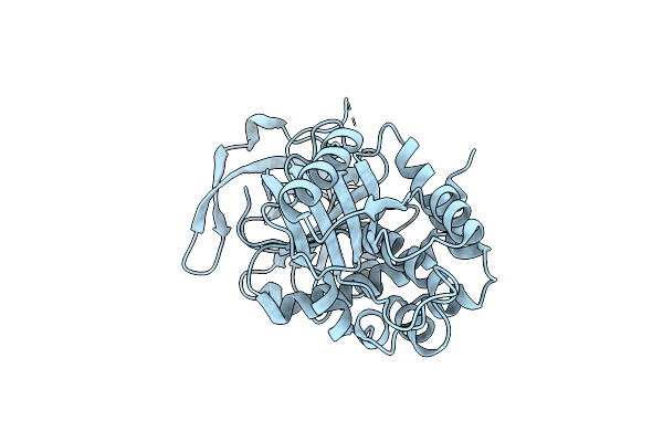
Crystal Structure Analysis Of Coagulation Factor Viii
Organism: Homo sapiens
Method: X-RAY DIFFRACTION Resolution:3.70 Å Release Date: 2008-04-15 Classification: BLOOD CLOTTING Ligands: CA, CU |
Organism: Homo sapiens
Method: X-RAY DIFFRACTION
Release Date: 2008-04-15
Ligands: CA, CU
 |
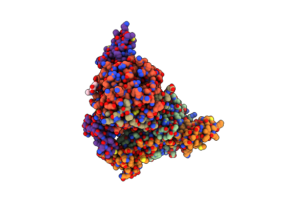
The Crystal Structure Of Mouse Protein-Z Dependent Protease Inhibitor(Mzpi)
Organism: Mus musculus
Method: X-RAY DIFFRACTION Resolution:2.50 Å Release Date: 2012-12-05 Classification: BLOOD CLOTTING |
Organism: Mus musculus
Method: X-RAY DIFFRACTION
Release Date: 2012-12-05
 |
Thrombin-Bound Boophilin Displays A Functional And Accessible Reactive-Site Loop
Organism: Rhipicephalus microplus, Bos taurus
Method: X-RAY DIFFRACTION Resolution:2.35 Å Release Date: 2008-01-22 Classification: BLOOD CLOTTING/BLOOD CLOTTING INHIBITOR Ligands: PO4, NA, NAG |
Organism: Rhipicephalus microplus, Bos taurus
Method: X-RAY DIFFRACTION
Release Date: 2008-01-22
Ligands: PO4, NA, NAG