

Search Count: 31
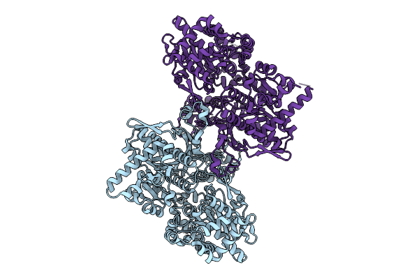 |
Structure Of Glycogen Phosphorylase - Dimeric Form - From Escherichia Coli
Organism: Escherichia coli
Method: ELECTRON MICROSCOPY Resolution:3.10 Å Release Date: 2026-04-08 Classification: TRANSFERASE |
Organism: Escherichia coli
Method: ELECTRON MICROSCOPY
Release Date: 2026-04-08
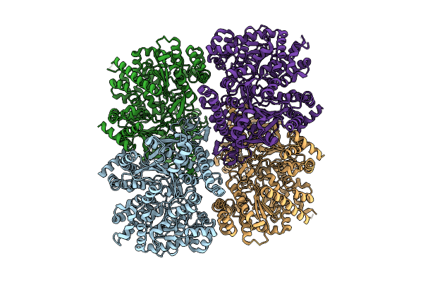 |
Structure Of Glycogen Phosphorylase - Tetrameric Form - From Escherichia Coli
Organism: Escherichia coli
Method: ELECTRON MICROSCOPY Resolution:2.97 Å Release Date: 2026-04-08 Classification: TRANSFERASE |
Organism: Escherichia coli
Method: ELECTRON MICROSCOPY
Release Date: 2026-04-08
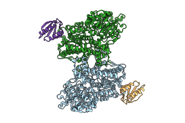 |
Structure Of Glycogen Phosphorylase - Dimeric Form - In Complex With Hpr From Escherichia Coli
Organism: Escherichia coli
Method: ELECTRON MICROSCOPY Resolution:2.79 Å Release Date: 2026-04-08 Classification: TRANSFERASE |
Organism: Escherichia coli
Method: ELECTRON MICROSCOPY
Release Date: 2026-04-08
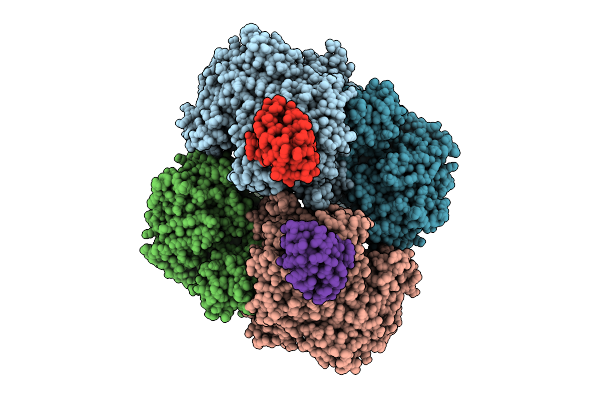 |
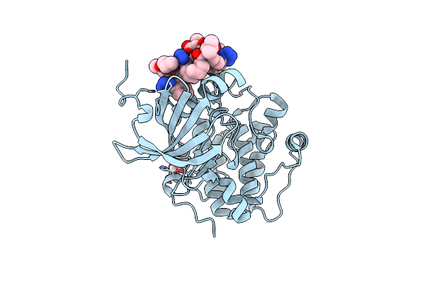
Structure Of Glycogen Phosphorylase - Tetrameric Form - In Complex With Hpr From Escherichia Coli
Organism: Escherichia coli
Method: ELECTRON MICROSCOPY Resolution:2.06 Å Release Date: 2026-04-08 Classification: TRANSFERASE |
Organism: Escherichia coli
Method: ELECTRON MICROSCOPY
Release Date: 2026-04-08
 |
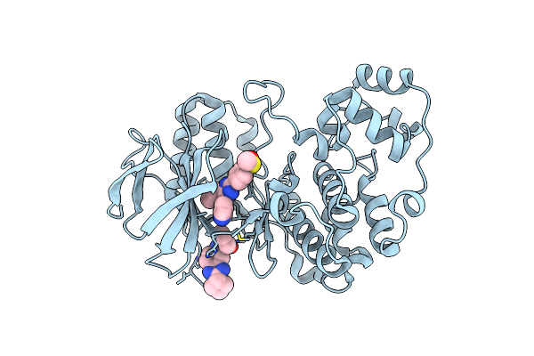
Human Pdk1 Kinase Domain In Complex With Valsartan
Organism: Homo sapiens
Method: X-RAY DIFFRACTION Resolution:1.31 Å Release Date: 2023-06-21 Classification: TRANSFERASE Ligands: U35, GOL |
Organism: Homo sapiens
Method: X-RAY DIFFRACTION
Release Date: 2023-06-21
Ligands: U35, GOL
 |
Crystal Structure Of P38Alpha C162S In Complex With Sb20358 And Cas 2094667-81-7 (Behind Catalytic Site; Y35 In), P 21 21 21
Organism: Mus musculus
Method: X-RAY DIFFRACTION Resolution:2.25 Å Release Date: 2023-06-14 Classification: ONCOPROTEIN Ligands: IIM, 87B |
Organism: Mus musculus
Method: X-RAY DIFFRACTION
Release Date: 2023-06-14
Ligands: IIM, 87B
 |
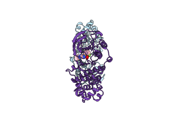
Crystal Structure Of P38Alpha C162S In Complex With Atpgs And Cas 2094667-81-7 (In Catalytic Site, Y35 Out), P 1 21 1
Organism: Mus musculus
Method: X-RAY DIFFRACTION Resolution:2.60 Å Release Date: 2023-06-14 Classification: ONCOPROTEIN Ligands: 87B, ATP |
Organism: Mus musculus
Method: X-RAY DIFFRACTION
Release Date: 2023-06-14
Ligands: 87B, ATP
 |
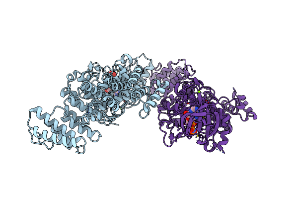
Crystal Structure Of P38Alpha C162S In Complex With Cas2094511-69-8, P 1 21 1
Organism: Mus musculus
Method: X-RAY DIFFRACTION Resolution:2.15 Å Release Date: 2023-05-17 Classification: ONCOPROTEIN Ligands: 8DI |
Organism: Mus musculus
Method: X-RAY DIFFRACTION
Release Date: 2023-05-17
Ligands: 8DI
 |
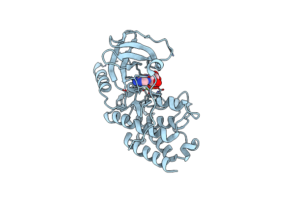
Crystal Structure Of The S/T Protein Kinase Pkng From Corynebacterium Glutamicum In Complex With Amp-Pnp
Organism: Corynebacterium glutamicum (strain atcc 13032 / dsm 20300 / bcrc 11384 / jcm 1318 / lmg 3730 / ncimb 10025)
Method: X-RAY DIFFRACTION Resolution:2.20 Å Release Date: 2021-10-13 Classification: TRANSFERASE Ligands: MG, ANP |
 |
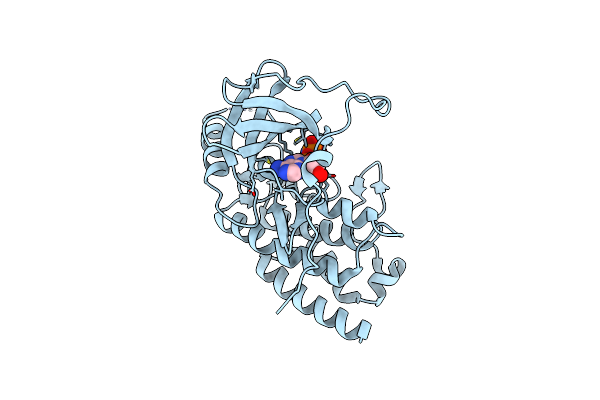
Crystal Structure Of The S/T Protein Kinase Pkng From Corynebacterium Glutamicum (Residues 130-433) In Complex With Amp-Pnp, Isoform 1
Organism: Corynebacterium glutamicum (strain atcc 13032 / dsm 20300 / bcrc 11384 / jcm 1318 / lmg 3730 / ncimb 10025)
Method: X-RAY DIFFRACTION Resolution:1.92 Å Release Date: 2021-10-13 Classification: TRANSFERASE Ligands: MG, ANP |
 |
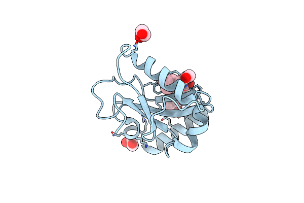
Crystal Structure Of The S/T Protein Kinase Pkng From Corynebacterium Glutamicum (Residues 130-433) In Complex With Amp-Pnp, Isoform 2
Organism: Corynebacterium glutamicum (strain atcc 13032 / dsm 20300 / bcrc 11384 / jcm 1318 / lmg 3730 / ncimb 10025)
Method: X-RAY DIFFRACTION Resolution:1.99 Å Release Date: 2021-10-13 Classification: TRANSFERASE Ligands: MG, ANP |
 |
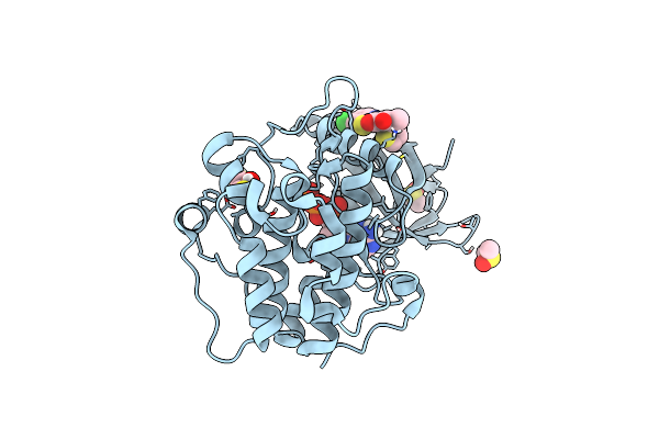
Lc3A In Complex With (3R,4S,5R,6R)-5-Hydroxy-6-((4-Hydroxy-3-(4-Hydroxy-3-Isopentylbenzamido)-8-Methyl-2-Oxo-2H-Chromen-7-Yl)Oxy)-3-Methoxy-2,2-Dimethyltetrahydro-2H-Pyran-4-Yl Carbamate
Organism: Homo sapiens
Method: X-RAY DIFFRACTION Resolution:1.67 Å Release Date: 2020-11-18 Classification: HYDROLASE Ligands: NOV, EDO |
Organism: Homo sapiens
Method: X-RAY DIFFRACTION
Release Date: 2020-11-18
Ligands: NOV, EDO
 |
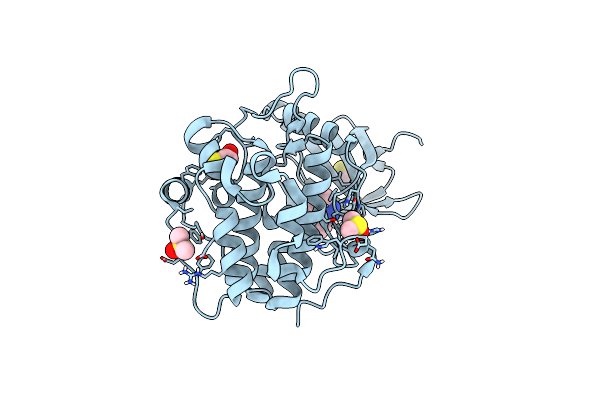
Human Pdk1-Pkciota Kinase Chimera In Complex With Allosteric Compound Ps267 Bound To The Pif-Pocket
Organism: Homo sapiens
Method: X-RAY DIFFRACTION Resolution:1.41 Å Release Date: 2017-01-18 Classification: TRANSFERASE Ligands: S26, DTT, DMS, ATP |
Organism: Homo sapiens
Method: X-RAY DIFFRACTION
Release Date: 2017-01-18
Ligands: S26, DTT, DMS, ATP
 |
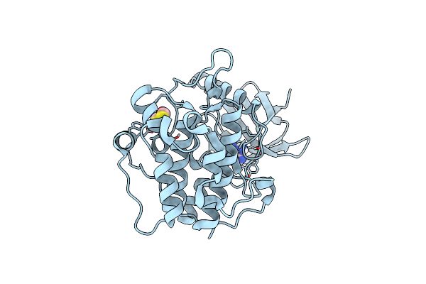
Human Pdk1 Kinase Domain In Complex With Compound Ps653 Bound To The Atp-Binding Site
Organism: Homo sapiens
Method: X-RAY DIFFRACTION Resolution:1.40 Å Release Date: 2016-10-19 Classification: TRANSFERASE Ligands: 537, DTD, DMS |
Organism: Homo sapiens
Method: X-RAY DIFFRACTION
Release Date: 2016-10-19
Ligands: 537, DTD, DMS
 |
Human Pdk1 Kinase Domain In Complex With Adenine Bound To The Atp-Binding Site
Organism: Homo sapiens
Method: X-RAY DIFFRACTION Resolution:1.26 Å Release Date: 2016-10-19 Classification: TRANSFERASE Ligands: ADE, DTD |
Organism: Homo sapiens
Method: X-RAY DIFFRACTION
Release Date: 2016-10-19
Ligands: ADE, DTD
 |
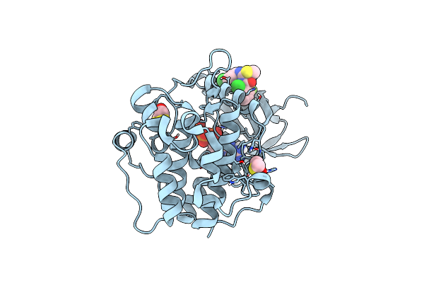
Human Pdk1 Kinase Domain In Complex With Adenosine Bound To The Atp-Binding Site
Organism: Homo sapiens
Method: X-RAY DIFFRACTION Resolution:1.38 Å Release Date: 2016-10-19 Classification: TRANSFERASE Ligands: DTD, ADN, CL |
Organism: Homo sapiens
Method: X-RAY DIFFRACTION
Release Date: 2016-10-19
Ligands: DTD, ADN, CL
 |
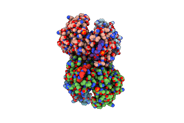
Human Pdk1 Kinase Domain In Complex With Allosteric Compound Pse10 Bound To The Pif-Pocket
Organism: Homo sapiens
Method: X-RAY DIFFRACTION Resolution:1.09 Å Release Date: 2016-10-19 Classification: TRANSFERASE Ligands: ATP, 78W, DMS, DTD, CL |
Organism: Homo sapiens
Method: X-RAY DIFFRACTION
Release Date: 2016-10-19
Ligands: ATP, 78W, DMS, DTD, CL
 |
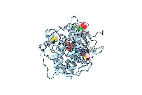
Human Pdk1 Kinase Domain In Complex With An Hm-Peptide Bound To The Pif-Pocket
Organism: Homo sapiens
Method: X-RAY DIFFRACTION Resolution:2.50 Å Release Date: 2016-10-19 Classification: TRANSFERASE Ligands: ATP, CL |
Organism: Homo sapiens
Method: X-RAY DIFFRACTION
Release Date: 2016-10-19
Ligands: ATP, CL
 |
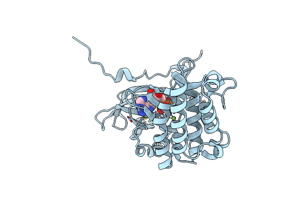
Human Pdk1 Kinase Domain In Complex With Allosteric Compound 7 Bound To The Pif-Pocket
Organism: Homo sapiens
Method: X-RAY DIFFRACTION Resolution:1.24 Å Release Date: 2015-10-21 Classification: TRANSFERASE Ligands: ATP, SVQ, DTD, DMS, NA |
Organism: Homo sapiens
Method: X-RAY DIFFRACTION
Release Date: 2015-10-21
Ligands: ATP, SVQ, DTD, DMS, NA
 |
Crystal Structure Of The S/T Protein Kinase Pkng From Mycobacterium Tuberculosis In Complex With Adp
Organism: Mycobacterium tuberculosis (strain atcc 25618 / h37rv)
Method: X-RAY DIFFRACTION Resolution:1.74 Å Release Date: 2015-05-20 Classification: TRANSFERASE Ligands: ADP, ZN, MG |
Organism: Mycobacterium tuberculosis (strain atcc 25618 / h37rv)
Method: X-RAY DIFFRACTION
Release Date: 2015-05-20
Ligands: ADP, ZN, MG