

Search Count: 25
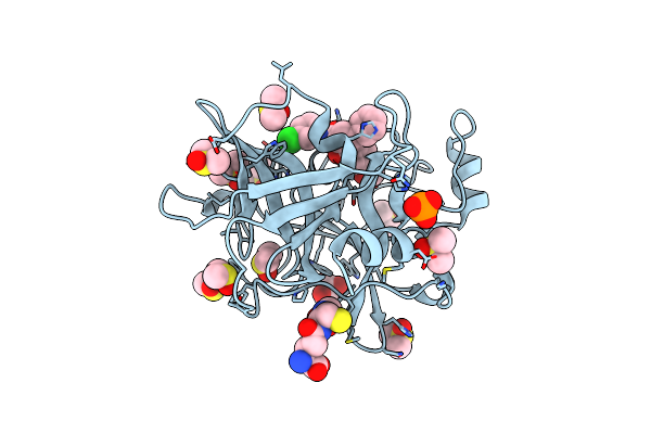 |
Human Plasmakallikrein Protease Domain In Complex With Active Site Directed Inhibitor
Organism: Homo sapiens
Method: X-RAY DIFFRACTION Resolution:1.42 Å Release Date: 2020-07-08 Classification: HYDROLASE Ligands: GSH, DMS, PO4, GOL, MU8 |
Organism: Homo sapiens
Method: X-RAY DIFFRACTION
Release Date: 2020-07-08
Ligands: GSH, DMS, PO4, GOL, MU8
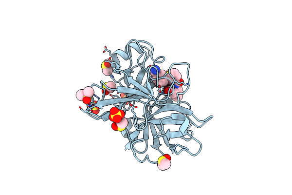 |
Coagulation Factor Xi Protease Domain In Complex With Active Site Inhibitor
Organism: Homo sapiens
Method: X-RAY DIFFRACTION Resolution:1.17 Å Release Date: 2020-07-08 Classification: HYDROLASE Ligands: SO4, DMS, NA, J7B |
Organism: Homo sapiens
Method: X-RAY DIFFRACTION
Release Date: 2020-07-08
Ligands: SO4, DMS, NA, J7B
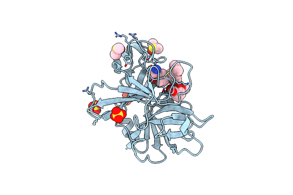 |
Coagulation Factor Xi Protease Domain In Complex With Active Site Inhibitor
Organism: Homo sapiens
Method: X-RAY DIFFRACTION Resolution:1.29 Å Release Date: 2020-07-08 Classification: HYDROLASE Ligands: NW2, SO4, DMS |
Organism: Homo sapiens
Method: X-RAY DIFFRACTION
Release Date: 2020-07-08
Ligands: NW2, SO4, DMS
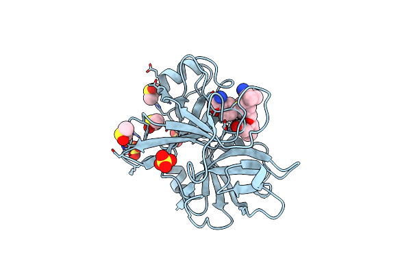 |
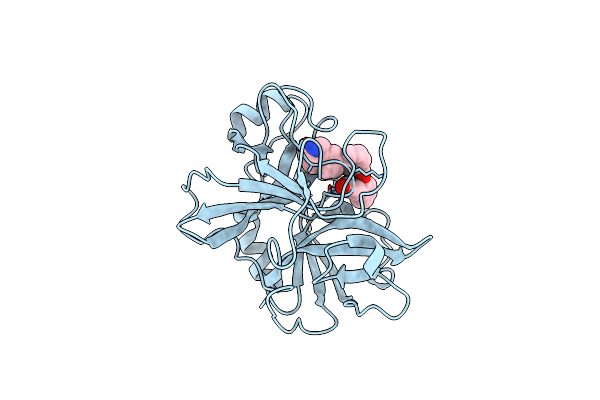
Coagulation Factor Xi Protease Domain In Complex With Active Site Inhibitor
Organism: Homo sapiens
Method: X-RAY DIFFRACTION Resolution:1.33 Å Release Date: 2020-07-08 Classification: HYDROLASE Ligands: SO4, DMS, NW5 |
Organism: Homo sapiens
Method: X-RAY DIFFRACTION
Release Date: 2020-07-08
Ligands: SO4, DMS, NW5
 |
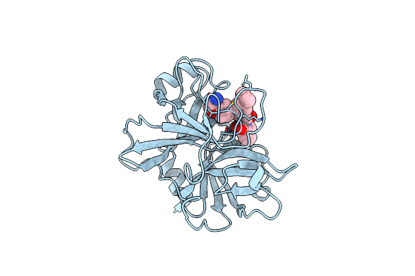
Coagulation Factor Xi Protease Domain In Complex With Active Site Inhibitor
Organism: Homo sapiens
Method: X-RAY DIFFRACTION Resolution:2.63 Å Release Date: 2020-07-08 Classification: HYDROLASE Ligands: NWE |
Organism: Homo sapiens
Method: X-RAY DIFFRACTION
Release Date: 2020-07-08
Ligands: NWE
 |
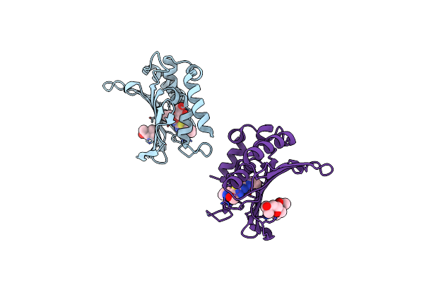
Coagulation Factor Xi Catalytic Domain (C123S) In Complex With Nvp-Xiv936
Organism: Homo sapiens
Method: X-RAY DIFFRACTION Resolution:1.26 Å Release Date: 2020-07-01 Classification: HYDROLASE Ligands: QGS |
Organism: Homo sapiens
Method: X-RAY DIFFRACTION
Release Date: 2020-07-01
Ligands: QGS
 |
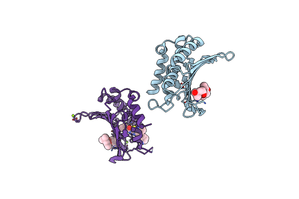
Crystal Structure Of The Atp Binding Domain Of S. Aureus Gyrb Complexed With A Ligand
Organism: Staphylococcus aureus
Method: X-RAY DIFFRACTION Resolution:2.05 Å Release Date: 2015-11-25 Classification: ISOMERASE/ISOMERASE INHIBITOR Ligands: 57U, MPD, GOL |
Organism: Staphylococcus aureus
Method: X-RAY DIFFRACTION
Release Date: 2015-11-25
Ligands: 57U, MPD, GOL
 |
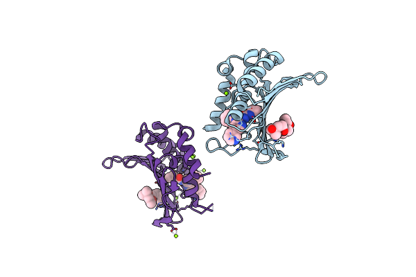
Crystal Structure Of The Atp Binding Domain Of S. Aureus Gyrb Complexed With A Ligand
Organism: Staphylococcus aureus
Method: X-RAY DIFFRACTION Resolution:1.50 Å Release Date: 2015-11-25 Classification: ISOMERASE/ISOMERASE INHIBITOR Ligands: MPD, 57V, MG |
Organism: Staphylococcus aureus
Method: X-RAY DIFFRACTION
Release Date: 2015-11-25
Ligands: MPD, 57V, MG
 |
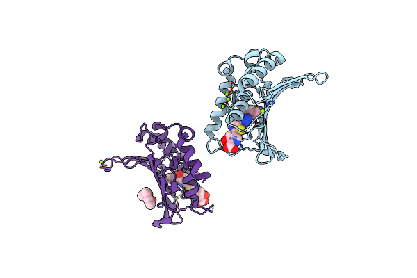
Crystal Structure Of The Atp Binding Domain Of S. Aureus Gyrb Complexed With A Ligand
Organism: Staphylococcus aureus
Method: X-RAY DIFFRACTION Resolution:1.55 Å Release Date: 2015-11-25 Classification: ISOMERASE/ISOMERASE INHIBITOR Ligands: MPD, 57W, MG |
Organism: Staphylococcus aureus
Method: X-RAY DIFFRACTION
Release Date: 2015-11-25
Ligands: MPD, 57W, MG
 |
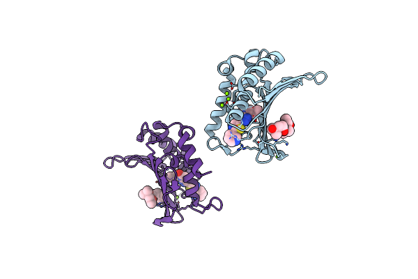
Crystal Structure Of The Atp Binding Domain Of S. Aureus Gyrb Complexed With A Ligand
Organism: Staphylococcus aureus
Method: X-RAY DIFFRACTION Resolution:1.55 Å Release Date: 2015-11-18 Classification: ISOMERASE/ISOMERASE INHIBITOR Ligands: MG, CL, 57Y, MPD |
Organism: Staphylococcus aureus
Method: X-RAY DIFFRACTION
Release Date: 2015-11-18
Ligands: MG, CL, 57Y, MPD
 |
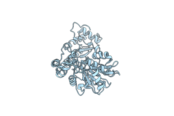
Crystal Structure Of The Atp Binding Domain Of S. Aureus Gyrb Complexed With A Ligand
Organism: Staphylococcus aureus
Method: X-RAY DIFFRACTION Resolution:1.60 Å Release Date: 2015-11-11 Classification: ISOMERASE/ISOMERASE INHIBITOR Ligands: MPD, CL, MG, 57X |
Organism: Staphylococcus aureus
Method: X-RAY DIFFRACTION
Release Date: 2015-11-11
Ligands: MPD, CL, MG, 57X
 |
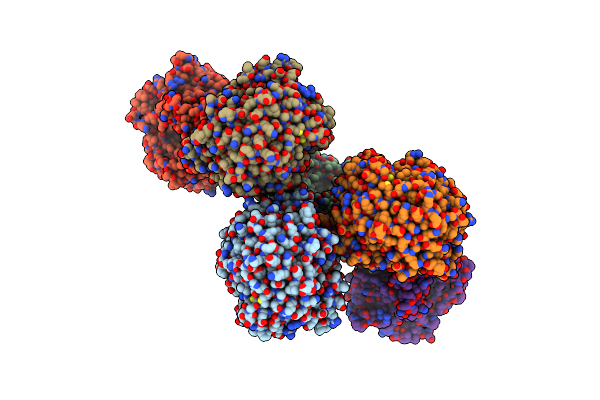
Crystal Structural Of Mouse Tyrosine Aminotransferase
Organism: Mus musculus
Method: X-RAY DIFFRACTION Resolution:2.91 Å Release Date: 2010-11-03 Classification: TRANSFERASE |
Organism: Mus musculus
Method: X-RAY DIFFRACTION
Release Date: 2010-11-03
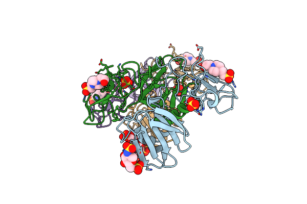 |
Crystal Structure Of 1K1 Mutant Of Hepatocyte Growth Factor/Scatter Factor Fragment Nk1 In Complex With Heparin
Organism: Homo sapiens
Method: X-RAY DIFFRACTION Resolution:2.81 Å Release Date: 2010-08-18 Classification: HORMONE Ligands: SO4, EPE, SGN |
Organism: Homo sapiens
Method: X-RAY DIFFRACTION
Release Date: 2010-08-18
Ligands: SO4, EPE, SGN
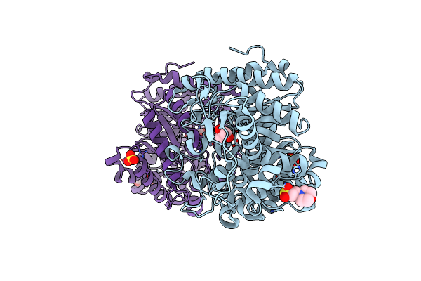 |
Rice Bglu1 Beta-Glucosidase, A Plant Exoglucanase/Beta-Glucosidase
Organism: Oryza sativa japonica group
Method: X-RAY DIFFRACTION Resolution:2.20 Å Release Date: 2008-02-12 Classification: HYDROLASE Ligands: ZN, SO4, MES, GOL |
Organism: Oryza sativa japonica group
Method: X-RAY DIFFRACTION
Release Date: 2008-02-12
Ligands: ZN, SO4, MES, GOL
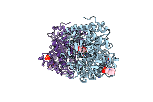 |
Rice Bglu1 Beta-Glucosidase, A Plant Exoglucanase/Beta-Glucosidase
Organism: Oryza sativa japonica group
Method: X-RAY DIFFRACTION Resolution:1.55 Å Release Date: 2008-02-12 Classification: HYDROLASE Ligands: G2F, ZN, SO4, MES, GOL |
Organism: Oryza sativa japonica group
Method: X-RAY DIFFRACTION
Release Date: 2008-02-12
Ligands: G2F, ZN, SO4, MES, GOL
 |
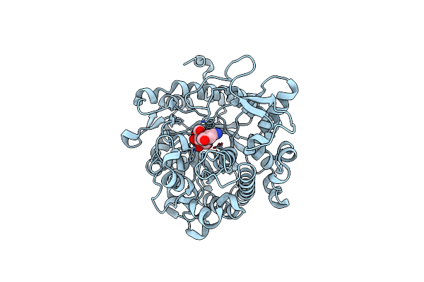
Crystal Structure Of The Sorghum Bicolor Dhurrinase 1
Organism: Sorghum bicolor
Method: X-RAY DIFFRACTION Resolution:1.80 Å Release Date: 2004-05-20 Classification: HYDROLASE |
Organism: Sorghum bicolor
Method: X-RAY DIFFRACTION
Release Date: 2004-05-20
 |
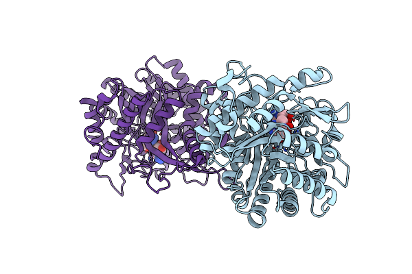
Crystal Structure Of The Sorghum Bicolor Dhurrinase 1
Organism: Sorghum bicolor milo
Method: X-RAY DIFFRACTION Resolution:2.00 Å Release Date: 2004-05-20 Classification: HYDROLASE Ligands: BGC, CCN, IPH |
Organism: Sorghum bicolor milo
Method: X-RAY DIFFRACTION
Release Date: 2004-05-20
Ligands: BGC, CCN, IPH
 |
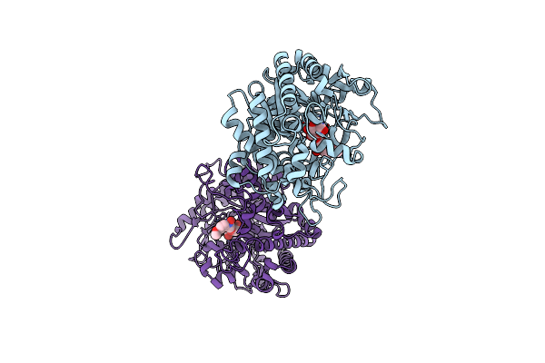
Crystal Structure Of The Zea Maze Beta-Glucosidase-1 In Complex With Gluco-Tetrazole
Organism: Zea mays
Method: X-RAY DIFFRACTION Resolution:1.90 Å Release Date: 2004-05-20 Classification: HYDROLASE Ligands: NTZ |
Organism: Zea mays
Method: X-RAY DIFFRACTION
Release Date: 2004-05-20
Ligands: NTZ
 |
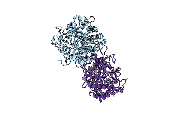
Crystal Structure Of The Inactive Double Mutant Of The Maize Beta-Glucosidase Zmglu1-E191D-F198V In Complex With Dimboa-Glucoside
Organism: Zea mays
Method: X-RAY DIFFRACTION Resolution:1.90 Å Release Date: 2003-03-11 Classification: HYDROLASE Ligands: HBO, BGC |
Organism: Zea mays
Method: X-RAY DIFFRACTION
Release Date: 2003-03-11
Ligands: HBO, BGC
 |
Crystal Structure Of A Monocot (Maize Zmglu1) Beta-Glucosidase
Organism: Zea mays
Method: X-RAY DIFFRACTION Resolution:2.50 Å Release Date: 2001-02-19 Classification: HYDROLASE |
Organism: Zea mays
Method: X-RAY DIFFRACTION
Release Date: 2001-02-19