

Search Count: 30
 |
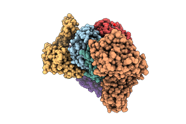
Human Carboxyhemoglobin Bound To Full-Length Staphylococcus Aureus Isdh - Isdh:Hbdim Complex
Organism: Staphylococcus aureus, Homo sapiens
Method: ELECTRON MICROSCOPY Release Date: 2026-05-13 Classification: METAL TRANSPORT Ligands: HEM |
Organism: Staphylococcus aureus, Homo sapiens
Method: ELECTRON MICROSCOPY
Release Date: 2026-05-13
Ligands: HEM
 |
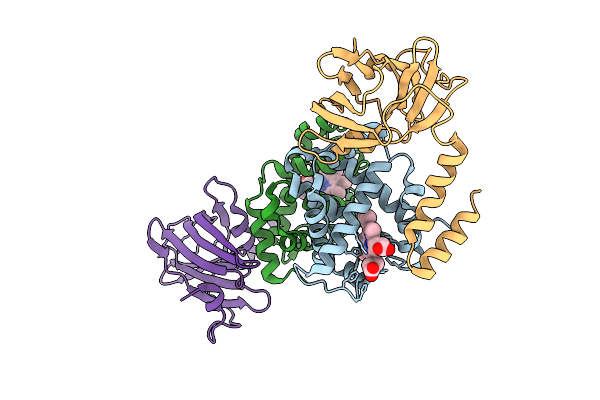
Human Carboxyhemoglobin Bound To Staphylococcus Aureus Isdh-N2N3 - 3Isdh(Alpha2Beta):Hbtet Complex
Organism: Staphylococcus aureus, Homo sapiens
Method: ELECTRON MICROSCOPY Release Date: 2025-11-12 Classification: METAL TRANSPORT Ligands: HEM |
Organism: Staphylococcus aureus, Homo sapiens
Method: ELECTRON MICROSCOPY
Release Date: 2025-11-12
Ligands: HEM
 |
Human Carboxyhemoglobin Bound To Staphylococcus Aureus Isdh-N2N3 - 2Isdh:Hbdim Complex - 3Dva Component 4 Right Tail
Organism: Staphylococcus aureus, Homo sapiens
Method: ELECTRON MICROSCOPY Resolution:4.20 Å Release Date: 2025-11-12 Classification: METAL TRANSPORT Ligands: HEM |
Organism: Staphylococcus aureus, Homo sapiens
Method: ELECTRON MICROSCOPY
Release Date: 2025-11-12
Ligands: HEM
 |
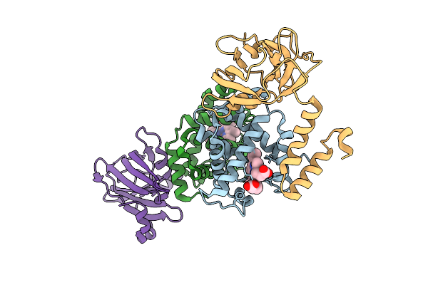
Human Carboxyhemoglobin Bound To Staphylococcus Aureus Isdh-N2N3 - 2Isdh:Hbdim Complex - 3Dva Component 0 Left Tail
Organism: Staphylococcus aureus, Homo sapiens
Method: ELECTRON MICROSCOPY Resolution:4.00 Å Release Date: 2025-11-12 Classification: METAL TRANSPORT Ligands: HEM |
Organism: Staphylococcus aureus, Homo sapiens
Method: ELECTRON MICROSCOPY
Release Date: 2025-11-12
Ligands: HEM
 |
Human Carboxyhemoglobin Bound To Staphylococcus Aureus Isdh-N2N3 - 2Isdh:Hbdim Complex - 3Dva Component 0 Right Tail
Organism: Staphylococcus aureus, Homo sapiens
Method: ELECTRON MICROSCOPY Resolution:4.10 Å Release Date: 2025-11-12 Classification: METAL TRANSPORT Ligands: HEM |
Organism: Staphylococcus aureus, Homo sapiens
Method: ELECTRON MICROSCOPY
Release Date: 2025-11-12
Ligands: HEM
 |
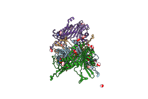
Human Carboxyhemoglobin Bound To Staphylococcus Aureus Isdh-N2N3 - 2Isdh:Hbdim Complex - 3Dva Component 4 Left Tail
Organism: Staphylococcus aureus, Homo sapiens
Method: ELECTRON MICROSCOPY Resolution:4.00 Å Release Date: 2025-11-12 Classification: METAL TRANSPORT Ligands: HEM |
Organism: Staphylococcus aureus, Homo sapiens
Method: ELECTRON MICROSCOPY
Release Date: 2025-11-12
Ligands: HEM
 |
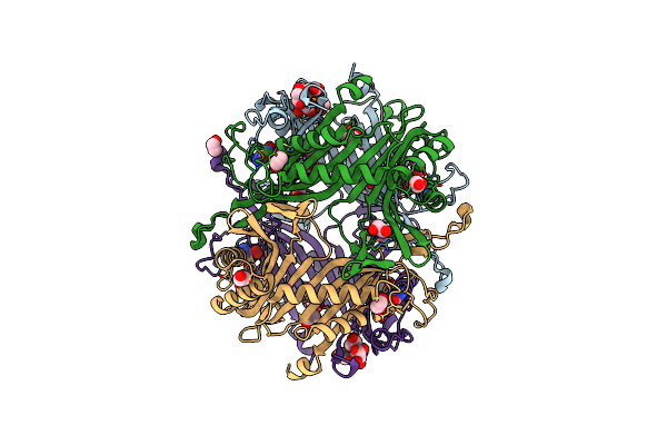
Crystal Structure Of The Cysteine-Rich Gallus Gallus Urate Oxidase In Complex With The 8-Azaxanthine Inhibitor Under Reducing Conditions (Space Group C 2 2 21)
Organism: Gallus gallus
Method: X-RAY DIFFRACTION Resolution:1.71 Å Release Date: 2024-01-17 Classification: OXIDOREDUCTASE Ligands: AZA, OXY, EDO, CL |
Organism: Gallus gallus
Method: X-RAY DIFFRACTION
Release Date: 2024-01-17
Ligands: AZA, OXY, EDO, CL
 |
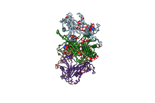
Crystal Structure Of The Cysteine-Rich Gallus Gallus Urate Oxidase In Complex With The 8-Azaxanthine Inhibitor Under Reducing Conditions (Space Group P 21 21 21)
Organism: Gallus gallus
Method: X-RAY DIFFRACTION Resolution:2.12 Å Release Date: 2024-01-17 Classification: OXIDOREDUCTASE Ligands: AZA, EDO, TAR, OXY, CL |
Organism: Gallus gallus
Method: X-RAY DIFFRACTION
Release Date: 2024-01-17
Ligands: AZA, EDO, TAR, OXY, CL
 |
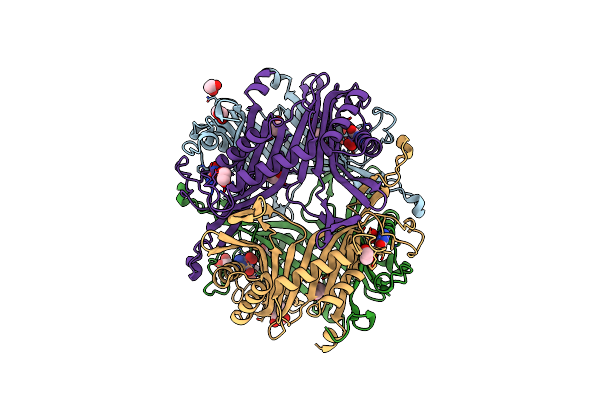
Crystal Structure Of The Cysteine-Rich Gallus Gallus Urate Oxidase In Complex With The 8-Azaxanthine Inhibitor Under Oxidising Conditions (Space Group C 2 2 21)
Organism: Gallus gallus
Method: X-RAY DIFFRACTION Resolution:1.86 Å Release Date: 2024-01-17 Classification: OXIDOREDUCTASE Ligands: AZA, OXY, EDO, CL, BR |
Organism: Gallus gallus
Method: X-RAY DIFFRACTION
Release Date: 2024-01-17
Ligands: AZA, OXY, EDO, CL, BR
 |
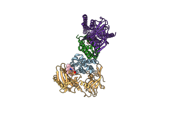
Crystal Structure Of The Cysteine-Rich Gallus Gallus Urate Oxidase In Complex With The 8-Azaxanthine Inhibitor Under Oxidising Conditions (Space Group P 21 21 21)
Organism: Gallus gallus
Method: X-RAY DIFFRACTION Resolution:1.89 Å Release Date: 2024-01-17 Classification: OXIDOREDUCTASE Ligands: AZA, OXY, EDO, CL |
Organism: Gallus gallus
Method: X-RAY DIFFRACTION
Release Date: 2024-01-17
Ligands: AZA, OXY, EDO, CL
 |
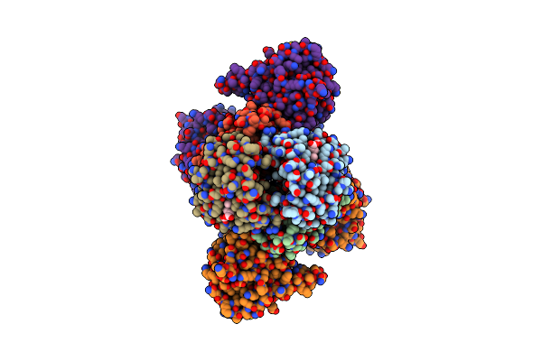
Human Methemoglobin Bound To Staphylococcus Aureus Hemophore Isdb
Organism: Staphylococcus aureus subsp. aureus mw2, Homo sapiens
Method: ELECTRON MICROSCOPY Release Date: 2022-04-13 Classification: METAL TRANSPORT Ligands: HEM |
Organism: Staphylococcus aureus subsp. aureus mw2, Homo sapiens
Method: ELECTRON MICROSCOPY
Release Date: 2022-04-13
Ligands: HEM
 |
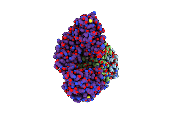
Human Carboxyhemoglobin Bound To Staphylococcus Aureus Hemophore Isdb - 1:2 Complex
Organism: Staphylococcus aureus subsp. aureus mw2, Homo sapiens
Method: ELECTRON MICROSCOPY Release Date: 2022-04-13 Classification: METAL TRANSPORT Ligands: HEM |
Organism: Staphylococcus aureus subsp. aureus mw2, Homo sapiens
Method: ELECTRON MICROSCOPY
Release Date: 2022-04-13
Ligands: HEM
 |
Human Carboxyhemoglobin Bound To Staphylococcus Aureus Hemophore Isdb - 1:1 Complex
Organism: Staphylococcus aureus subsp. aureus mw2, Homo sapiens
Method: ELECTRON MICROSCOPY Release Date: 2022-04-13 Classification: METAL TRANSPORT Ligands: HEM |
Organism: Staphylococcus aureus subsp. aureus mw2, Homo sapiens
Method: ELECTRON MICROSCOPY
Release Date: 2022-04-13
Ligands: HEM
 |
Crystal Structure Of Oass Complexed With Upar Inhibitor
Organism: Salmonella enterica subsp. enterica serovar typhimurium str. lt2
Method: X-RAY DIFFRACTION Resolution:1.20 Å Release Date: 2021-04-07 Classification: LYASE Ligands: PLP, Q7B, DMS, GOL, CO |
Organism: Salmonella enterica subsp. enterica serovar typhimurium str. lt2
Method: X-RAY DIFFRACTION
Release Date: 2021-04-07
Ligands: PLP, Q7B, DMS, GOL, CO
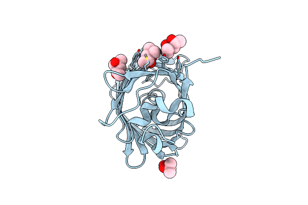 |
Structure Of Gfpmut2 Crystallized At Ph 6
Organism: Aequorea victoria
Method: X-RAY DIFFRACTION Resolution:1.65 Å Release Date: 2018-12-19 Classification: FLUORESCENT PROTEIN Ligands: MPD, MRD |
Organism: Aequorea victoria
Method: X-RAY DIFFRACTION
Release Date: 2018-12-19
Ligands: MPD, MRD
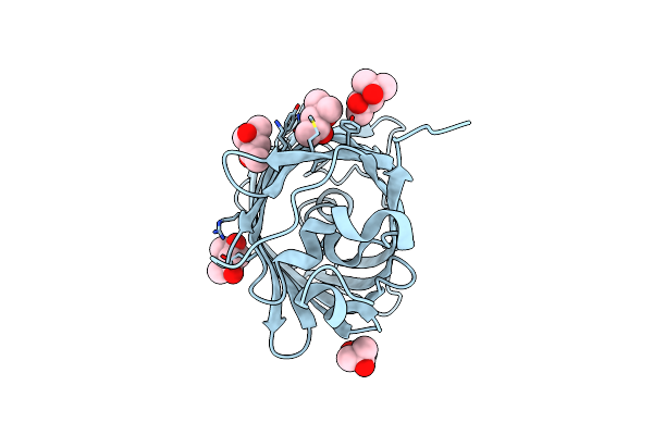 |
Structure Of Gfpmut2 Crystallized At Ph 6 And Transferred To Ph 7
Organism: Aequorea victoria
Method: X-RAY DIFFRACTION Resolution:1.67 Å Release Date: 2018-12-19 Classification: FLUORESCENT PROTEIN Ligands: MPD, MRD |
Organism: Aequorea victoria
Method: X-RAY DIFFRACTION
Release Date: 2018-12-19
Ligands: MPD, MRD
 |
Structure Of Gfpmut2 Crystallized At Ph 8.5
Organism: Aequorea victoria
Method: X-RAY DIFFRACTION Resolution:1.79 Å Release Date: 2018-12-19 Classification: FLUORESCENT PROTEIN Ligands: CA |
Organism: Aequorea victoria
Method: X-RAY DIFFRACTION
Release Date: 2018-12-19
Ligands: CA
 |
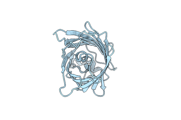
Structure Of Gfpmut2 Crystallized At Ph 8.5 And Transferred To Ph 6
Organism: Aequorea victoria
Method: X-RAY DIFFRACTION Resolution:2.40 Å Release Date: 2018-12-19 Classification: FLUORESCENT PROTEIN Ligands: CA |
Organism: Aequorea victoria
Method: X-RAY DIFFRACTION
Release Date: 2018-12-19
Ligands: CA
 |
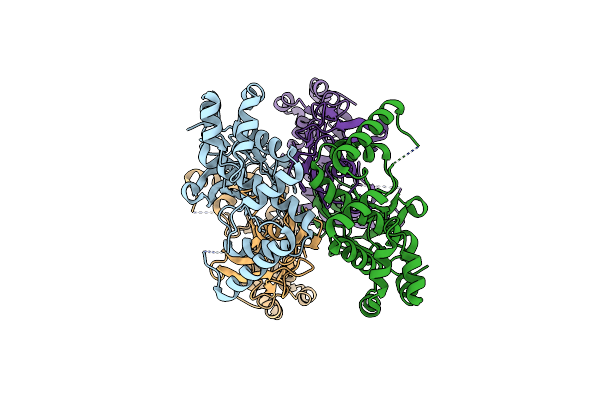
Structure Of Gfpmut2 Crystallized At Ph 6 And Transferred To Ph 9
Organism: Aequorea victoria
Method: X-RAY DIFFRACTION Resolution:2.30 Å Release Date: 2018-12-19 Classification: FLUORESCENT PROTEIN |
Organism: Aequorea victoria
Method: X-RAY DIFFRACTION
Release Date: 2018-12-19
 |
Crystal Structure Of (S)-Allantoin Synthase
Organism: Phaeodactylum tricornutum ccap 1055/1
Method: X-RAY DIFFRACTION Resolution:1.85 Å Release Date: 2018-11-21 Classification: LYASE |
Organism: Phaeodactylum tricornutum ccap 1055/1
Method: X-RAY DIFFRACTION
Release Date: 2018-11-21