

Search Count: 23
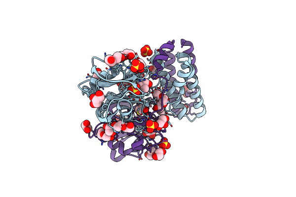 |
Crystal Structure Of Udp-X Diphosphatase
Organism: Streptococcus pneumoniae
Method: X-RAY DIFFRACTION Resolution:1.39 Å Release Date: 2013-08-07 Classification: HYDROLASE Ligands: EDO, TRS, SO4, PEG, GOL, PGE |
Organism: Streptococcus pneumoniae
Method: X-RAY DIFFRACTION
Release Date: 2013-08-07
Ligands: EDO, TRS, SO4, PEG, GOL, PGE
 |
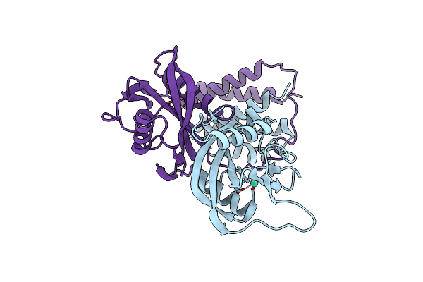
Crystal Structure Of Cdp-Chase
Organism: Bacillus cereus
Method: X-RAY DIFFRACTION Resolution:1.80 Å Release Date: 2011-07-13 Classification: HYDROLASE Ligands: SO4 |
Organism: Bacillus cereus
Method: X-RAY DIFFRACTION
Release Date: 2011-07-13
Ligands: SO4
 |
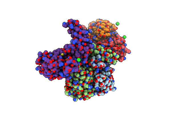
Crystal Structure Of Cdp-Chase In Complex With Gd3+
Organism: Bacillus cereus
Method: X-RAY DIFFRACTION Resolution:2.50 Å Release Date: 2011-07-13 Classification: HYDROLASE Ligands: GD3 |
Organism: Bacillus cereus
Method: X-RAY DIFFRACTION
Release Date: 2011-07-13
Ligands: GD3
 |
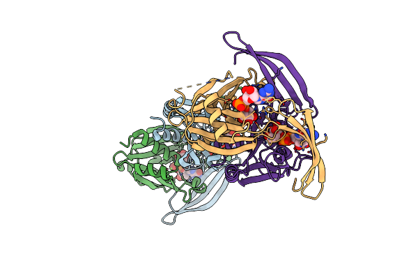
Structure Of The E.Coli Gdp-Mannose Hydrolase (Yffh) In Complex With Tartrate
Organism: Escherichia coli
Method: X-RAY DIFFRACTION Resolution:2.50 Å Release Date: 2011-05-11 Classification: HYDROLASE Ligands: TAR, NA, CL |
Organism: Escherichia coli
Method: X-RAY DIFFRACTION
Release Date: 2011-05-11
Ligands: TAR, NA, CL
 |
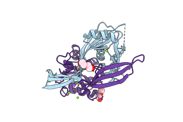
Structure Of The E100A E.Coli Gdp-Mannose Hydrolase (Yffh) In Complex With Gdp-Mannose And Mg++
Organism: Escherichia coli
Method: X-RAY DIFFRACTION Resolution:2.45 Å Release Date: 2011-05-11 Classification: HYDROLASE Ligands: GDD, MG, NA, CL |
Organism: Escherichia coli
Method: X-RAY DIFFRACTION
Release Date: 2011-05-11
Ligands: GDD, MG, NA, CL
 |
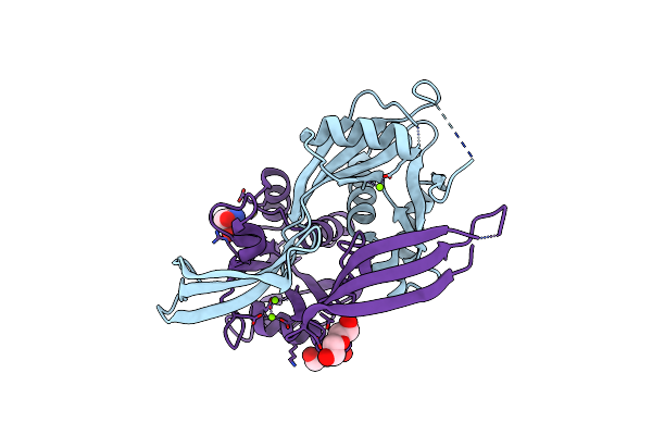
Structure Of The E100A E.Coli Gdp-Mannose Hydrolase (Yffh) In Complex With Mg++
Organism: Escherichia coli
Method: X-RAY DIFFRACTION Resolution:2.10 Å Release Date: 2011-05-11 Classification: HYDROLASE Ligands: MG, PEG, NA, CL |
Organism: Escherichia coli
Method: X-RAY DIFFRACTION
Release Date: 2011-05-11
Ligands: MG, PEG, NA, CL
 |
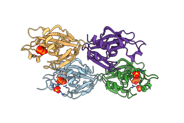
Structure Of The D152A E.Coli Gdp-Mannose Hydrolase (Yffh) In Complex With Mg++
Organism: Escherichia coli
Method: X-RAY DIFFRACTION Resolution:2.05 Å Release Date: 2011-05-11 Classification: HYDROLASE Ligands: MG, CL, PEG, TRS |
Organism: Escherichia coli
Method: X-RAY DIFFRACTION
Release Date: 2011-05-11
Ligands: MG, CL, PEG, TRS
 |
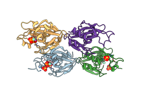
Structure Of The E. Coli Dihydroneopterin Triphosphate Pyrophosphohydrolase
Organism: Escherichia coli
Method: X-RAY DIFFRACTION Resolution:1.80 Å Release Date: 2007-08-28 Classification: HYDROLASE Ligands: SO4, PPV |
Organism: Escherichia coli
Method: X-RAY DIFFRACTION
Release Date: 2007-08-28
Ligands: SO4, PPV
 |
Structure Of The E. Coli Dihydroneopterin Triphosphate Pyrophosphohydrolase In Complex With Sm+3 And Pyrophosphate
Organism: Escherichia coli
Method: X-RAY DIFFRACTION Resolution:2.60 Å Release Date: 2007-08-28 Classification: HYDROLASE Ligands: SO4, SM, NA, PPV |
Organism: Escherichia coli
Method: X-RAY DIFFRACTION
Release Date: 2007-08-28
Ligands: SO4, SM, NA, PPV
 |
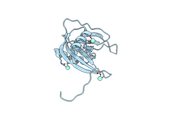
Crystal Structure Of Nudix Hydrolase Dr1025 From Deinococcus Radiodurans
Organism: Deinococcus radiodurans
Method: X-RAY DIFFRACTION Resolution:1.39 Å Release Date: 2004-05-11 Classification: HYDROLASE |
Organism: Deinococcus radiodurans
Method: X-RAY DIFFRACTION
Release Date: 2004-05-11
 |
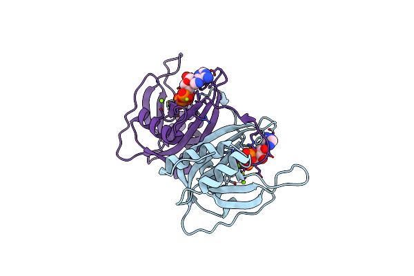
Crystal Structure Of Nudix Hydrolase Dr1025 In Complex With Sm+3
Organism: Deinococcus radiodurans
Method: X-RAY DIFFRACTION Resolution:1.80 Å Release Date: 2004-05-11 Classification: HYDROLASE Ligands: SM |
Organism: Deinococcus radiodurans
Method: X-RAY DIFFRACTION
Release Date: 2004-05-11
Ligands: SM
 |
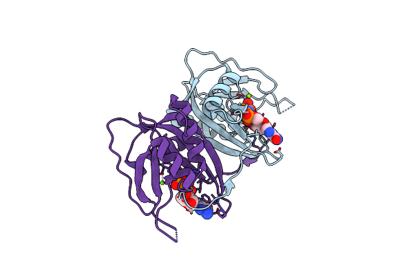
Crystal Structure Of The Nudix Hydrolase Dr1025 In Complex With Atp
Organism: Deinococcus radiodurans
Method: X-RAY DIFFRACTION Resolution:1.60 Å Release Date: 2004-05-11 Classification: HYDROLASE Ligands: MG, ATP |
Organism: Deinococcus radiodurans
Method: X-RAY DIFFRACTION
Release Date: 2004-05-11
Ligands: MG, ATP
 |
Crystal Structure Of Nudix Hydrolase Dr1025 In Complexed With Gnp And Mg+2
Organism: Deinococcus radiodurans
Method: X-RAY DIFFRACTION Resolution:1.60 Å Release Date: 2004-05-11 Classification: HYDROLASE Ligands: MG, GNP |
Organism: Deinococcus radiodurans
Method: X-RAY DIFFRACTION
Release Date: 2004-05-11
Ligands: MG, GNP
 |
The Structure Of A Coa Pyrophosphatase From D. Radiodurans
Organism: Deinococcus radiodurans
Method: X-RAY DIFFRACTION Resolution:2.09 Å Release Date: 2003-05-13 Classification: HYDROLASE |
Organism: Deinococcus radiodurans
Method: X-RAY DIFFRACTION
Release Date: 2003-05-13
 |
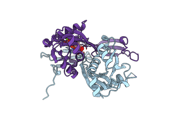
The Structure Of A Coa Pyrophosphatase From D. Radiodurans Complexed With A Magnesium Ion
Organism: Deinococcus radiodurans
Method: X-RAY DIFFRACTION Resolution:1.70 Å Release Date: 2003-05-13 Classification: HYDROLASE Ligands: MG |
Organism: Deinococcus radiodurans
Method: X-RAY DIFFRACTION
Release Date: 2003-05-13
Ligands: MG
 |
Structure Of The Adpr-Ase In Complex With Ampcpr And Mg
Organism: Escherichia coli
Method: X-RAY DIFFRACTION Resolution:2.04 Å Release Date: 2002-10-09 Classification: HYDROLASE Ligands: CL, MG, ADV |
Organism: Escherichia coli
Method: X-RAY DIFFRACTION
Release Date: 2002-10-09
Ligands: CL, MG, ADV
 |
The Crystal Structure Of The E.Coli Adp-Ribose Pyrophosphatase
Organism: Escherichia coli
Method: X-RAY DIFFRACTION Resolution:1.90 Å Release Date: 2001-05-02 Classification: HYDROLASE |
Organism: Escherichia coli
Method: X-RAY DIFFRACTION
Release Date: 2001-05-02
 |
Complex Structure Of The Adpr-Ase And Its Substrate Adp-Ribose
Organism: Escherichia coli
Method: X-RAY DIFFRACTION Resolution:2.30 Å Release Date: 2001-05-02 Classification: HYDROLASE Ligands: APR |
Organism: Escherichia coli
Method: X-RAY DIFFRACTION
Release Date: 2001-05-02
Ligands: APR
 |
Crystal Structure Of The Adp-Ribose Pyrophosphatase In Complex With Gd+3
Organism: Escherichia coli
Method: X-RAY DIFFRACTION Resolution:2.71 Å Release Date: 2001-05-02 Classification: HYDROLASE Ligands: GD3 |
Organism: Escherichia coli
Method: X-RAY DIFFRACTION
Release Date: 2001-05-02
Ligands: GD3
 |
Mutt Pyrophosphohydrolase-Metal-Nucleotide-Metal Complex, Nmr, 16 Structures
Organism: Escherichia coli
Method: SOLUTION NMR Release Date: 1997-05-15 Classification: MUTATOR PROTEIN Ligands: MG, APC, CON |
Organism: Escherichia coli
Method: SOLUTION NMR
Release Date: 1997-05-15
Ligands: MG, APC, CON