

Search Count: 143
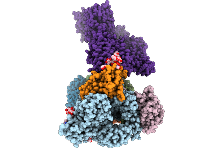 |
Cryo-Em Structure Of Yeast Emc:Spf1 Insertase:Dislocase Complex In Digitonin
Organism: Saccharomyces cerevisiae
Method: ELECTRON MICROSCOPY Release Date: 2026-08-12 Classification: MEMBRANE PROTEIN Ligands: NAG, AJP |
Organism: Saccharomyces cerevisiae
Method: ELECTRON MICROSCOPY
Release Date: 2026-08-12
Ligands: NAG, AJP
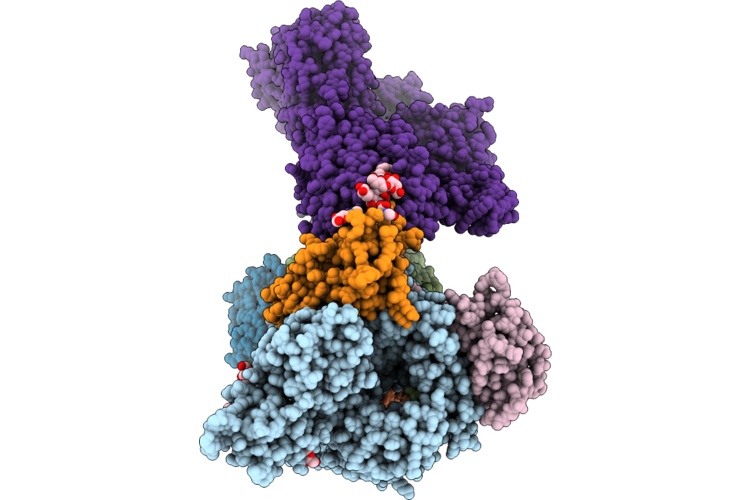 |
Cryo-Em Structure Of Yeast Emc:Spf1 Insertase:Dislocase Complex In E1-Atp Conformation In Digitonin
Organism: Saccharomyces cerevisiae
Method: ELECTRON MICROSCOPY Release Date: 2026-08-12 Classification: MEMBRANE PROTEIN Ligands: NAG, AJP |
Organism: Saccharomyces cerevisiae
Method: ELECTRON MICROSCOPY
Release Date: 2026-08-12
Ligands: NAG, AJP
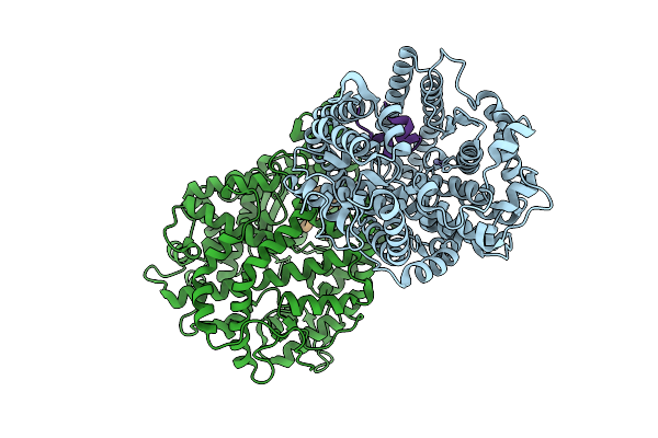 |
Ace2 Extracellular Domain In Complex With The Macrocyclic Peptide Gr3.1.2
Organism: Homo sapiens, Synthetic plasmid
Method: X-RAY DIFFRACTION Resolution:2.02 Å Release Date: 2026-04-08 Classification: HYDROLASE Ligands: ZN |
Organism: Homo sapiens, Synthetic plasmid
Method: X-RAY DIFFRACTION
Release Date: 2026-04-08
Ligands: ZN
 |
Ace2 Extracellular Domain In Complex With The Macrocyclic Peptide Gr1.4
Organism: Homo sapiens, Synthetic construct
Method: X-RAY DIFFRACTION Resolution:2.39 Å Release Date: 2026-04-08 Classification: HYDROLASE |
Organism: Homo sapiens, Synthetic construct
Method: X-RAY DIFFRACTION
Release Date: 2026-04-08
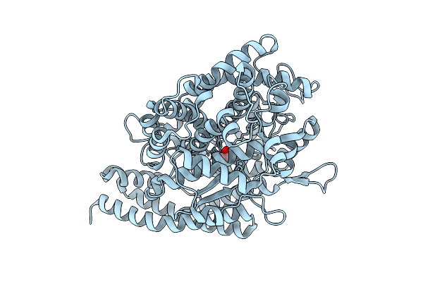 |
Apo Ace2 Extracellular Domain
Organism: Homo sapiens
Method: X-RAY DIFFRACTION Resolution:1.79 Å Release Date: 2026-04-08 Classification: HYDROLASE Ligands: MLA, ZN |
Organism: Homo sapiens
Method: X-RAY DIFFRACTION
Release Date: 2026-04-08
Ligands: MLA, ZN
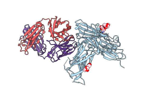 |
X-Ray Crystal Structure Of Atx-350-2 Fab Bound To Epstein-Barr Virus Glycoprotein 350
Organism: Mus musculus, Human gammaherpesvirus 4
Method: X-RAY DIFFRACTION Resolution:3.93 Å Release Date: 2025-12-24 Classification: IMMUNE SYSTEM Ligands: NAG |
Organism: Mus musculus, Human gammaherpesvirus 4
Method: X-RAY DIFFRACTION
Release Date: 2025-12-24
Ligands: NAG
 |
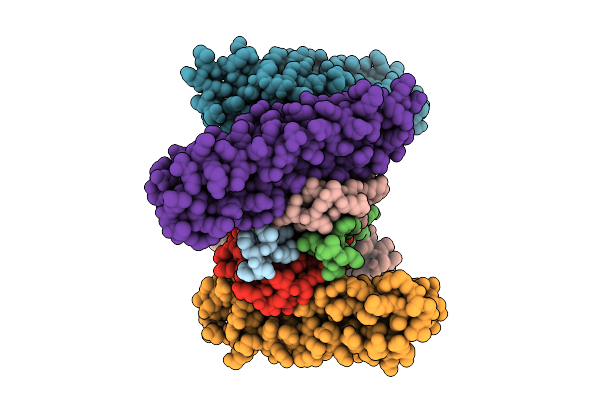
Cryo-Em Structure Of Kshv Glycoprotein Ghgl In Complex With Mlkh3 And Mlkh10 Fabs
Organism: Mus musculus, Human gammaherpesvirus 8
Method: ELECTRON MICROSCOPY Release Date: 2025-12-10 Classification: IMMUNE SYSTEM |
Organism: Mus musculus, Human gammaherpesvirus 8
Method: ELECTRON MICROSCOPY
Release Date: 2025-12-10
 |
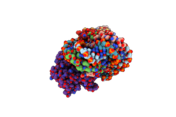
Crystal Structure Of Bak Bound To The Inhibitory Abak
Organism: Homo sapiens, Synthetic construct
Method: X-RAY DIFFRACTION Resolution:2.86 Å Release Date: 2025-07-16 Classification: APOPTOSIS |
Organism: Homo sapiens, Synthetic construct
Method: X-RAY DIFFRACTION
Release Date: 2025-07-16
 |
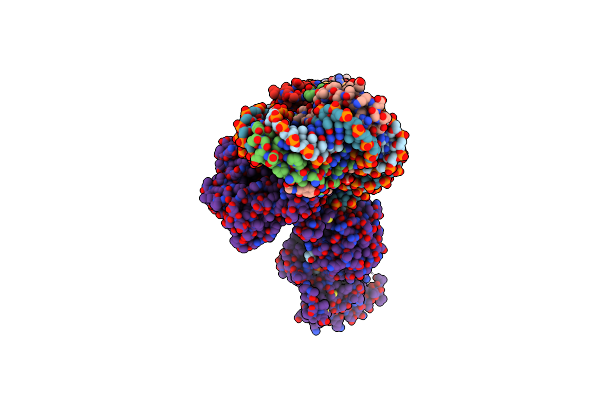
Cryoem Structure Of The F Plasmid Relaxosome In Its Pre-Initiation State, Derived From The Ds-27_+143-R Locally-Refined Map 3.76 A
Organism: Escherichia coli k-12
Method: ELECTRON MICROSCOPY Release Date: 2025-06-04 Classification: DNA BINDING PROTEIN |
Organism: Escherichia coli k-12
Method: ELECTRON MICROSCOPY
Release Date: 2025-06-04
 |
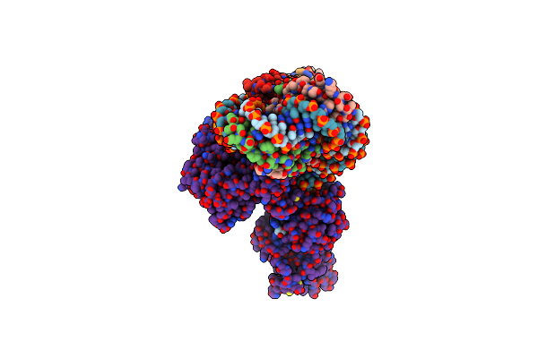
Cryoem Structure Of The F Plasmid Relaxosome With Trai In Its Te Mode, Derived From The Ss-27_+8Ds+9_+143-R Locally-Refined 3.45 A Map.
Organism: Escherichia coli k-12
Method: ELECTRON MICROSCOPY Release Date: 2025-06-04 Classification: DNA BINDING PROTEIN Ligands: MG |
Organism: Escherichia coli k-12
Method: ELECTRON MICROSCOPY
Release Date: 2025-06-04
Ligands: MG
 |
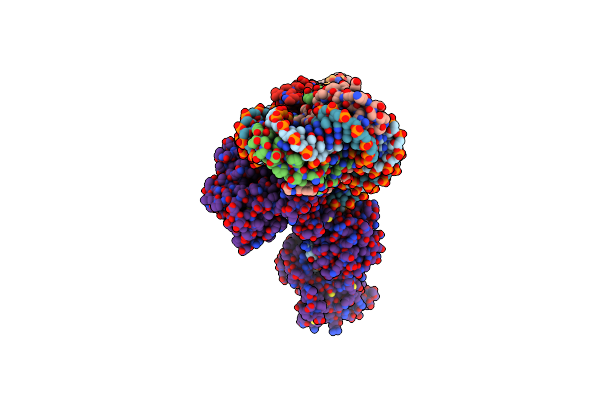
Cryoem Structure Of The F Plasmid Relaxosome With Truncated Trai1-863 In Its Te Mode, Derived From The Ss-27_+8Ds+9_+143-R_Deltaah+Ctd Locally-Refined 3.42 A Map
Organism: Escherichia coli k-12
Method: ELECTRON MICROSCOPY Release Date: 2025-06-04 Classification: DNA BINDING PROTEIN Ligands: MG |
Organism: Escherichia coli k-12
Method: ELECTRON MICROSCOPY
Release Date: 2025-06-04
Ligands: MG
 |
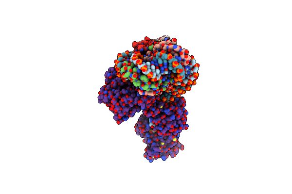
Cryoem Structure Of The F Plasmid Relaxosome With Trai In Its Te Mode, Without Accessory Protein Tram. Derived From The Ss-27_+8Ds+9_+143-R_Deltatram Locally-Refined 2.94 A Map.
Organism: Escherichia coli k-12
Method: ELECTRON MICROSCOPY Release Date: 2025-06-04 Classification: DNA BINDING PROTEIN Ligands: MG |
Organism: Escherichia coli k-12
Method: ELECTRON MICROSCOPY
Release Date: 2025-06-04
Ligands: MG
 |
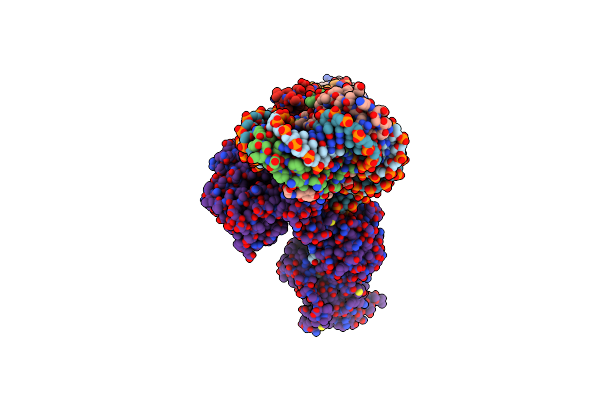
Cryoem Structure Of The F Plasmid Relaxosome With Orit Dna Ss-27_+3Ds+4_+143 And Trai Its Te Mode, Derived From Ss-27_+3Ds+4_+143-R Locally-Refined 3.68 A Map.
Organism: Escherichia coli k-12
Method: ELECTRON MICROSCOPY Release Date: 2025-06-04 Classification: DNA BINDING PROTEIN Ligands: MG |
Organism: Escherichia coli k-12
Method: ELECTRON MICROSCOPY
Release Date: 2025-06-04
Ligands: MG
 |
Cryoem Structure Of The F Plasmid Relaxosome With Orit Dna Ss-27_-3Ds-2_+143 And Trai Its Te Mode, Derived From Ss-27_-3Ds-2_+143-R Locally-Refined 3.42 A Map.
Organism: Escherichia coli k-12
Method: ELECTRON MICROSCOPY Release Date: 2025-06-04 Classification: DNA BINDING PROTEIN Ligands: MG |
Organism: Escherichia coli k-12
Method: ELECTRON MICROSCOPY
Release Date: 2025-06-04
Ligands: MG
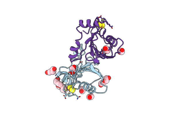 |
[2Fe:2S] Ferredoxin Fesii From Azotobacter Vinelandii, Reduced Form
Organism: Azotobacter vinelandii dj
Method: X-RAY DIFFRACTION Resolution:1.45 Å Release Date: 2025-01-01 Classification: METAL BINDING PROTEIN Ligands: FES, EDO, P6G, PEG |
Organism: Azotobacter vinelandii dj
Method: X-RAY DIFFRACTION
Release Date: 2025-01-01
Ligands: FES, EDO, P6G, PEG
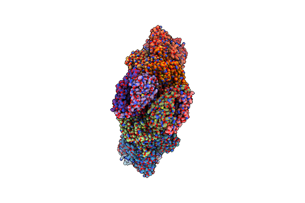 |
Cryo-Em Structure Of The Molybdenum Nitrogenase Complexed With Iron Protein (Nifh) And Shethna Protein Ii (Fesii)
Organism: Azotobacter vinelandii
Method: ELECTRON MICROSCOPY Release Date: 2025-01-01 Classification: OXIDOREDUCTASE Ligands: CLF, HCA, ICS, CA, FES, SF4 |
Organism: Azotobacter vinelandii
Method: ELECTRON MICROSCOPY
Release Date: 2025-01-01
Ligands: CLF, HCA, ICS, CA, FES, SF4
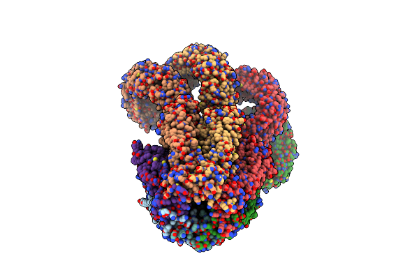 |
Structure Of A Homomeric Human Lrrc8C Volume-Regulated Anion Channel
Organism: Homo sapiens
Method: ELECTRON MICROSCOPY Release Date: 2024-11-13 Classification: MEMBRANE PROTEIN |
Organism: Homo sapiens
Method: ELECTRON MICROSCOPY
Release Date: 2024-11-13
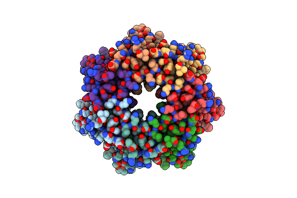 |
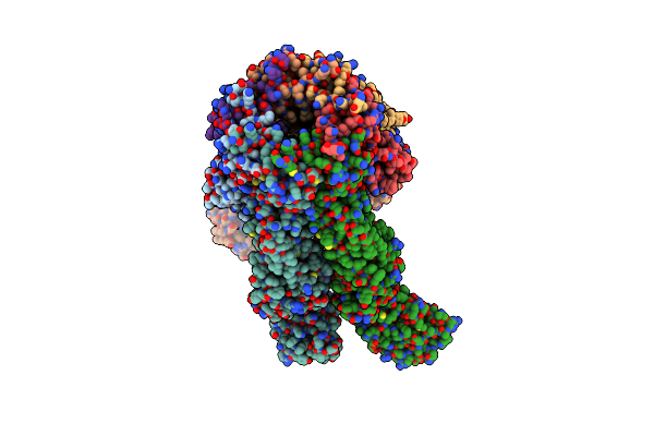
Structure Of The Extracellular Subdomain Of A Homomeric Lrrc8C Truncation Disease Mutant
Organism: Homo sapiens
Method: ELECTRON MICROSCOPY Release Date: 2024-11-13 Classification: MEMBRANE PROTEIN |
Organism: Homo sapiens
Method: ELECTRON MICROSCOPY
Release Date: 2024-11-13
 |
Structure Of A Homomeric Lrrc8C Point Mutation Disease Mutant
Organism: Homo sapiens
Method: ELECTRON MICROSCOPY Release Date: 2024-11-13 Classification: MEMBRANE PROTEIN |
Organism: Homo sapiens
Method: ELECTRON MICROSCOPY
Release Date: 2024-11-13
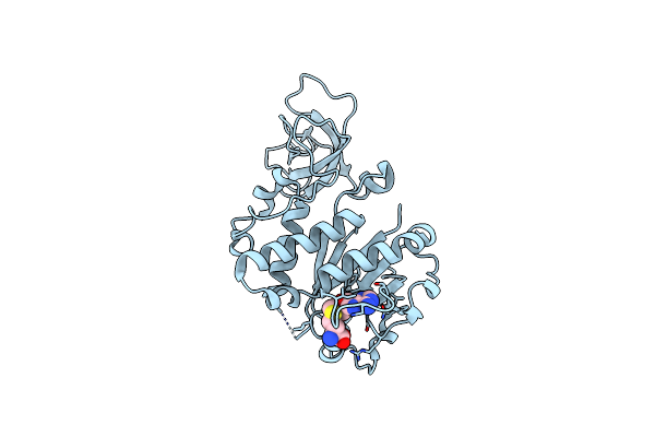 |
Crystal Structure Of Spout1/Cenp-32 Bound To Sah
Organism: Homo sapiens
Method: X-RAY DIFFRACTION Resolution:2.38 Å Release Date: 2024-10-23 Classification: CELL CYCLE Ligands: SAH |
Organism: Homo sapiens
Method: X-RAY DIFFRACTION
Release Date: 2024-10-23
Ligands: SAH