

Search Count: 25
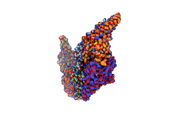 |
Crystal Structure Of Gmppnp-Bound Human Gimap7 L100Q Variant
Organism: Homo sapiens
Method: X-RAY DIFFRACTION Resolution:3.15 Å Release Date: 2013-03-13 Classification: HYDROLASE Ligands: MG, GNP |
Organism: Homo sapiens
Method: X-RAY DIFFRACTION
Release Date: 2013-03-13
Ligands: MG, GNP
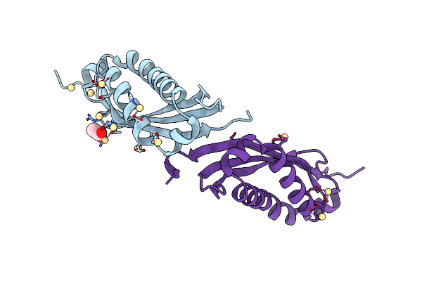 |
Spfh Domain Of Mouse Stomatin (Crystal Form 1)
Organism: Mus musculus
Method: X-RAY DIFFRACTION Resolution:2.46 Å Release Date: 2012-08-15 Classification: MEMBRANE PROTEIN Ligands: CD, ACT |
Organism: Mus musculus
Method: X-RAY DIFFRACTION
Release Date: 2012-08-15
Ligands: CD, ACT
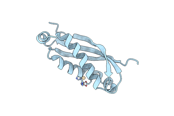 |
Spfh Domain Of Mouse Stomatin (Crystal Form 3)
Organism: Mus musculus
Method: X-RAY DIFFRACTION Resolution:1.80 Å Release Date: 2012-08-15 Classification: MEMBRANE PROTEIN Ligands: CD |
Organism: Mus musculus
Method: X-RAY DIFFRACTION
Release Date: 2012-08-15
Ligands: CD
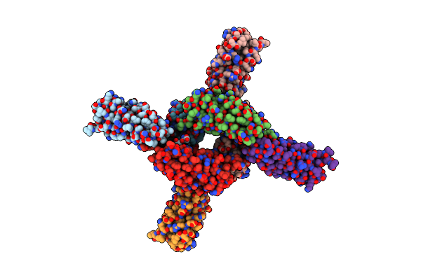 |
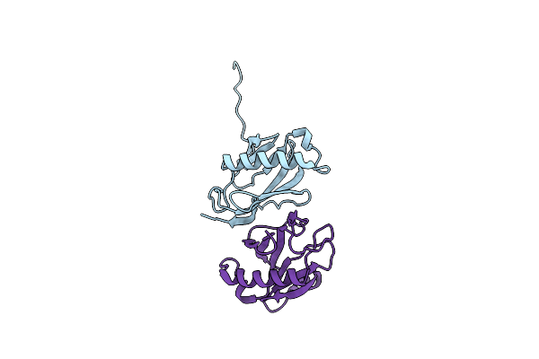
Spfh Domain Of The Mouse Stomatin (Crystal Form 2)
Organism: Mus musculus
Method: X-RAY DIFFRACTION Resolution:2.69 Å Release Date: 2012-08-15 Classification: MEMBRANE PROTEIN |
Organism: Mus musculus
Method: X-RAY DIFFRACTION
Release Date: 2012-08-15
 |
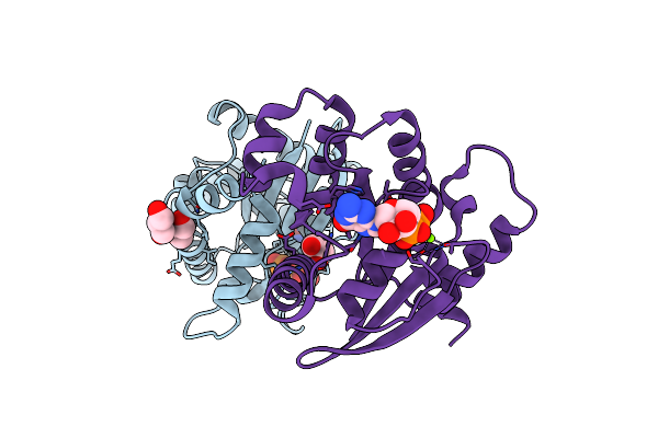
X-Ray Structure Of The Yos9 Dimerization Domain
Organism: Saccharomyces cerevisiae
Method: X-RAY DIFFRACTION Resolution:2.55 Å Release Date: 2012-01-25 Classification: CARBOHYDRATE BINDING PROTEIN |
Organism: Saccharomyces cerevisiae
Method: X-RAY DIFFRACTION
Release Date: 2012-01-25
 |
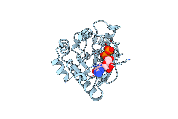
Crystal Structure Of Gdp-Bound Human Gimap2, Amino Acid Residues 1- 234
Organism: Homo sapiens
Method: X-RAY DIFFRACTION Resolution:1.70 Å Release Date: 2010-10-20 Classification: IMMUNE SYSTEM Ligands: GDP, MG, GOL |
Organism: Homo sapiens
Method: X-RAY DIFFRACTION
Release Date: 2010-10-20
Ligands: GDP, MG, GOL
 |
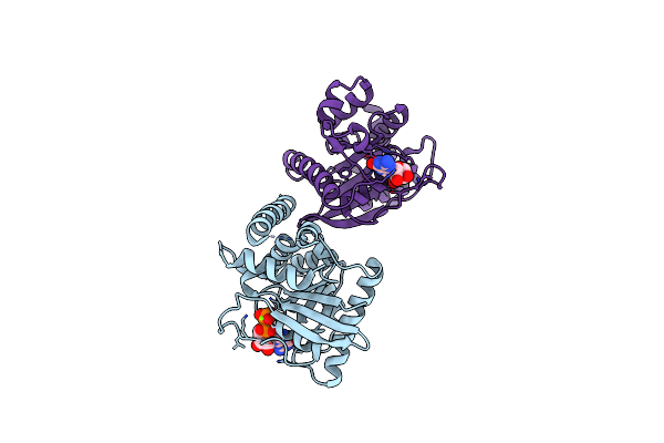
Crystal Structure Of Gtp-Bound Human Gimap2, Amino Acid Residues 1- 234
Organism: Homo sapiens
Method: X-RAY DIFFRACTION Resolution:1.90 Å Release Date: 2010-10-20 Classification: IMMUNE SYSTEM Ligands: GTP, MG |
Organism: Homo sapiens
Method: X-RAY DIFFRACTION
Release Date: 2010-10-20
Ligands: GTP, MG
 |
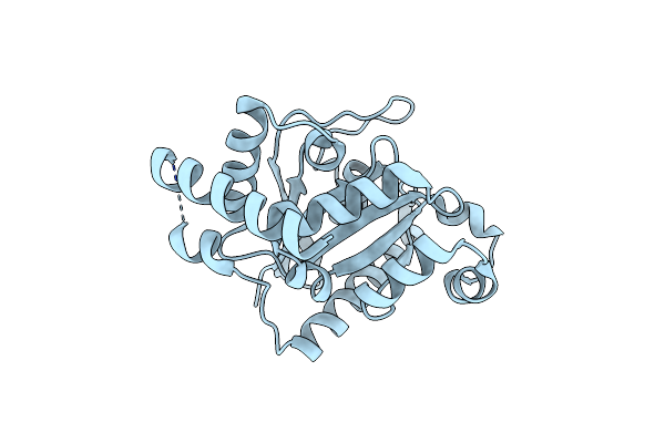
Crystal Structure Of Gdp-Bound Human Gimap2, Amino Acid Residues 21- 260
Organism: Homo sapiens
Method: X-RAY DIFFRACTION Resolution:2.80 Å Release Date: 2010-10-20 Classification: IMMUNE SYSTEM Ligands: GDP, MG |
Organism: Homo sapiens
Method: X-RAY DIFFRACTION
Release Date: 2010-10-20
Ligands: GDP, MG
 |
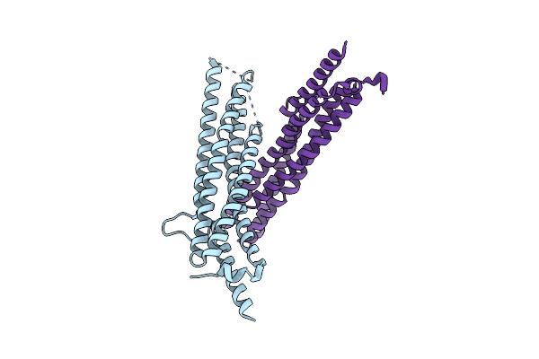
Crystal Structure Of Nucleotide-Free Human Gimap2, Amino Acid Residues 1-260
Organism: Homo sapiens
Method: X-RAY DIFFRACTION Resolution:1.50 Å Release Date: 2010-10-20 Classification: IMMUNE SYSTEM |
Organism: Homo sapiens
Method: X-RAY DIFFRACTION
Release Date: 2010-10-20
 |
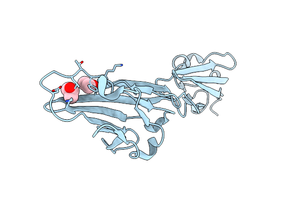
Structural Basis Of Oligomerisation In The Mxa Stalk
Organism: Homo sapiens
Method: X-RAY DIFFRACTION Resolution:2.40 Å Release Date: 2010-05-05 Classification: ANTIVIRAL PROTEIN |
Organism: Homo sapiens
Method: X-RAY DIFFRACTION
Release Date: 2010-05-05
 |
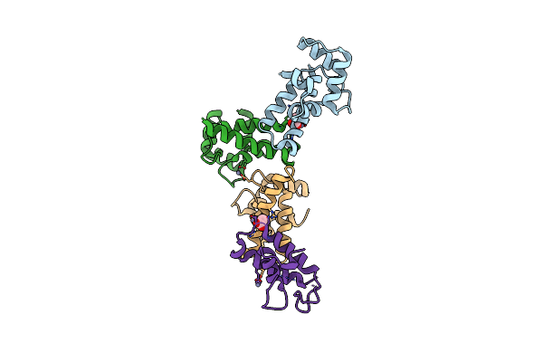
Crystal Structure Of The Extracellular Domains Of Coxsackie & Adenovirus Receptor From Mouse (Mcar)
Organism: Mus musculus
Method: X-RAY DIFFRACTION Resolution:2.19 Å Release Date: 2010-03-16 Classification: CELL ADHESION Ligands: IPA |
Organism: Mus musculus
Method: X-RAY DIFFRACTION
Release Date: 2010-03-16
Ligands: IPA
 |
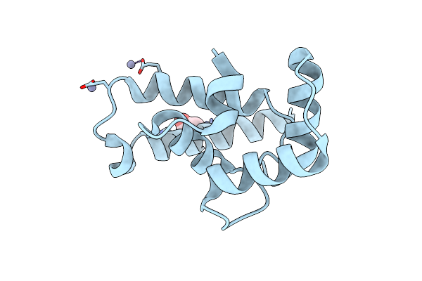
Crystal Structure Of Human Iba2, Orthorhombic Crystal Form
Organism: Homo sapiens
Method: X-RAY DIFFRACTION Resolution:2.15 Å Release Date: 2009-07-14 Classification: METAL BINDING PROTEIN Ligands: ZN, ACT, CL, NA |
Organism: Homo sapiens
Method: X-RAY DIFFRACTION
Release Date: 2009-07-14
Ligands: ZN, ACT, CL, NA
 |
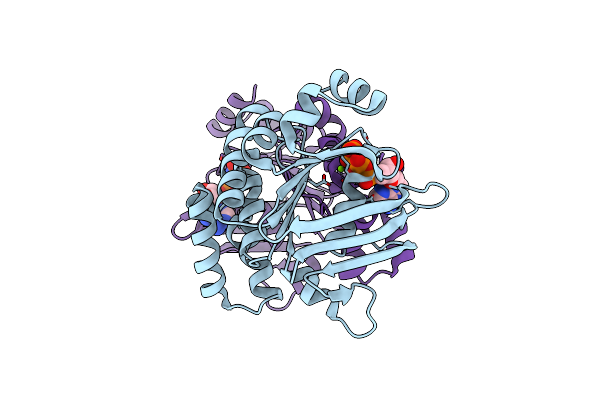
Crystal Structure Of Human Iba2, Trigonal Crystal Form
Organism: Homo sapiens
Method: X-RAY DIFFRACTION Resolution:2.45 Å Release Date: 2009-07-14 Classification: METAL BINDING PROTEIN Ligands: ZN, ACT |
Organism: Homo sapiens
Method: X-RAY DIFFRACTION
Release Date: 2009-07-14
Ligands: ZN, ACT
 |
Abc Protein Artp In Complex With Amp-Pnp/Mg2+
Organism: Geobacillus stearothermophilus
Method: X-RAY DIFFRACTION Resolution:2.25 Å Release Date: 2009-02-03 Classification: HYDROLASE Ligands: MG, ANP |
Organism: Geobacillus stearothermophilus
Method: X-RAY DIFFRACTION
Release Date: 2009-02-03
Ligands: MG, ANP
 |
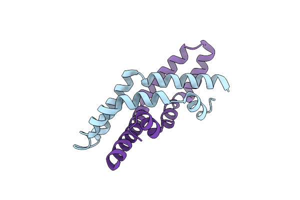
Abc Protein Artp In Complex With Atp-Gamma-S
Organism: Geobacillus stearothermophilus
Method: X-RAY DIFFRACTION Resolution:2.33 Å Release Date: 2009-02-03 Classification: HYDROLASE Ligands: MG, AGS |
Organism: Geobacillus stearothermophilus
Method: X-RAY DIFFRACTION
Release Date: 2009-02-03
Ligands: MG, AGS
 |
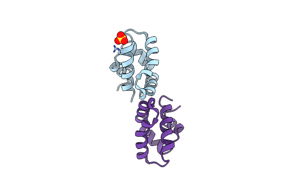
Orthorhombic Crystal Structure Of Human Saposin C Dimer In Open Conformation
Organism: Homo sapiens
Method: X-RAY DIFFRACTION Resolution:2.45 Å Release Date: 2008-04-29 Classification: LIPID BINDING PROTEIN |
Organism: Homo sapiens
Method: X-RAY DIFFRACTION
Release Date: 2008-04-29
 |
Crystal Structure Of Human Saposin D Variant Sapd K9E
Organism: Homo sapiens
Method: X-RAY DIFFRACTION Resolution:2.50 Å Release Date: 2008-04-29 Classification: LIPID BINDING PROTEIN Ligands: SO4 |
Organism: Homo sapiens
Method: X-RAY DIFFRACTION
Release Date: 2008-04-29
Ligands: SO4
 |
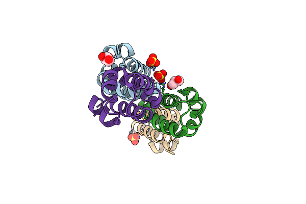
Crystal Structure Of Iodinated Human Saposin D In Space Group C2221
Organism: Homo sapiens
Method: X-RAY DIFFRACTION Resolution:2.50 Å Release Date: 2008-04-29 Classification: LIPID BINDING PROTEIN |
Organism: Homo sapiens
Method: X-RAY DIFFRACTION
Release Date: 2008-04-29
 |
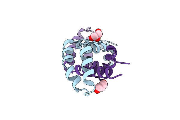
Crystal Structure Of Human Saposin D
Organism: Homo sapiens
Method: X-RAY DIFFRACTION Resolution:2.10 Å Release Date: 2008-04-29 Classification: LIPID BINDING PROTEIN Ligands: SO4, GOL |
Organism: Homo sapiens
Method: X-RAY DIFFRACTION
Release Date: 2008-04-29
Ligands: SO4, GOL
 |
Crystal Structure Of Human Saposin C Dimer In Open Conformation
Organism: Homo sapiens
Method: X-RAY DIFFRACTION Resolution:2.50 Å Release Date: 2008-04-29 Classification: LIPID BINDING PROTEIN Ligands: GOL |
Organism: Homo sapiens
Method: X-RAY DIFFRACTION
Release Date: 2008-04-29
Ligands: GOL