

Search Count: 19
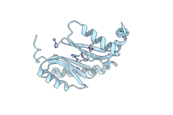 |
The Crystal Structure Of Bona From Acinetobacter Baumannii
Organism: Acinetobacter baumannii
Method: X-RAY DIFFRACTION Resolution:1.65 Å Release Date: 2021-06-02 Classification: LIPID BINDING PROTEIN Ligands: ZN |
Organism: Acinetobacter baumannii
Method: X-RAY DIFFRACTION
Release Date: 2021-06-02
Ligands: ZN
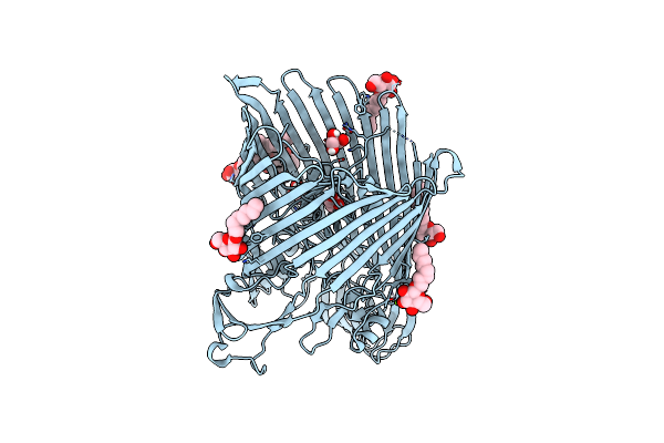 |
The Crystal Structure Of The Outer Membrane Transporter Yddb From Escherichia Coli
Organism: Escherichia coli
Method: X-RAY DIFFRACTION Resolution:2.40 Å Release Date: 2019-10-02 Classification: TRANSPORT PROTEIN Ligands: BOG, MG, GOL |
Organism: Escherichia coli
Method: X-RAY DIFFRACTION
Release Date: 2019-10-02
Ligands: BOG, MG, GOL
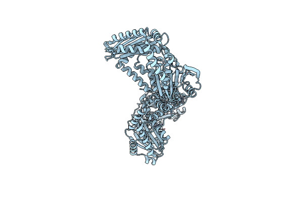 |
The Crystal Structure Of The Periplasmic Protease Pqql From Escherichia Coli
Organism: Escherichia coli (strain k12)
Method: X-RAY DIFFRACTION Resolution:2.60 Å Release Date: 2019-10-02 Classification: HYDROLASE Ligands: ZN, CL |
Organism: Escherichia coli (strain k12)
Method: X-RAY DIFFRACTION
Release Date: 2019-10-02
Ligands: ZN, CL
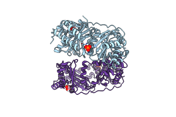 |
The Crystal Structure Of The First Half Of The Periplasmic Protease Pqql From Escherichia Coli
Organism: Escherichia coli (strain k12)
Method: X-RAY DIFFRACTION Resolution:2.00 Å Release Date: 2019-10-02 Classification: HYDROLASE Ligands: GOL, PO4, CL |
Organism: Escherichia coli (strain k12)
Method: X-RAY DIFFRACTION
Release Date: 2019-10-02
Ligands: GOL, PO4, CL
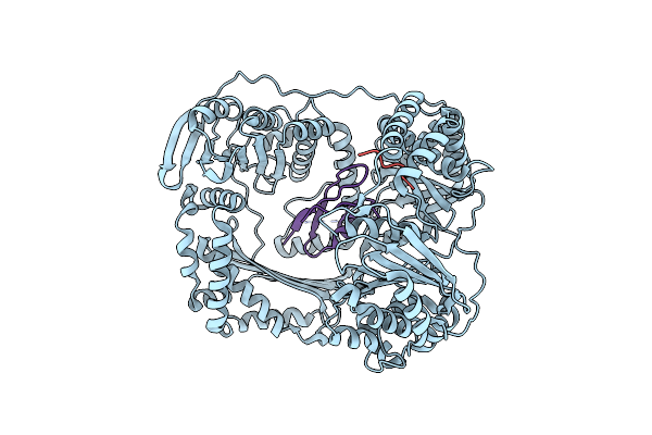 |
The Crystal Structure Of The Ferredoxin Protease Fusc In Complex With Its Substrate Plant Ferredoxin
Organism: Pectobacterium atrosepticum scri1043, Arabidopsis thaliana
Method: X-RAY DIFFRACTION Resolution:2.70 Å Release Date: 2018-06-20 Classification: HYDROLASE |
Organism: Pectobacterium atrosepticum scri1043, Arabidopsis thaliana
Method: X-RAY DIFFRACTION
Release Date: 2018-06-20
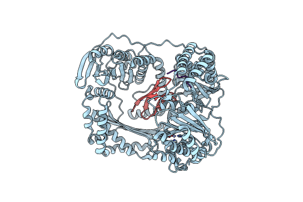 |
The Crystal Structure Of The Ferredoxin Protease Fusc E83A Mutant In Complex With Arabidopsis Ferredoxin
Organism: Pectobacterium atrosepticum (strain scri 1043 / atcc baa-672), Arabidopsis thaliana
Method: X-RAY DIFFRACTION Resolution:1.90 Å Release Date: 2018-06-20 Classification: HYDROLASE Ligands: ZN |
Organism: Pectobacterium atrosepticum (strain scri 1043 / atcc baa-672), Arabidopsis thaliana
Method: X-RAY DIFFRACTION
Release Date: 2018-06-20
Ligands: ZN
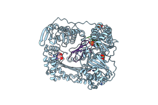 |
The Crystal Structure Of The Ferredoxin Protease Fusc In Complex With Arabidopsis Ferredoxin, Ethylmercury Phosphate Soaked Dataset
Organism: Pectobacterium atrosepticum (strain scri 1043 / atcc baa-672), Arabidopsis thaliana
Method: X-RAY DIFFRACTION Resolution:2.30 Å Release Date: 2018-06-20 Classification: HYDROLASE Ligands: PO4, HG |
Organism: Pectobacterium atrosepticum (strain scri 1043 / atcc baa-672), Arabidopsis thaliana
Method: X-RAY DIFFRACTION
Release Date: 2018-06-20
Ligands: PO4, HG
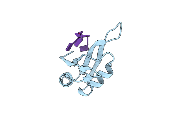 |
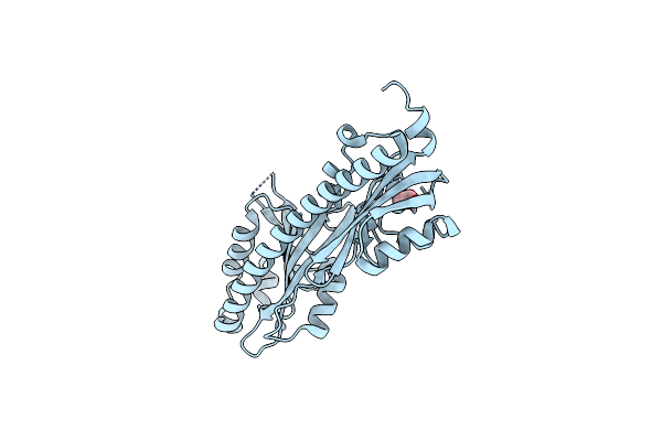
Tia-1 Rrm2 Recognition Of Target Oligonucleotide
Organism: Homo sapiens
Method: X-RAY DIFFRACTION Resolution:2.31 Å Release Date: 2017-02-22 Classification: rna binding protein/dna |
Organism: Homo sapiens
Method: X-RAY DIFFRACTION
Release Date: 2017-02-22
 |
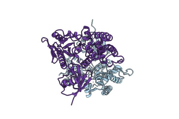
Crystal Structure Of The Periplasmic Sensory Domain Of The Campylobacter Jejuni Chemoreceptor Tlp1
Organism: Campylobacter jejuni subsp. jejuni serotype o:2
Method: X-RAY DIFFRACTION Resolution:1.40 Å Release Date: 2016-03-09 Classification: SIGNALING PROTEIN Ligands: ACT, CL |
Organism: Campylobacter jejuni subsp. jejuni serotype o:2
Method: X-RAY DIFFRACTION
Release Date: 2016-03-09
Ligands: ACT, CL
 |
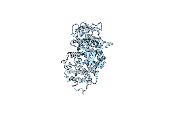
Crystal Structure Of Human Ezrin In Space Group P21
Organism: Homo sapiens
Method: X-RAY DIFFRACTION Resolution:1.90 Å Release Date: 2015-12-09 Classification: PEPTIDE BINDING PROTEIN |
Organism: Homo sapiens
Method: X-RAY DIFFRACTION
Release Date: 2015-12-09
 |
Crystal Structure Of Human Ezrin In Space Group C2221
Organism: Homo sapiens
Method: X-RAY DIFFRACTION Resolution:2.00 Å Release Date: 2015-12-09 Classification: PEPTIDE BINDING PROTEIN |
Organism: Homo sapiens
Method: X-RAY DIFFRACTION
Release Date: 2015-12-09
 |
Crystal Structure Of The Ferm Domain Of Human Ezrin
Organism: Homo sapiens
Method: X-RAY DIFFRACTION Resolution:1.75 Å Release Date: 2015-12-09 Classification: PEPTIDE BINDING PROTEIN Ligands: SO4 |
Organism: Homo sapiens
Method: X-RAY DIFFRACTION
Release Date: 2015-12-09
Ligands: SO4
 |
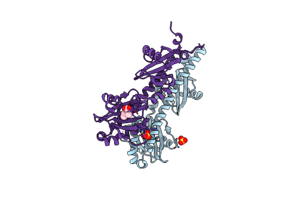
Crystal Structure Of The Sensory Domain Of The Campylobacter Jejuni Chemoreceptor Tlp3 (Ccml)
Organism: Campylobacter jejuni subsp. jejuni serotype o:2 (strain nctc 11168)
Method: X-RAY DIFFRACTION Resolution:1.50 Å Release Date: 2015-11-04 Classification: SIGNALING PROTEIN Ligands: SO4 |
Organism: Campylobacter jejuni subsp. jejuni serotype o:2 (strain nctc 11168)
Method: X-RAY DIFFRACTION
Release Date: 2015-11-04
Ligands: SO4
 |
Crystal Structure Of The Sensory Domain Of The Campylobacter Jejuni Chemoreceptor Tlp3 (Ccml) With Isoleucine Bound.
Organism: Campylobacter jejuni subsp. jejuni serotype o:2 (strain nctc 11168)
Method: X-RAY DIFFRACTION Resolution:1.30 Å Release Date: 2015-11-04 Classification: SIGNALING PROTEIN Ligands: ILE, SO4 |
Organism: Campylobacter jejuni subsp. jejuni serotype o:2 (strain nctc 11168)
Method: X-RAY DIFFRACTION
Release Date: 2015-11-04
Ligands: ILE, SO4
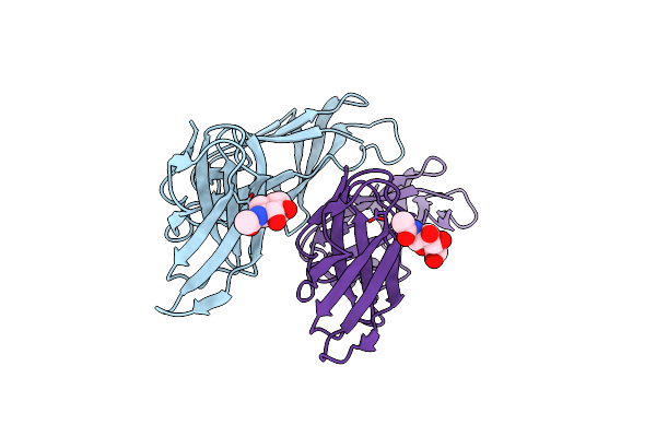 |
Crystal Structure Of Killer Cell Immunoglobulin-Like Receptor 2Dl4
Organism: Homo sapiens
Method: X-RAY DIFFRACTION Resolution:2.80 Å Release Date: 2015-03-18 Classification: IMMUNE SYSTEM Ligands: NAG |
Organism: Homo sapiens
Method: X-RAY DIFFRACTION
Release Date: 2015-03-18
Ligands: NAG
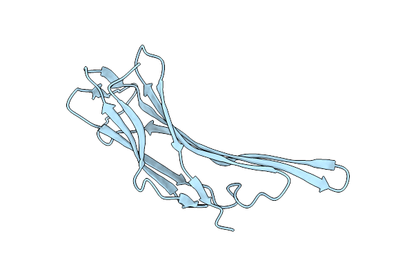 |
N-Terminal Domain Of Vdup-1
Organism: Homo sapiens
Method: X-RAY DIFFRACTION Resolution:1.50 Å Release Date: 2013-02-27 Classification: PROTEIN BINDING |
Organism: Homo sapiens
Method: X-RAY DIFFRACTION
Release Date: 2013-02-27
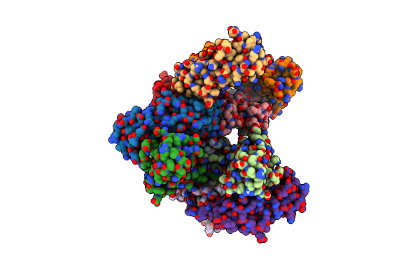 |
N-Terminal Domain Of Vdup-1
Organism: Homo sapiens
Method: X-RAY DIFFRACTION Resolution:2.90 Å Release Date: 2013-02-27 Classification: PROTEIN BINDING Ligands: CA |
Organism: Homo sapiens
Method: X-RAY DIFFRACTION
Release Date: 2013-02-27
Ligands: CA
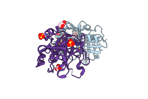 |
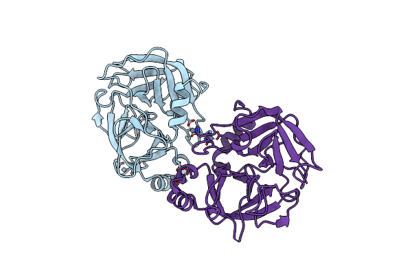
Crystal Structure Of Scabies Mite Inactivated Protease Paralogue S-I1 (Smipp-S-I1)
Organism: Sarcoptes scabiei type hominis
Method: X-RAY DIFFRACTION Resolution:1.85 Å Release Date: 2009-05-12 Classification: HYDROLASE Ligands: SO4, GOL |
Organism: Sarcoptes scabiei type hominis
Method: X-RAY DIFFRACTION
Release Date: 2009-05-12
Ligands: SO4, GOL
 |
Crystal Structure Of Scabies Mite Inactivated Protease Paralogue S-D1 (Smipp-S-D1)
Organism: Sarcoptes scabiei type hominis
Method: X-RAY DIFFRACTION Resolution:2.00 Å Release Date: 2009-05-12 Classification: HYDROLASE Ligands: ZN |
Organism: Sarcoptes scabiei type hominis
Method: X-RAY DIFFRACTION
Release Date: 2009-05-12
Ligands: ZN