

Search Count: 31
 |
Serial Femtosecond X-Ray Structure Of A Fluorescence Optimized Bathy Phytochrome Pairfp2 Derived From Wild-Type Agp2 In Its Pfr State (I0A).
Organism: Agrobacterium fabrum str. c58
Method: X-RAY DIFFRACTION Resolution:2.15 Å Release Date: 2025-05-14 Classification: SIGNALING PROTEIN Ligands: EL5, SO4, CL, EDO |
Organism: Agrobacterium fabrum str. c58
Method: X-RAY DIFFRACTION
Release Date: 2025-05-14
Ligands: EL5, SO4, CL, EDO
 |
Serial Femtosecond X-Ray Structure Of A Fluorescence Optimized Bathy Phytochrome Pairfp2 Derived From Wild-Type Agp2 In Its Pfr State (I0B).
Organism: Agrobacterium fabrum str. c58
Method: X-RAY DIFFRACTION Resolution:2.20 Å Release Date: 2025-05-14 Classification: SIGNALING PROTEIN Ligands: EL5, SO4 |
Organism: Agrobacterium fabrum str. c58
Method: X-RAY DIFFRACTION
Release Date: 2025-05-14
Ligands: EL5, SO4
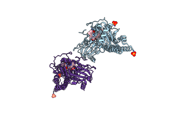 |
Serial Femtosecond X-Ray Structure Of A Fluorescence Optimized Bathy Phytochrome Pairfp2 Derived From Wild-Type Agp2 In I1 Intermediate State.
Organism: Agrobacterium fabrum str. c58
Method: X-RAY DIFFRACTION Resolution:2.54 Å Release Date: 2025-05-14 Classification: SIGNALING PROTEIN Ligands: EL5, SO4 |
Organism: Agrobacterium fabrum str. c58
Method: X-RAY DIFFRACTION
Release Date: 2025-05-14
Ligands: EL5, SO4
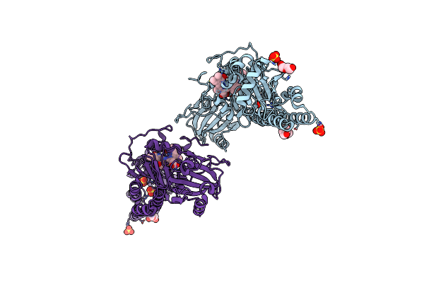 |
Serial Femtosecond X-Ray Structure Of A Fluorescence Optimized Bathy Phytochrome Pairfp2 Derived From Wild-Type Agp2 In I2 Intermediate State.
Organism: Agrobacterium fabrum str. c58
Method: X-RAY DIFFRACTION Resolution:2.43 Å Release Date: 2025-05-14 Classification: SIGNALING PROTEIN Ligands: EL5, SO4, PGE, PEG, CL |
Organism: Agrobacterium fabrum str. c58
Method: X-RAY DIFFRACTION
Release Date: 2025-05-14
Ligands: EL5, SO4, PGE, PEG, CL
 |
Serial Femtosecond X-Ray Structure Of A Fluorescence Optimized Bathy Phytochrome Pairfp2 Derived From Wild-Type Agp2 In I3 Intermediate State.
Organism: Agrobacterium fabrum str. c58
Method: X-RAY DIFFRACTION Resolution:2.40 Å Release Date: 2025-05-14 Classification: SIGNALING PROTEIN Ligands: EL5, SO4, GOL, PEG |
Organism: Agrobacterium fabrum str. c58
Method: X-RAY DIFFRACTION
Release Date: 2025-05-14
Ligands: EL5, SO4, GOL, PEG
 |
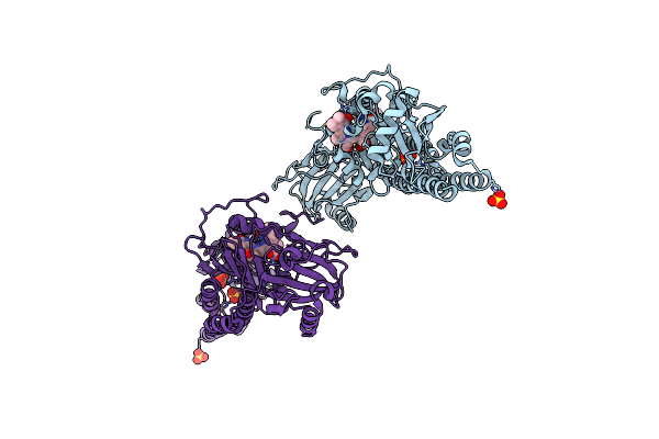
Serial Femtosecond X-Ray Structure Of A Fluorescence Optimized Bathy Phytochrome Pairfp2 Derived From Wild-Type Agp2 In I4 Intermediate State.
Organism: Agrobacterium fabrum str. c58
Method: X-RAY DIFFRACTION Resolution:2.30 Å Release Date: 2025-05-14 Classification: SIGNALING PROTEIN Ligands: EL5, SO4, CL, PEG |
Organism: Agrobacterium fabrum str. c58
Method: X-RAY DIFFRACTION
Release Date: 2025-05-14
Ligands: EL5, SO4, CL, PEG
 |
Serial Femtosecond X-Ray Structure Of A Fluorescence Optimized Bathy Phytochrome Pairfp2 Derived From Wild-Type Agp2 In I5 Intermediate State.
Organism: Agrobacterium fabrum str. c58
Method: X-RAY DIFFRACTION Resolution:2.43 Å Release Date: 2025-05-14 Classification: SIGNALING PROTEIN Ligands: EL5, SO4, CL |
Organism: Agrobacterium fabrum str. c58
Method: X-RAY DIFFRACTION
Release Date: 2025-05-14
Ligands: EL5, SO4, CL
 |
Serial Femtosecond X-Ray Structure Of A Fluorescence Optimized Bathy Phytochrome Pairfp2 Derived From Wild-Type Agp2 In I6 Intermediate State.
Organism: Agrobacterium fabrum str. c58
Method: X-RAY DIFFRACTION Resolution:2.49 Å Release Date: 2025-05-14 Classification: SIGNALING PROTEIN Ligands: EL5, SO4, CL |
Organism: Agrobacterium fabrum str. c58
Method: X-RAY DIFFRACTION
Release Date: 2025-05-14
Ligands: EL5, SO4, CL
 |
Serial Femtosecond X-Ray Structure Of A Fluorescence Optimized Bathy Phytochrome Pairfp2 Derived From Wild-Type Agp2 In I7 Intermediate State.
Organism: Agrobacterium fabrum str. c58
Method: X-RAY DIFFRACTION Resolution:2.80 Å Release Date: 2025-05-14 Classification: SIGNALING PROTEIN Ligands: EL5, SO4, PEG |
Organism: Agrobacterium fabrum str. c58
Method: X-RAY DIFFRACTION
Release Date: 2025-05-14
Ligands: EL5, SO4, PEG
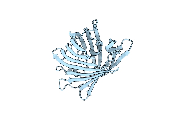 |
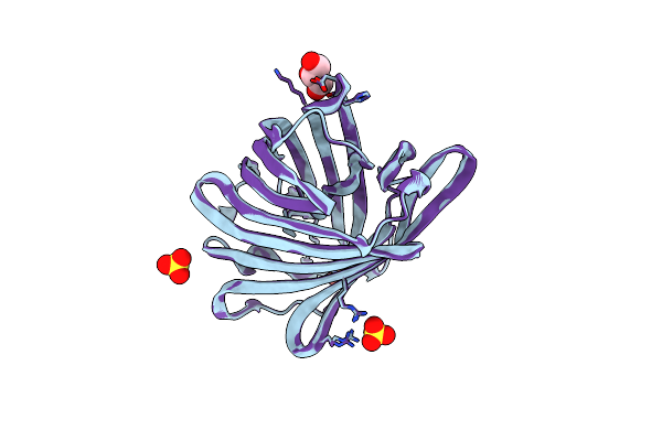
Structure Of Rskiiro Using Ssx
Organism: Lobophyllia hemprichii
Method: X-RAY DIFFRACTION Resolution:1.75 Å Release Date: 2024-11-27 Classification: FLUORESCENT PROTEIN Ligands: SO4 |
Organism: Lobophyllia hemprichii
Method: X-RAY DIFFRACTION
Release Date: 2024-11-27
Ligands: SO4
 |
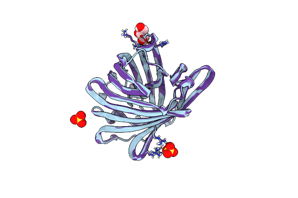
Structure Of Rskiiro Using Ssx After Illumination With 0.1 Mj/Mm^2 Of 405 Nm Light
Organism: Lobophyllia hemprichii
Method: X-RAY DIFFRACTION Resolution:1.75 Å Release Date: 2024-11-27 Classification: FLUORESCENT PROTEIN Ligands: SO4, GOL |
Organism: Lobophyllia hemprichii
Method: X-RAY DIFFRACTION
Release Date: 2024-11-27
Ligands: SO4, GOL
 |
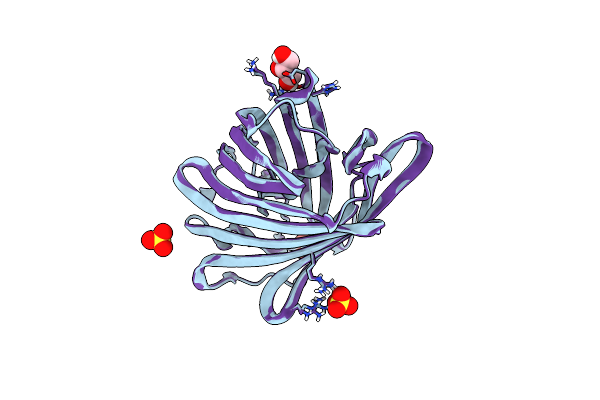
Structure Of Rskiiro Using Ssx After Illumination With 0.53 Mj/Mm^2 Of 405 Nm Light
Organism: Lobophyllia hemprichii
Method: X-RAY DIFFRACTION Resolution:1.70 Å Release Date: 2024-11-27 Classification: FLUORESCENT PROTEIN Ligands: SO4, GOL |
Organism: Lobophyllia hemprichii
Method: X-RAY DIFFRACTION
Release Date: 2024-11-27
Ligands: SO4, GOL
 |
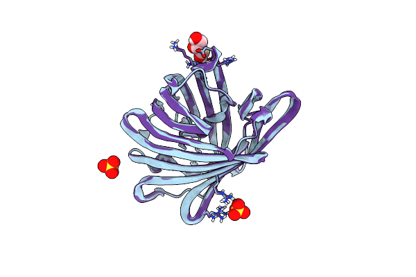
Structure Of Rskiiro Using Ssx After Illumination With 1.78 Mj/Mm^2 Of 405 Nm Light
Organism: Lobophyllia hemprichii
Method: X-RAY DIFFRACTION Resolution:1.75 Å Release Date: 2024-11-27 Classification: FLUORESCENT PROTEIN Ligands: SO4, GOL |
Organism: Lobophyllia hemprichii
Method: X-RAY DIFFRACTION
Release Date: 2024-11-27
Ligands: SO4, GOL
 |
Structure Of Rskiiro Using Ssx After Illumination With 6.74 Mj/Mm^2 Of 405 Nm Light
Organism: Lobophyllia hemprichii
Method: X-RAY DIFFRACTION Resolution:1.75 Å Release Date: 2024-11-27 Classification: FLUORESCENT PROTEIN Ligands: SO4, GOL |
Organism: Lobophyllia hemprichii
Method: X-RAY DIFFRACTION
Release Date: 2024-11-27
Ligands: SO4, GOL
 |
Structure Of Rskiiro Using Ssx After Illumination With 14.43 Mj/Mm^2 Of 405 Nm Light
Organism: Lobophyllia hemprichii
Method: X-RAY DIFFRACTION Resolution:1.75 Å Release Date: 2024-11-27 Classification: FLUORESCENT PROTEIN Ligands: SO4, GOL |
Organism: Lobophyllia hemprichii
Method: X-RAY DIFFRACTION
Release Date: 2024-11-27
Ligands: SO4, GOL
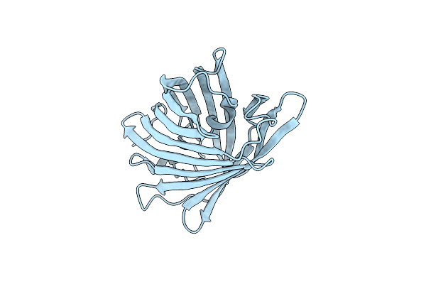 |
Rskiiro Pump Probe Structure By Tr-Sfx
Organism: Lobophyllia hemprichii
Method: X-RAY DIFFRACTION Resolution:1.50 Å Release Date: 2023-10-18 Classification: FLUORESCENT PROTEIN |
Organism: Lobophyllia hemprichii
Method: X-RAY DIFFRACTION
Release Date: 2023-10-18
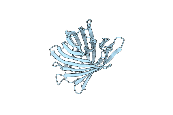 |
Rskiiro Pump Dump Probe Structure By Tr-Sfx
Organism: Lobophyllia hemprichii
Method: X-RAY DIFFRACTION Resolution:1.31 Å Release Date: 2023-10-18 Classification: FLUORESCENT PROTEIN |
Organism: Lobophyllia hemprichii
Method: X-RAY DIFFRACTION
Release Date: 2023-10-18
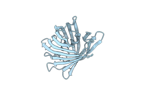 |
Rskiiro Trans Chromophore Dark Structure By Sfx
Organism: Lobophyllia hemprichii
Method: X-RAY DIFFRACTION Resolution:1.30 Å Release Date: 2023-09-27 Classification: FLUORESCENT PROTEIN |
Organism: Lobophyllia hemprichii
Method: X-RAY DIFFRACTION
Release Date: 2023-09-27
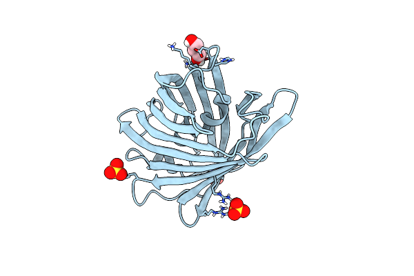 |
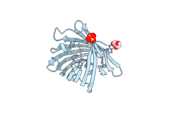
Cis Structure Of Rskiiro At 290 K
Organism: Lobophyllia hemprichii
Method: X-RAY DIFFRACTION Resolution:1.16 Å Release Date: 2023-07-05 Classification: FLUORESCENT PROTEIN Ligands: GOL, SO4 |
Organism: Lobophyllia hemprichii
Method: X-RAY DIFFRACTION
Release Date: 2023-07-05
Ligands: GOL, SO4
 |
Cis Structure Intermediate Of Rskiiro Illuminated At 200 K
Organism: Lobophyllia hemprichii
Method: X-RAY DIFFRACTION Resolution:1.46 Å Release Date: 2023-07-05 Classification: FLUORESCENT PROTEIN Ligands: GOL, SO4 |
Organism: Lobophyllia hemprichii
Method: X-RAY DIFFRACTION
Release Date: 2023-07-05
Ligands: GOL, SO4