

Search Count: 26
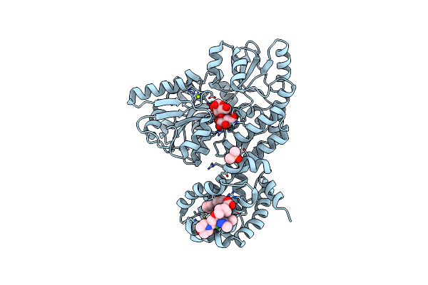 |
Crystal Structure Of Human Mbp-Myeloid Cell Leukemia 1 (Mcl-1) In Complex With Brd810 Inhibitor
Organism: Escherichia coli k-12, Homo sapiens
Method: X-RAY DIFFRACTION Resolution:1.56 Å Release Date: 2024-06-19 Classification: APOPTOSIS Ligands: MG, YI7, DMS, PGE |
Organism: Escherichia coli k-12, Homo sapiens
Method: X-RAY DIFFRACTION
Release Date: 2024-06-19
Ligands: MG, YI7, DMS, PGE
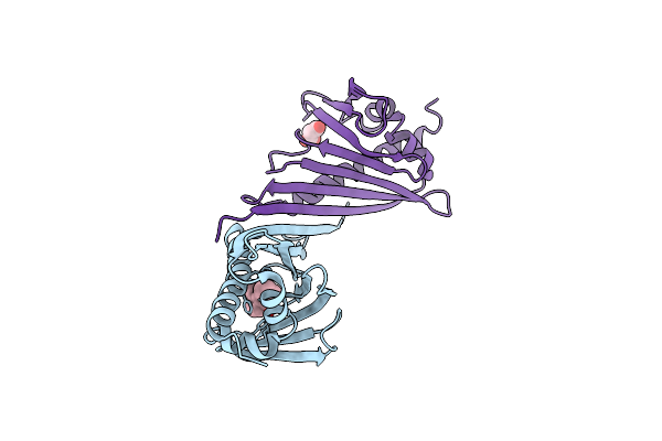 |
Crystal Structure Of Medicago Truncatula Nodulin 13 (Mtn13) In Complex With 3-Carboxybenzophenone
Organism: Medicago truncatula
Method: X-RAY DIFFRACTION Resolution:2.52 Å Release Date: 2022-10-26 Classification: PLANT PROTEIN Ligands: A8I, MLI |
Organism: Medicago truncatula
Method: X-RAY DIFFRACTION
Release Date: 2022-10-26
Ligands: A8I, MLI
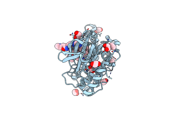 |
Crystal Structure Of Jnk3 In Complex With Light-Activated Covalent Inhibitor Mr-Ii-249 With Both Non-Covalent And Covalent Binding Modes (Compound 4)
Organism: Homo sapiens
Method: X-RAY DIFFRACTION Resolution:2.18 Å Release Date: 2021-07-21 Classification: TRANSFERASE Ligands: 0EI, EDO |
Organism: Homo sapiens
Method: X-RAY DIFFRACTION
Release Date: 2021-07-21
Ligands: 0EI, EDO
 |
Crystal Structure Of Jnk3 In Complex With Fmu-001-367 (Compound 1)
Organism: Homo sapiens
Method: X-RAY DIFFRACTION Resolution:1.70 Å Release Date: 2021-07-21 Classification: TRANSFERASE Ligands: 0G3, EDO, MES |
Organism: Homo sapiens
Method: X-RAY DIFFRACTION
Release Date: 2021-07-21
Ligands: 0G3, EDO, MES
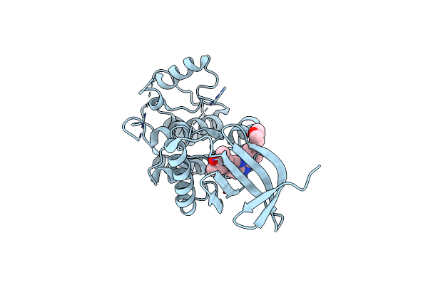 |
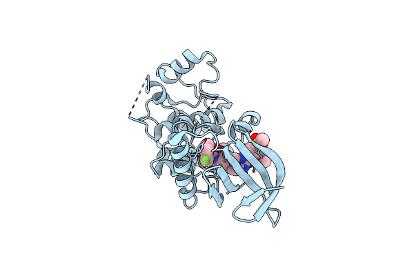
X-Ray Structure Of Mps1 In Complex With Compound 16
Organism: Homo sapiens
Method: X-RAY DIFFRACTION Resolution:2.60 Å Release Date: 2020-05-13 Classification: CELL CYCLE Ligands: NN5 |
Organism: Homo sapiens
Method: X-RAY DIFFRACTION
Release Date: 2020-05-13
Ligands: NN5
 |
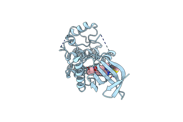
X-Ray Structure Of Mps1 In Complex With Compound 41
Organism: Homo sapiens
Method: X-RAY DIFFRACTION Resolution:2.65 Å Release Date: 2020-05-13 Classification: CELL CYCLE Ligands: 8QW, CL |
Organism: Homo sapiens
Method: X-RAY DIFFRACTION
Release Date: 2020-05-13
Ligands: 8QW, CL
 |
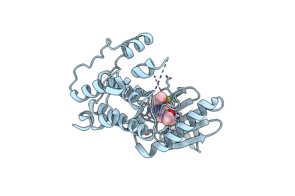
X-Ray Structure Of Mps1 In Complex With Compound 46
Organism: Homo sapiens
Method: X-RAY DIFFRACTION Resolution:2.30 Å Release Date: 2020-05-13 Classification: CELL CYCLE Ligands: O23, CL |
Organism: Homo sapiens
Method: X-RAY DIFFRACTION
Release Date: 2020-05-13
Ligands: O23, CL
 |
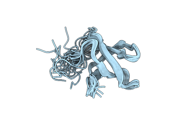
X-Ray Structure Of Mps1 In Complex With Compound 79
Organism: Homo sapiens
Method: X-RAY DIFFRACTION Resolution:2.58 Å Release Date: 2020-05-13 Classification: CELL CYCLE Ligands: 8RH |
Organism: Homo sapiens
Method: X-RAY DIFFRACTION
Release Date: 2020-05-13
Ligands: 8RH
 |
Solution Structure Of The Tudor Domain Of Pshcp
Organism: Prochlorococcus marinus (strain mit 9303)
Method: SOLUTION NMR Release Date: 2019-08-21 Classification: RNA BINDING PROTEIN |
Organism: Prochlorococcus marinus (strain mit 9303)
Method: SOLUTION NMR
Release Date: 2019-08-21
 |
Crystal Structure Phgdh In Complex With Nadh And 3-Phosphoglycerate At 1.77 A Resolution
Organism: Homo sapiens
Method: X-RAY DIFFRACTION Resolution:1.77 Å Release Date: 2019-08-07 Classification: OXIDOREDUCTASE Ligands: 3PG, NAI |
Organism: Homo sapiens
Method: X-RAY DIFFRACTION
Release Date: 2019-08-07
Ligands: 3PG, NAI
 |
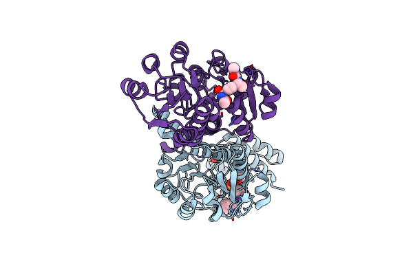
Crystal Structure Of Phgdh In Complex With Compound 9
Organism: Homo sapiens
Method: X-RAY DIFFRACTION Resolution:2.15 Å Release Date: 2019-08-07 Classification: OXIDOREDUCTASE Ligands: K4T, SO4 |
Organism: Homo sapiens
Method: X-RAY DIFFRACTION
Release Date: 2019-08-07
Ligands: K4T, SO4
 |
Crystal Structure Of Phgdh In Complex With Compound 40
Organism: Homo sapiens
Method: X-RAY DIFFRACTION Resolution:2.00 Å Release Date: 2019-08-07 Classification: OXIDOREDUCTASE Ligands: SO4, K52 |
Organism: Homo sapiens
Method: X-RAY DIFFRACTION
Release Date: 2019-08-07
Ligands: SO4, K52
 |
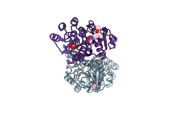
Crystal Structure Of Phgdh In Complex With Compound 15
Organism: Homo sapiens
Method: X-RAY DIFFRACTION Resolution:1.42 Å Release Date: 2019-08-07 Classification: OXIDOREDUCTASE Ligands: SO4, K58 |
Organism: Homo sapiens
Method: X-RAY DIFFRACTION
Release Date: 2019-08-07
Ligands: SO4, K58
 |
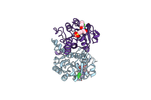
Crystal Structure Of Phgdh In Complex With Compound 39
Organism: Homo sapiens
Method: X-RAY DIFFRACTION Resolution:1.89 Å Release Date: 2019-08-07 Classification: OXIDOREDUCTASE Ligands: SO4, K5N |
Organism: Homo sapiens
Method: X-RAY DIFFRACTION
Release Date: 2019-08-07
Ligands: SO4, K5N
 |
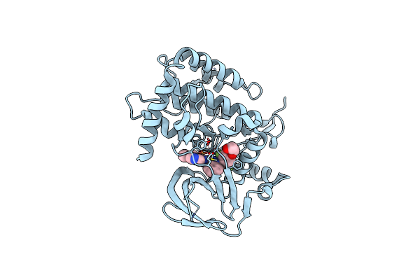
Crystal Structure Of Phgdh In Complex With Bi-4924
Organism: Homo sapiens
Method: X-RAY DIFFRACTION Resolution:1.98 Å Release Date: 2019-08-07 Classification: OXIDOREDUCTASE Ligands: K5K |
Organism: Homo sapiens
Method: X-RAY DIFFRACTION
Release Date: 2019-08-07
Ligands: K5K
 |
Crystal Structure Of The Human Bub1 Kinase Domain In Complex With Bay 1816032
Organism: Homo sapiens
Method: X-RAY DIFFRACTION Resolution:2.00 Å Release Date: 2018-12-19 Classification: TRANSFERASE Ligands: CVQ, MG |
Organism: Homo sapiens
Method: X-RAY DIFFRACTION
Release Date: 2018-12-19
Ligands: CVQ, MG
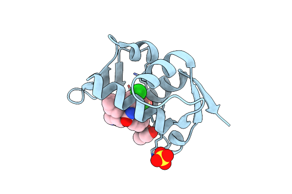 |
Crystal Structure Of Mdm2 In Complex With Compound 13.
Organism: Homo sapiens
Method: X-RAY DIFFRACTION Resolution:1.77 Å Release Date: 2018-12-19 Classification: LIGASE Ligands: H28, GOL, SO4 |
Organism: Homo sapiens
Method: X-RAY DIFFRACTION
Release Date: 2018-12-19
Ligands: H28, GOL, SO4
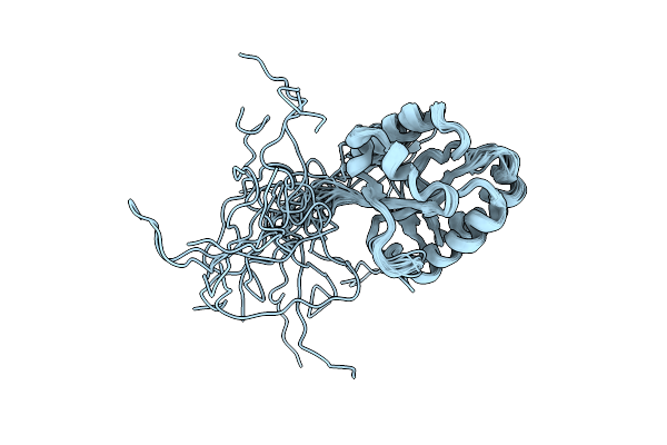 |
Solution Structure Of The Pseudo-Receiver Domain Of Atg32
Organism: Saccharomyces cerevisiae
Method: SOLUTION NMR Release Date: 2018-07-04 Classification: PROTEIN TRANSPORT |
Organism: Saccharomyces cerevisiae
Method: SOLUTION NMR
Release Date: 2018-07-04
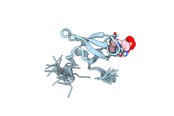 |
Solution Structure Of Smn Tudor Domain In Complex With Symmetrically Dimethylated Arginine
Organism: Homo sapiens
Method: SOLUTION NMR Release Date: 2011-11-30 Classification: RNA BINDING PROTEIN Ligands: 2MR |
Organism: Homo sapiens
Method: SOLUTION NMR
Release Date: 2011-11-30
Ligands: 2MR
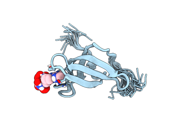 |
Solution Structure Of Spf30 Tudor Domain In Complex With Symmetrically Dimethylated Arginine
Organism: Homo sapiens
Method: SOLUTION NMR Release Date: 2011-11-30 Classification: RNA BINDING PROTEIN Ligands: 2MR |
Organism: Homo sapiens
Method: SOLUTION NMR
Release Date: 2011-11-30
Ligands: 2MR