

Search Count: 40
 |
Human Alpha7 Nicotinic Receptor In Complex With The F1 Nanobody
Organism: Homo sapiens, Vicugna pacos
Method: ELECTRON MICROSCOPY Release Date: 2026-02-18 Classification: MEMBRANE PROTEIN |
Organism: Homo sapiens, Vicugna pacos
Method: ELECTRON MICROSCOPY
Release Date: 2026-02-18
 |
Human Alpha7 Nicotinic Receptor In Complex With The E6 Nanobody
Organism: Homo sapiens, Vicugna pacos
Method: ELECTRON MICROSCOPY Resolution:2.16 Å Release Date: 2026-02-18 Classification: MEMBRANE PROTEIN |
Organism: Homo sapiens, Vicugna pacos
Method: ELECTRON MICROSCOPY
Release Date: 2026-02-18
 |
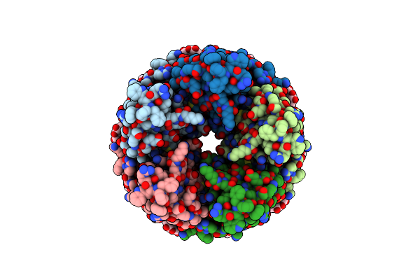
Human Alpha7 Nicotinic Receptor In Complex With The C4 Nanobody
Organism: Homo sapiens, Vicugna pacos huacaya
Method: ELECTRON MICROSCOPY Release Date: 2023-10-11 Classification: MEMBRANE PROTEIN Ligands: NAG |
Organism: Homo sapiens, Vicugna pacos huacaya
Method: ELECTRON MICROSCOPY
Release Date: 2023-10-11
Ligands: NAG
 |
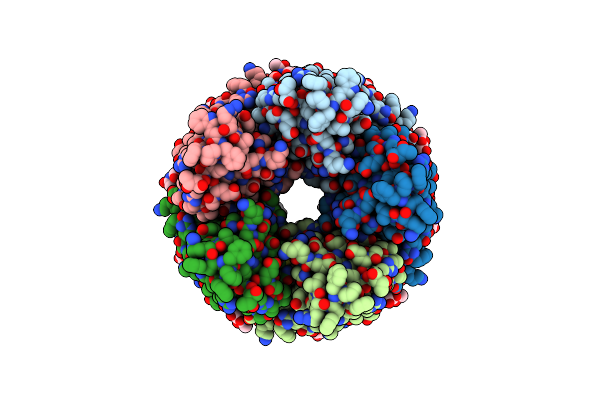
Human Alpha7 Nicotinic Receptor In Complex With The C4 Nanobody And Nicotine
Organism: Homo sapiens, Vicugna pacos
Method: ELECTRON MICROSCOPY Release Date: 2023-10-11 Classification: MEMBRANE PROTEIN Ligands: NAG, NCT |
Organism: Homo sapiens, Vicugna pacos
Method: ELECTRON MICROSCOPY
Release Date: 2023-10-11
Ligands: NAG, NCT
 |
Human Alpha7 Nicotinic Receptor In Complex With The E3 Nanobody
Organism: Homo sapiens, Vicugna pacos
Method: ELECTRON MICROSCOPY Release Date: 2023-10-11 Classification: MEMBRANE PROTEIN Ligands: NAG |
Organism: Homo sapiens, Vicugna pacos
Method: ELECTRON MICROSCOPY
Release Date: 2023-10-11
Ligands: NAG
 |
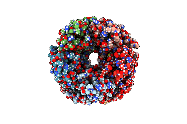
Human Alpha7 Nicotinic Receptor In Complex With The E3 Nanobody And Nicotine
Organism: Homo sapiens
Method: ELECTRON MICROSCOPY Release Date: 2023-10-11 Classification: MEMBRANE PROTEIN Ligands: NAG, NCT |
Organism: Homo sapiens
Method: ELECTRON MICROSCOPY
Release Date: 2023-10-11
Ligands: NAG, NCT
 |
Human Alpha7 Nicotinic Receptor In Complex With The C4 Nanobody Under Sub-Saturating Conditions
Organism: Homo sapiens, Vicugna pacos
Method: ELECTRON MICROSCOPY Release Date: 2023-10-11 Classification: MEMBRANE PROTEIN Ligands: NAG |
Organism: Homo sapiens, Vicugna pacos
Method: ELECTRON MICROSCOPY
Release Date: 2023-10-11
Ligands: NAG
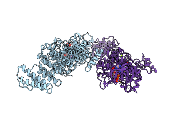 |
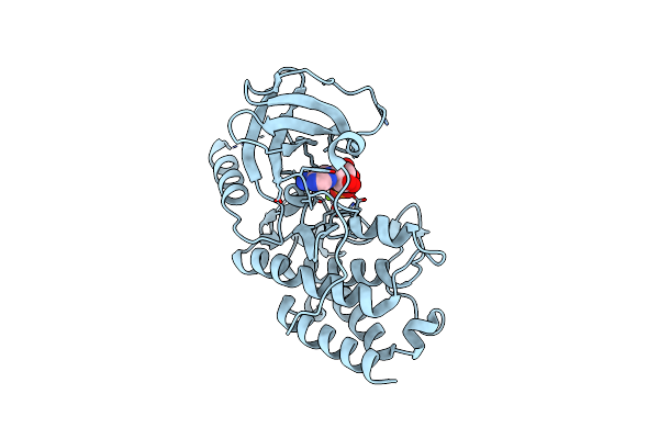
Crystal Structure Of The S/T Protein Kinase Pkng From Corynebacterium Glutamicum In Complex With Amp-Pnp
Organism: Corynebacterium glutamicum (strain atcc 13032 / dsm 20300 / bcrc 11384 / jcm 1318 / lmg 3730 / ncimb 10025)
Method: X-RAY DIFFRACTION Resolution:2.20 Å Release Date: 2021-10-13 Classification: TRANSFERASE Ligands: MG, ANP |
 |
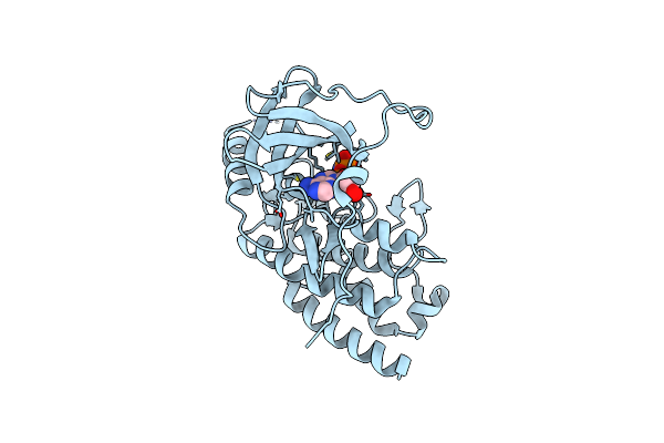
Crystal Structure Of The S/T Protein Kinase Pkng From Corynebacterium Glutamicum (Residues 130-433) In Complex With Amp-Pnp, Isoform 1
Organism: Corynebacterium glutamicum (strain atcc 13032 / dsm 20300 / bcrc 11384 / jcm 1318 / lmg 3730 / ncimb 10025)
Method: X-RAY DIFFRACTION Resolution:1.92 Å Release Date: 2021-10-13 Classification: TRANSFERASE Ligands: MG, ANP |
 |
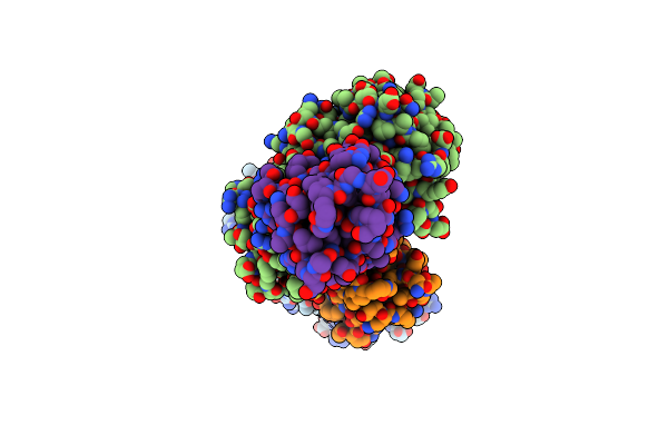
Crystal Structure Of The S/T Protein Kinase Pkng From Corynebacterium Glutamicum (Residues 130-433) In Complex With Amp-Pnp, Isoform 2
Organism: Corynebacterium glutamicum (strain atcc 13032 / dsm 20300 / bcrc 11384 / jcm 1318 / lmg 3730 / ncimb 10025)
Method: X-RAY DIFFRACTION Resolution:1.99 Å Release Date: 2021-10-13 Classification: TRANSFERASE Ligands: MG, ANP |
 |
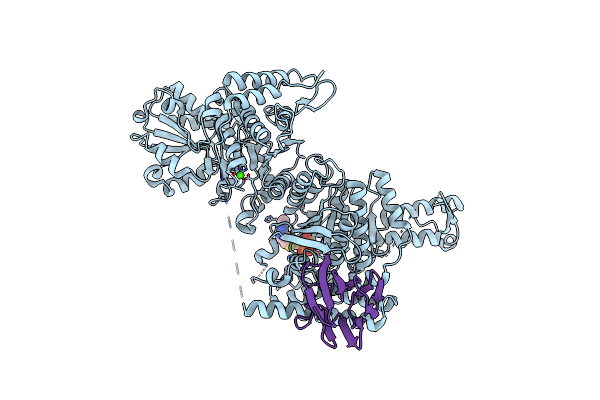
Crystal Structure Of The Mycobacterium Tuberculosis Pknb Kinase Domain (L33E Mutant) In Complex With Its Substrate Gara
Organism: Mycobacterium tuberculosis (strain atcc 25618 / h37rv)
Method: X-RAY DIFFRACTION Resolution:2.37 Å Release Date: 2019-05-22 Classification: SIGNALING PROTEIN Ligands: ACP, MG, SO4 |
Organism: Mycobacterium tuberculosis (strain atcc 25618 / h37rv)
Method: X-RAY DIFFRACTION
Release Date: 2019-05-22
Ligands: ACP, MG, SO4
 |
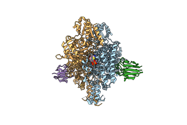
Crystal Structure Of The Wild-Type Suca Domain Of Mycobacterium Smegmatis Kgd (Alpha-Ketoglutarate Decarboxylase), In Complex With Gara
Organism: Mycobacterium smegmatis (strain atcc 700084 / mc(2)155)
Method: X-RAY DIFFRACTION Resolution:2.15 Å Release Date: 2019-05-22 Classification: OXIDOREDUCTASE Ligands: TPP, MG, CA |
Organism: Mycobacterium smegmatis (strain atcc 700084 / mc(2)155)
Method: X-RAY DIFFRACTION
Release Date: 2019-05-22
Ligands: TPP, MG, CA
 |
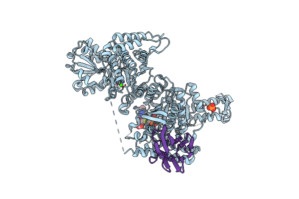
Crystal Structure Of The Suca Domain Of Mycobacterium Smegmatis Kgd (Alpha-Ketoglutarate Decarboxylase), Mutant R802A, In Complex With Gara
Organism: Mycobacterium smegmatis (strain atcc 700084 / mc(2)155)
Method: X-RAY DIFFRACTION Resolution:2.20 Å Release Date: 2019-05-22 Classification: OXIDOREDUCTASE Ligands: TPP, MG, CA |
Organism: Mycobacterium smegmatis (strain atcc 700084 / mc(2)155)
Method: X-RAY DIFFRACTION
Release Date: 2019-05-22
Ligands: TPP, MG, CA
 |
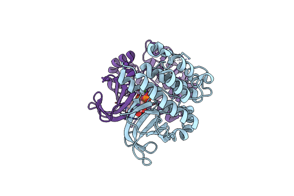
Crystal Structure Of The Suca Domain Of Mycobacterium Smegmatis Kgd (R802A) In Complex With Gara, Following 2-Oxoglutarate Soak
Organism: Mycobacterium smegmatis (strain atcc 700084 / mc(2)155)
Method: X-RAY DIFFRACTION Resolution:2.40 Å Release Date: 2019-05-22 Classification: OXIDOREDUCTASE Ligands: MG, CA, PO4, TD6 |
Organism: Mycobacterium smegmatis (strain atcc 700084 / mc(2)155)
Method: X-RAY DIFFRACTION
Release Date: 2019-05-22
Ligands: MG, CA, PO4, TD6
 |
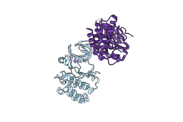
Crystal Structure Of Mycobacterium Tuberculosis Pkni Kinase Domain
Organism: Mycobacterium tuberculosis (strain atcc 25618 / h37rv)
Method: X-RAY DIFFRACTION Resolution:2.00 Å Release Date: 2017-01-11 Classification: SIGNALING PROTEIN Ligands: ADP, CA |
Organism: Mycobacterium tuberculosis (strain atcc 25618 / h37rv)
Method: X-RAY DIFFRACTION
Release Date: 2017-01-11
Ligands: ADP, CA
 |
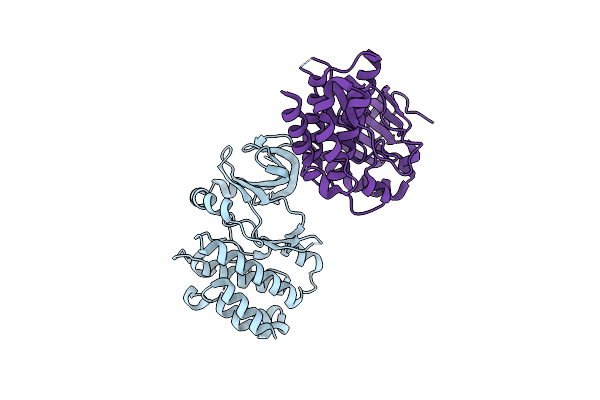
Crystal Structure Of Mycobacterium Tuberculosis Pkni Kinase Domain, C20A Mutant
Organism: Mycobacterium tuberculosis h37rv
Method: X-RAY DIFFRACTION Resolution:2.50 Å Release Date: 2017-01-11 Classification: SIGNALING PROTEIN |
Organism: Mycobacterium tuberculosis h37rv
Method: X-RAY DIFFRACTION
Release Date: 2017-01-11
 |
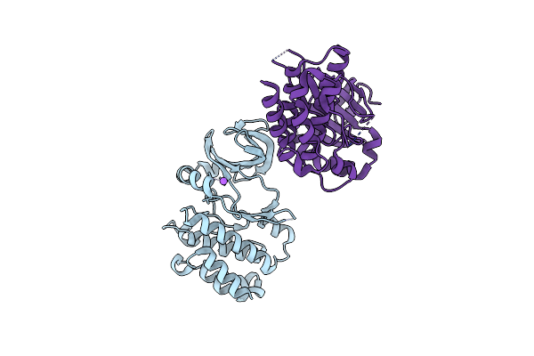
Crystal Structure Of Mycobacterium Tuberculosis Pkni Kinase Domain, C20A_R136A Double Mutant
Organism: Mycobacterium tuberculosis (strain atcc 25618 / h37rv)
Method: X-RAY DIFFRACTION Resolution:3.03 Å Release Date: 2017-01-11 Classification: SIGNALING PROTEIN |
Organism: Mycobacterium tuberculosis (strain atcc 25618 / h37rv)
Method: X-RAY DIFFRACTION
Release Date: 2017-01-11
 |
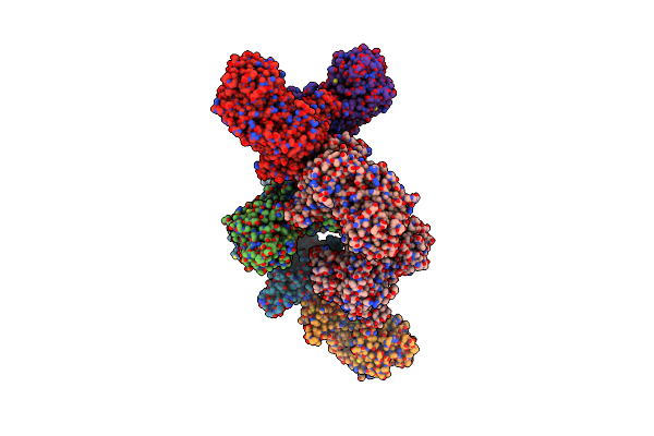
Crystal Structure Of Mycobacterium Tuberculosis Pkni Kinase Domain, C20A_R136N Double Mutant
Organism: Mycobacterium tuberculosis (strain atcc 25618 / h37rv)
Method: X-RAY DIFFRACTION Resolution:2.98 Å Release Date: 2017-01-11 Classification: SIGNALING PROTEIN |
Organism: Mycobacterium tuberculosis (strain atcc 25618 / h37rv)
Method: X-RAY DIFFRACTION
Release Date: 2017-01-11
 |
Crystal Structure Of The M. Tuberculosis Ctp Synthase Pyrg (Apo Form)
Organism: Mycobacterium tuberculosis (strain atcc 25618 / h37rv)
Method: X-RAY DIFFRACTION Resolution:3.52 Å Release Date: 2015-07-01 Classification: LIGASE Ligands: CA |
Organism: Mycobacterium tuberculosis (strain atcc 25618 / h37rv)
Method: X-RAY DIFFRACTION
Release Date: 2015-07-01
Ligands: CA
 |
Crystal Structure Of The M. Tuberculosis Ctp Synthase Pyrg In Complex With Two Utp Molecules
Organism: Mycobacterium tuberculosis (strain atcc 25618 / h37rv)
Method: X-RAY DIFFRACTION Resolution:1.99 Å Release Date: 2015-07-01 Classification: LIGASE Ligands: UTP, MG, GOL |
Organism: Mycobacterium tuberculosis (strain atcc 25618 / h37rv)
Method: X-RAY DIFFRACTION
Release Date: 2015-07-01
Ligands: UTP, MG, GOL