

Search Count: 169
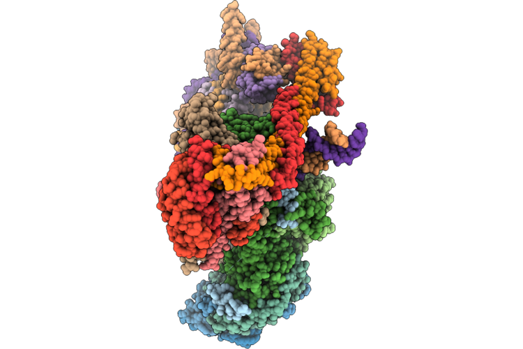 |
Structure Of The Human Inner Kinetochore Ccan And Cenp-C Bound To Dna
Organism: Homo sapiens, Trichoplusia ni
Method: ELECTRON MICROSCOPY Resolution:2.70 Å Release Date: 2026-05-20 Classification: CELL CYCLE |
Organism: Homo sapiens, Trichoplusia ni
Method: ELECTRON MICROSCOPY
Release Date: 2026-05-20
 |
Structure Of The Human Inner Kinetochore Ccan Bound To Dna
Organism: Homo sapiens
Method: ELECTRON MICROSCOPY Resolution:3.54 Å Release Date: 2026-05-20 Classification: CELL CYCLE |
Organism: Homo sapiens
Method: ELECTRON MICROSCOPY
Release Date: 2026-05-20
 |
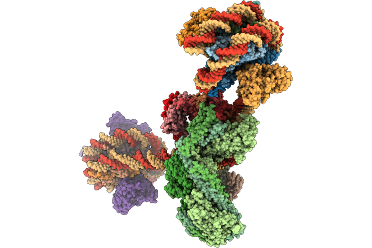
Structure Of The Human Inner Kinetochore Ccan Bound To A Mono-Cenp-A Nucleosome
Organism: Homo sapiens
Method: ELECTRON MICROSCOPY Resolution:4.50 Å Release Date: 2026-05-20 Classification: CELL CYCLE |
Organism: Homo sapiens
Method: ELECTRON MICROSCOPY
Release Date: 2026-05-20
 |
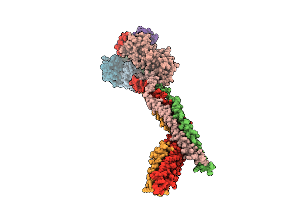
Structure Of The Human Inner Kinetochore Ccan Bound To A Di-Cenp-A Nucleosome
Organism: Homo sapiens, Mus musculus
Method: ELECTRON MICROSCOPY Resolution:15.50 Å Release Date: 2026-05-20 Classification: CELL CYCLE |
Organism: Homo sapiens, Mus musculus
Method: ELECTRON MICROSCOPY
Release Date: 2026-05-20
 |
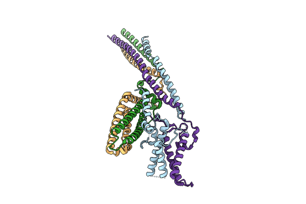
Cryo-Em Structure Of The Saccharomyces Cerevisiae Kmn Junction Complex Lacking The Mis12C(Mtw1C) Head 2 Domain
Organism: Saccharomyces cerevisiae s288c
Method: ELECTRON MICROSCOPY Release Date: 2026-04-08 Classification: CELL CYCLE |
Organism: Saccharomyces cerevisiae s288c
Method: ELECTRON MICROSCOPY
Release Date: 2026-04-08
 |
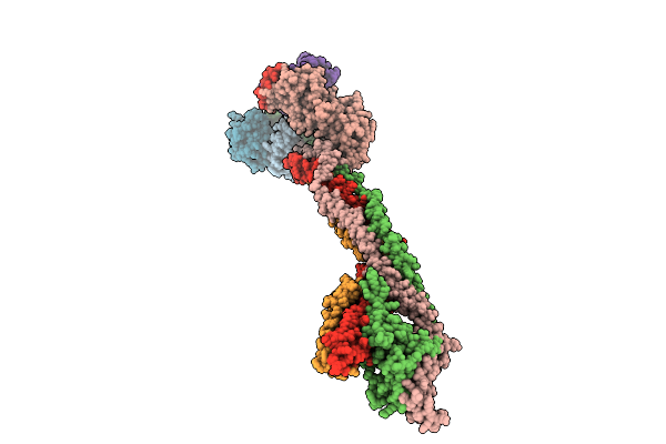
Cryo-Em Structure Of The Base Of The Saccharomyces Cerevisiae Kmn Junction Complex Containing The Mis12C(Mtw1C) Head 2 Domain
Organism: Saccharomyces cerevisiae s288c
Method: ELECTRON MICROSCOPY Release Date: 2026-04-08 Classification: CELL CYCLE |
Organism: Saccharomyces cerevisiae s288c
Method: ELECTRON MICROSCOPY
Release Date: 2026-04-08
 |
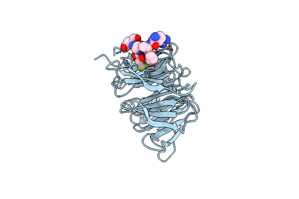
Cryo-Em Structure Of The Saccharomyces Cerevisiae Kmn Junction Complex Containing The Mis12C(Mtw1C) Head 2 Domain
Organism: Saccharomyces cerevisiae, Saccharomyces cerevisiae s288c
Method: ELECTRON MICROSCOPY Release Date: 2026-04-08 Classification: CELL CYCLE |
Organism: Saccharomyces cerevisiae, Saccharomyces cerevisiae s288c
Method: ELECTRON MICROSCOPY
Release Date: 2026-04-08
 |
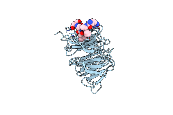
Crystal Structure Of Human Cdc20 Bound To Synthetic D-Box Peptide D21
Organism: Homo sapiens
Method: X-RAY DIFFRACTION Resolution:1.51 Å Release Date: 2025-02-12 Classification: LIGASE |
Organism: Homo sapiens
Method: X-RAY DIFFRACTION
Release Date: 2025-02-12
 |
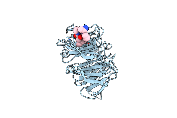
Crystal Structure Of Human Cdc20 Bound To Synthetic D-Box Peptide D20
Organism: Homo sapiens
Method: X-RAY DIFFRACTION Resolution:1.46 Å Release Date: 2025-02-12 Classification: LIGASE |
Organism: Homo sapiens
Method: X-RAY DIFFRACTION
Release Date: 2025-02-12
 |
Crystal Structure Of Human Cdc20 Bound To Synthetic D-Box Peptide D7
Organism: Homo sapiens
Method: X-RAY DIFFRACTION Resolution:1.92 Å Release Date: 2025-02-12 Classification: LIGASE |
Organism: Homo sapiens
Method: X-RAY DIFFRACTION
Release Date: 2025-02-12
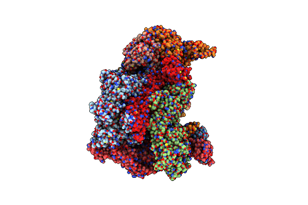 |
High-Resolution Structure Of The Anaphase-Promoting Complex/Cyclosome (Apc/C) Bound To Co-Activator Cdh1
Organism: Homo sapiens
Method: ELECTRON MICROSCOPY Release Date: 2024-08-14 Classification: CELL CYCLE Ligands: ZN |
Organism: Homo sapiens
Method: ELECTRON MICROSCOPY
Release Date: 2024-08-14
Ligands: ZN
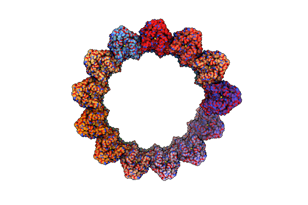 |
Structure Of The Native Microtubule Lattice Nucleated From The Yeast Spindle Pole Body
Organism: Saccharomyces cerevisiae
Method: ELECTRON MICROSCOPY Release Date: 2024-04-24 Classification: CELL CYCLE |
Organism: Saccharomyces cerevisiae
Method: ELECTRON MICROSCOPY
Release Date: 2024-04-24
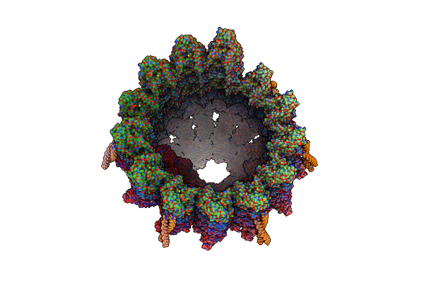 |
Structure Of The Native Y-Tubulin Ring Complex (Yturc) Capping Microtubule Minus Ends At The Spindle Pole Body
Organism: Saccharomyces cerevisiae
Method: ELECTRON MICROSCOPY Release Date: 2024-04-24 Classification: CELL CYCLE Ligands: GTP, GDP |
Organism: Saccharomyces cerevisiae
Method: ELECTRON MICROSCOPY
Release Date: 2024-04-24
Ligands: GTP, GDP
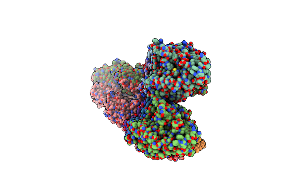 |
Structure Of The Y-Tubulin Small Complex (Ytusc) As Part Of The Native Y-Tubulin Ring Complex (Yturc) Capping Microtubule Minus Ends At The Spindle Pole Body
Organism: Saccharomyces cerevisiae
Method: ELECTRON MICROSCOPY Release Date: 2024-04-24 Classification: CELL CYCLE Ligands: GTP |
Organism: Saccharomyces cerevisiae
Method: ELECTRON MICROSCOPY
Release Date: 2024-04-24
Ligands: GTP
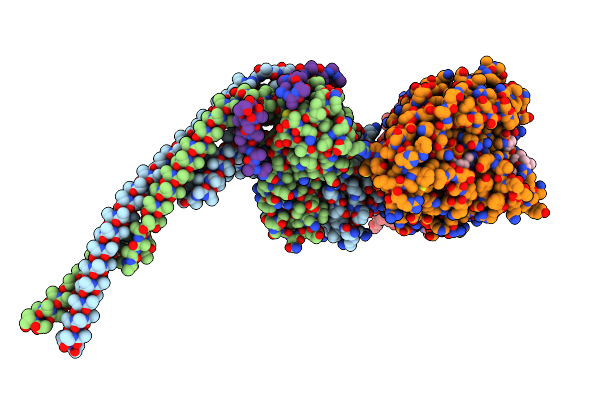 |
Outer Kinetochore Ndc80-Dam1 Alpha/Beta-Tubulin Complex
Organism: Saccharomyces cerevisiae, Sus scrofa
Method: ELECTRON MICROSCOPY Release Date: 2024-04-03 Classification: CELL CYCLE Ligands: GTP, MG, GDP, TA1 |
Organism: Saccharomyces cerevisiae, Sus scrofa
Method: ELECTRON MICROSCOPY
Release Date: 2024-04-03
Ligands: GTP, MG, GDP, TA1
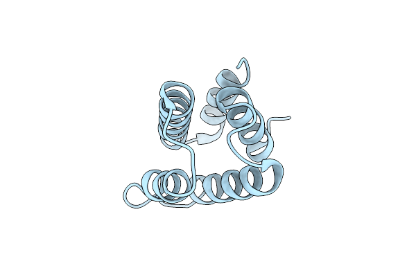 |
Crystal Structure Of The Yeast Spindle Body Component Spc98
Organism: Saccharomyces cerevisiae
Method: X-RAY DIFFRACTION Resolution:1.87 Å Release Date: 2024-04-03 Classification: CELL CYCLE |
Organism: Saccharomyces cerevisiae
Method: X-RAY DIFFRACTION
Release Date: 2024-04-03
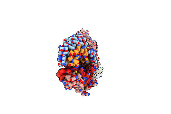 |
Structure Of The Human Outer Kinetochore Kmn Network Complex
Organism: Homo sapiens
Method: ELECTRON MICROSCOPY Release Date: 2024-03-20 Classification: CELL CYCLE Ligands: HOH |
Organism: Homo sapiens
Method: ELECTRON MICROSCOPY
Release Date: 2024-03-20
Ligands: HOH
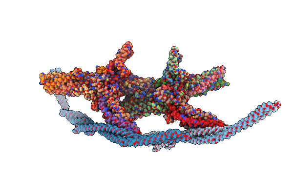 |
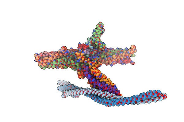
Outer Kinetochore Dam1 Protomer Dimer Ndc80-Nuf2 Coiled-Coil Complex
Organism: Saccharomyces cerevisiae
Method: ELECTRON MICROSCOPY Release Date: 2023-12-06 Classification: CELL CYCLE |
Organism: Saccharomyces cerevisiae
Method: ELECTRON MICROSCOPY
Release Date: 2023-12-06
 |
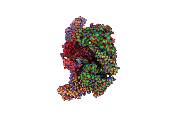
Outer Kinetochore Dam1 Protomer Monomer Ndc80-Nuf2 Coiled-Coil Complex
Organism: Saccharomyces cerevisiae
Method: ELECTRON MICROSCOPY Release Date: 2023-12-06 Classification: CELL CYCLE |
Organism: Saccharomyces cerevisiae
Method: ELECTRON MICROSCOPY
Release Date: 2023-12-06
 |
Cryo-Em Structure Of Cbf1-Ccan Bound Topologically To Centromeric Dna
Organism: Saccharomyces cerevisiae
Method: ELECTRON MICROSCOPY Release Date: 2023-08-09 Classification: CELL CYCLE |
Organism: Saccharomyces cerevisiae
Method: ELECTRON MICROSCOPY
Release Date: 2023-08-09