

Search Count: 90
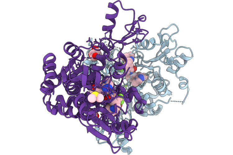 |
Structure Of Cbl-B Bound To Compound 33
Organism: Homo sapiens
Method: X-RAY DIFFRACTION Resolution:2.30 Å Release Date: 2026-07-01 Classification: LIGASE Ligands: GOL, IOD, EDO, MG, ZN, A1DCD, DMS |
Organism: Homo sapiens
Method: X-RAY DIFFRACTION
Release Date: 2026-07-01
Ligands: GOL, IOD, EDO, MG, ZN, A1DCD, DMS
 |
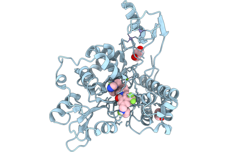
Structure Of C-Cbl Bound To Compound 33
Organism: Schistosoma japonicum, Homo sapiens
Method: X-RAY DIFFRACTION Resolution:2.39 Å Release Date: 2026-07-01 Classification: LIGASE Ligands: A1DCD, ZN, MG |
Organism: Schistosoma japonicum, Homo sapiens
Method: X-RAY DIFFRACTION
Release Date: 2026-07-01
Ligands: A1DCD, ZN, MG
 |
Structure Of Cbl-B Bound To Compound 8
Organism: Homo sapiens
Method: X-RAY DIFFRACTION Resolution:1.77 Å Release Date: 2026-07-01 Classification: LIGASE/INHIBITOR Ligands: GOL, ZN, EDO, MG, A1DCC |
Organism: Homo sapiens
Method: X-RAY DIFFRACTION
Release Date: 2026-07-01
Ligands: GOL, ZN, EDO, MG, A1DCC
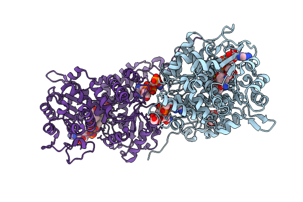 |
Human Malic Enzyme 2 Complex With Inhibitor Npd-389
Organism: Homo sapiens
Method: X-RAY DIFFRACTION Resolution:2.45 Å Release Date: 2025-10-29 Classification: OXIDOREDUCTASE Ligands: MG, A1AE5, SIN, NAD, PO4 |
Organism: Homo sapiens
Method: X-RAY DIFFRACTION
Release Date: 2025-10-29
Ligands: MG, A1AE5, SIN, NAD, PO4
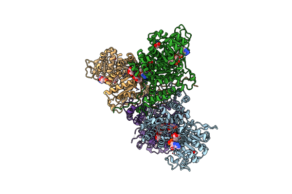 |
Human Malic Enzyme 2 Cofactor Complex At 1.89 Angstrom.
Organism: Homo sapiens
Method: X-RAY DIFFRACTION Resolution:1.89 Å Release Date: 2025-03-12 Classification: OXIDOREDUCTASE Ligands: NAD, EDO, CL, MG, TLA |
Organism: Homo sapiens
Method: X-RAY DIFFRACTION
Release Date: 2025-03-12
Ligands: NAD, EDO, CL, MG, TLA
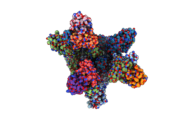 |
Ba.5 Spike Complex With Cr9
Organism: Severe acute respiratory syndrome coronavirus 2, Homo sapiens
Method: ELECTRON MICROSCOPY Release Date: 2025-02-12 Classification: VIRAL PROTEIN/IMMUNE SYSTEM |
Organism: Severe acute respiratory syndrome coronavirus 2, Homo sapiens
Method: ELECTRON MICROSCOPY
Release Date: 2025-02-12
 |
Ba.5 Rbd In Complex With Cr9
Organism: Severe acute respiratory syndrome coronavirus 2, Homo sapiens
Method: ELECTRON MICROSCOPY Release Date: 2025-02-12 Classification: ALLERGEN/IMMUNE SYSTEM |
Organism: Severe acute respiratory syndrome coronavirus 2, Homo sapiens
Method: ELECTRON MICROSCOPY
Release Date: 2025-02-12
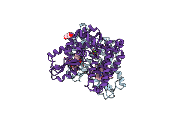 |
Crystal Structure Of Cbl-B Tkb Bound To Compound 26
Organism: Homo sapiens
Method: X-RAY DIFFRACTION Resolution:2.40 Å Release Date: 2024-07-03 Classification: TRANSFERASE/TRANSFERASE INHIBITOR Ligands: MG, A1AEG, GOL, PEG |
Organism: Homo sapiens
Method: X-RAY DIFFRACTION
Release Date: 2024-07-03
Ligands: MG, A1AEG, GOL, PEG
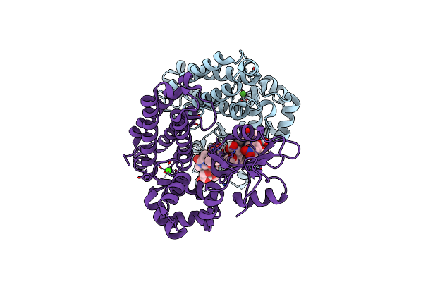 |
Crystal Structure Of Cbl-B Tkb Bound To Compound 2
Organism: Homo sapiens
Method: X-RAY DIFFRACTION Resolution:1.76 Å Release Date: 2024-07-03 Classification: TRANSFERASE/TRANSFERASE INHIBITOR Ligands: MG, CA, A1AD4 |
Organism: Homo sapiens
Method: X-RAY DIFFRACTION
Release Date: 2024-07-03
Ligands: MG, CA, A1AD4
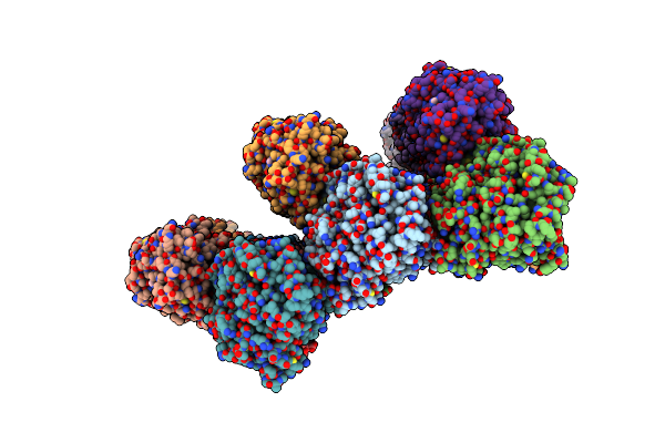 |
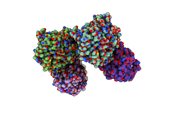
Gmpcpp-Alpha1A/Beta2A-Microtubule Decorated With Kinesin Non-Seam Region
Organism: Mus musculus, Homo sapiens
Method: ELECTRON MICROSCOPY Release Date: 2023-08-16 Classification: STRUCTURAL PROTEIN Ligands: GTP, G2P, ATP |
Organism: Mus musculus, Homo sapiens
Method: ELECTRON MICROSCOPY
Release Date: 2023-08-16
Ligands: GTP, G2P, ATP
 |
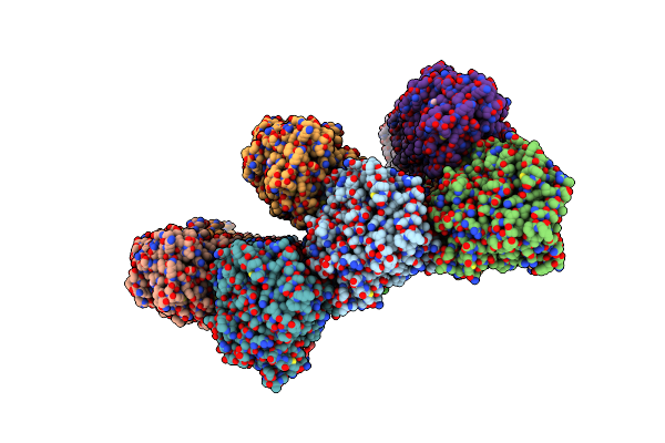
Gmpcpp-Alpha1A/Beta2A-Microtubule Decorated With Kinesin Seam Region
Organism: Mus musculus, Homo sapiens
Method: ELECTRON MICROSCOPY Release Date: 2023-08-16 Classification: STRUCTURAL PROTEIN Ligands: GTP, G2P, ATP |
Organism: Mus musculus, Homo sapiens
Method: ELECTRON MICROSCOPY
Release Date: 2023-08-16
Ligands: GTP, G2P, ATP
 |
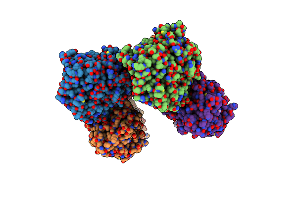
Gmpcpp-Alpha1C/Beta2A-Microtubule Decorated With Kinesin Non-Seam Region
Organism: Mus musculus, Homo sapiens
Method: ELECTRON MICROSCOPY Release Date: 2023-08-16 Classification: STRUCTURAL PROTEIN Ligands: GTP, G2P, ATP |
Organism: Mus musculus, Homo sapiens
Method: ELECTRON MICROSCOPY
Release Date: 2023-08-16
Ligands: GTP, G2P, ATP
 |
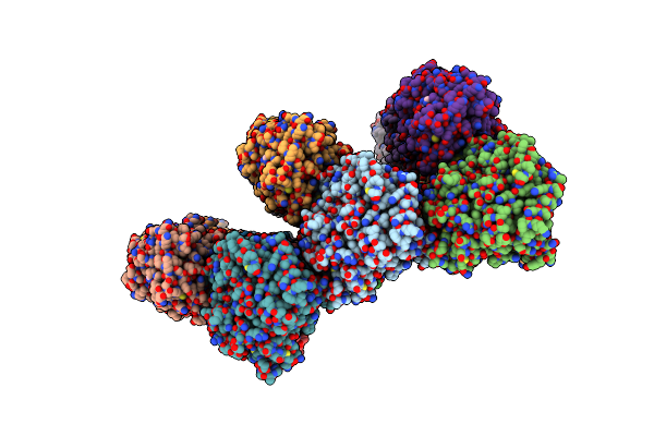
Gmpcpp-Alpha1C/Beta2A-Microtubule Decorated With Kinesin Seam Region
Organism: Mus musculus, Homo sapiens
Method: ELECTRON MICROSCOPY Release Date: 2023-08-16 Classification: STRUCTURAL PROTEIN Ligands: GTP, G2P, ATP |
Organism: Mus musculus, Homo sapiens
Method: ELECTRON MICROSCOPY
Release Date: 2023-08-16
Ligands: GTP, G2P, ATP
 |
Gmpcpp-Alpha4A/Beta2A-Microtubule Decorated With Kinesin Non-Seam Region
Organism: Mus musculus, Homo sapiens
Method: ELECTRON MICROSCOPY Release Date: 2023-08-16 Classification: STRUCTURAL PROTEIN Ligands: GTP, G2P, ATP |
Organism: Mus musculus, Homo sapiens
Method: ELECTRON MICROSCOPY
Release Date: 2023-08-16
Ligands: GTP, G2P, ATP
 |
Gmpcpp-Alpha4A/Beta2A-Microtubule Decorated With Kinesin Seam Region
Organism: Mus musculus, Homo sapiens
Method: ELECTRON MICROSCOPY Release Date: 2023-08-16 Classification: STRUCTURAL PROTEIN Ligands: GTP, G2P, ATP |
Organism: Mus musculus, Homo sapiens
Method: ELECTRON MICROSCOPY
Release Date: 2023-08-16
Ligands: GTP, G2P, ATP
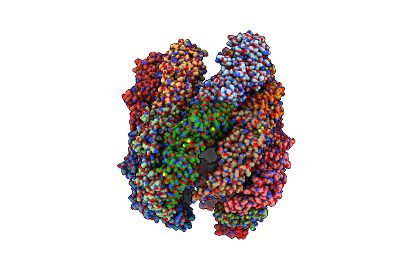 |
Cryo-Em Structure Of Human Tric-Tubulin-S2
Organism: Homo sapiens
Method: ELECTRON MICROSCOPY Release Date: 2023-06-07 Classification: STRUCTURAL PROTEIN Ligands: ATP |
Organism: Homo sapiens
Method: ELECTRON MICROSCOPY
Release Date: 2023-06-07
Ligands: ATP
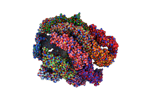 |
Cryo-Em Structure Of Human Tric-Adp
Organism: Homo sapiens
Method: ELECTRON MICROSCOPY Release Date: 2023-06-07 Classification: STRUCTURAL PROTEIN Ligands: ADP |
Organism: Homo sapiens
Method: ELECTRON MICROSCOPY
Release Date: 2023-06-07
Ligands: ADP
 |
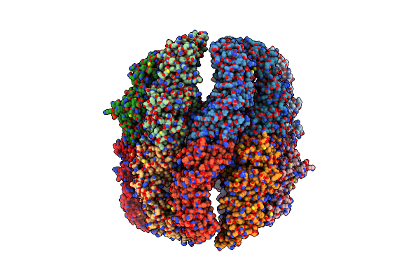
Cryo-Em Structure Of Human Tric-Atp-Closed State
Organism: Homo sapiens
Method: ELECTRON MICROSCOPY Release Date: 2023-06-07 Classification: STRUCTURAL PROTEIN |
Organism: Homo sapiens
Method: ELECTRON MICROSCOPY
Release Date: 2023-06-07
 |
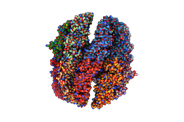
Cryo-Em Structure Of Human Tric-Atp-Open State
Organism: Homo sapiens
Method: ELECTRON MICROSCOPY Release Date: 2023-06-07 Classification: STRUCTURAL PROTEIN |
Organism: Homo sapiens
Method: ELECTRON MICROSCOPY
Release Date: 2023-06-07
 |
Cryo-Em Structure Of Human Tric-Npp State
Organism: Homo sapiens
Method: ELECTRON MICROSCOPY Release Date: 2023-04-12 Classification: STRUCTURAL PROTEIN |
Organism: Homo sapiens
Method: ELECTRON MICROSCOPY
Release Date: 2023-04-12