

Search Count: 37
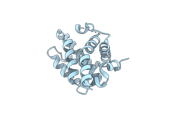 |
Structural Basis Of Bak Sequestration By Mcl-1 And Consequences For Apoptosis Initiation
Organism: Homo sapiens
Method: X-RAY DIFFRACTION Resolution:1.49 Å Release Date: 2025-06-04 Classification: APOPTOSIS |
Organism: Homo sapiens
Method: X-RAY DIFFRACTION
Release Date: 2025-06-04
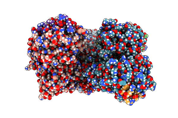 |
Structural Basis Of Bak Sequestration By Mcl-1 And Consequences For Apoptosis Initiation
Organism: Synthetic construct, Homo sapiens
Method: X-RAY DIFFRACTION Resolution:1.70 Å Release Date: 2025-06-04 Classification: APOPTOSIS |
Organism: Synthetic construct, Homo sapiens
Method: X-RAY DIFFRACTION
Release Date: 2025-06-04
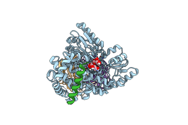 |
Structural Basis Of Bak Sequestration By Mcl-1 And Consequences For Apoptosis Initiation
Organism: Homo sapiens
Method: ELECTRON MICROSCOPY Release Date: 2025-06-04 Classification: APOPTOSIS |
Organism: Homo sapiens
Method: ELECTRON MICROSCOPY
Release Date: 2025-06-04
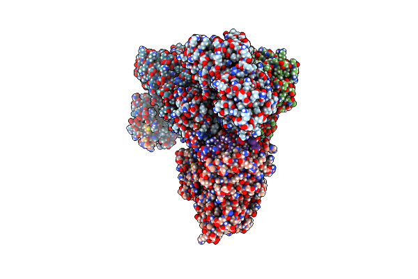 |
Structural Basis Of Bak Sequestration By Mcl-1 And Consequences For Apoptosis Initiation
Organism: Synthetic construct, Homo sapiens
Method: X-RAY DIFFRACTION Resolution:1.89 Å Release Date: 2025-06-04 Classification: APOPTOSIS |
Organism: Synthetic construct, Homo sapiens
Method: X-RAY DIFFRACTION
Release Date: 2025-06-04
 |
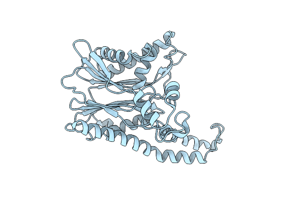
The Crystal Structure Of Carbonic Anhydrase Ii From Biortus.
Organism: Homo sapiens
Method: X-RAY DIFFRACTION Resolution:1.50 Å Release Date: 2024-07-03 Classification: LYASE Ligands: V7Z, ZN, EDO |
Organism: Homo sapiens
Method: X-RAY DIFFRACTION
Release Date: 2024-07-03
Ligands: V7Z, ZN, EDO
 |
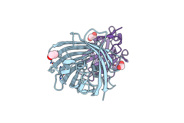
The Crystal Structure Of Tab1 From Biortus.
Organism: Homo sapiens
Method: X-RAY DIFFRACTION Resolution:3.35 Å Release Date: 2024-03-06 Classification: SIGNALING PROTEIN |
Organism: Homo sapiens
Method: X-RAY DIFFRACTION
Release Date: 2024-03-06
 |
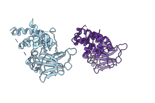
Structure Of The Gfp:Gfp-Nanobody Complex From Biortus.
Organism: Cytaeis uchidae, Synthetic construct
Method: X-RAY DIFFRACTION Resolution:2.10 Å Release Date: 2024-03-06 Classification: FLUORESCENT PROTEIN Ligands: EDO, ZN |
Organism: Cytaeis uchidae, Synthetic construct
Method: X-RAY DIFFRACTION
Release Date: 2024-03-06
Ligands: EDO, ZN
 |
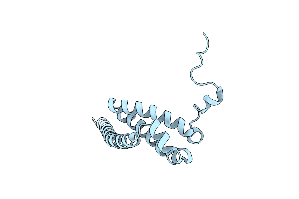
The Crystal Structure Of P53/Bcl-Xl Fusion Complex From Biortus.
Organism: Homo sapiens
Method: X-RAY DIFFRACTION Resolution:2.55 Å Release Date: 2024-03-06 Classification: PROTEIN BINDING Ligands: ZN |
Organism: Homo sapiens
Method: X-RAY DIFFRACTION
Release Date: 2024-03-06
Ligands: ZN
 |
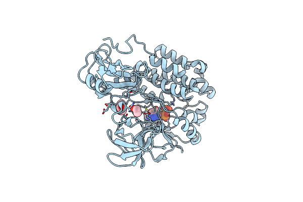
The Crystal Structure Of Usp8 From Biortus.
Organism: Homo sapiens
Method: X-RAY DIFFRACTION Resolution:3.10 Å Release Date: 2024-03-06 Classification: HYDROLASE |
Organism: Homo sapiens
Method: X-RAY DIFFRACTION
Release Date: 2024-03-06
 |
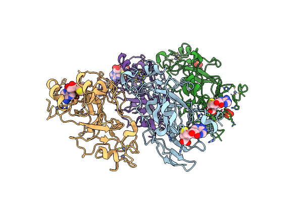
The Crystal Structure Of Src From Biortus.
Organism: Mus musculus
Method: X-RAY DIFFRACTION Resolution:1.95 Å Release Date: 2024-01-24 Classification: SIGNALING PROTEIN Ligands: ADP, MG, GOL, EDO |
Organism: Mus musculus
Method: X-RAY DIFFRACTION
Release Date: 2024-01-24
Ligands: ADP, MG, GOL, EDO
 |
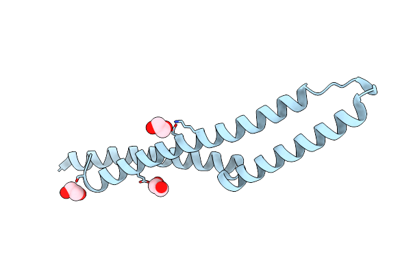
The Crystal Structure Of Ehmt1 From Biortus.
Organism: Homo sapiens
Method: X-RAY DIFFRACTION Resolution:3.35 Å Release Date: 2024-01-24 Classification: TRANSFERASE Ligands: SAH, ZN, SO4 |
Organism: Homo sapiens
Method: X-RAY DIFFRACTION
Release Date: 2024-01-24
Ligands: SAH, ZN, SO4
 |
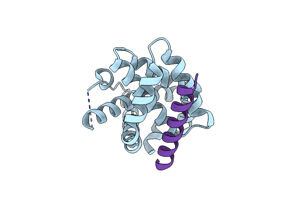
The Crystal Structure Of Apc From Biortus.
Organism: Homo sapiens
Method: X-RAY DIFFRACTION Resolution:2.00 Å Release Date: 2023-11-22 Classification: ANTITUMOR PROTEIN Ligands: EDO |
Organism: Homo sapiens
Method: X-RAY DIFFRACTION
Release Date: 2023-11-22
Ligands: EDO
 |
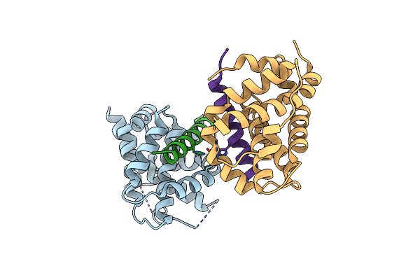
Crystal Structure Of Human Bak In Complex With W3W5_Bid
Organism: Homo sapiens
Method: X-RAY DIFFRACTION Resolution:1.50 Å Release Date: 2022-01-12 Classification: APOPTOSIS |
Organism: Homo sapiens
Method: X-RAY DIFFRACTION
Release Date: 2022-01-12
 |
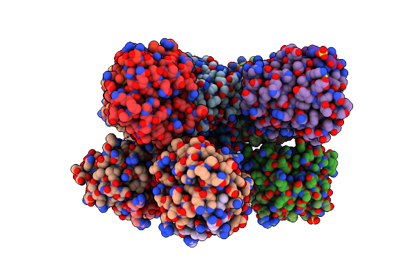
Crystal Structure Of Human Bak In Complex With M3W5_Bid
Organism: Homo sapiens
Method: X-RAY DIFFRACTION Resolution:1.85 Å Release Date: 2022-01-12 Classification: APOPTOSIS Ligands: CU |
Organism: Homo sapiens
Method: X-RAY DIFFRACTION
Release Date: 2022-01-12
Ligands: CU
 |
Crystal Structure Of Human Bak In Complex With Wt Bak Bh3 Peptide
Organism: Homo sapiens
Method: X-RAY DIFFRACTION Resolution:3.06 Å Release Date: 2022-01-12 Classification: APOPTOSIS Ligands: CU, SO4 |
Organism: Homo sapiens
Method: X-RAY DIFFRACTION
Release Date: 2022-01-12
Ligands: CU, SO4
 |
Structure Of Ttbk1 Kinase Domain In Complex With Compound 3
Organism: Homo sapiens
Method: X-RAY DIFFRACTION Resolution:1.56 Å Release Date: 2021-05-19 Classification: TRANSFERASE/INHIBITOR Ligands: VP7, NA |
Organism: Homo sapiens
Method: X-RAY DIFFRACTION
Release Date: 2021-05-19
Ligands: VP7, NA
 |
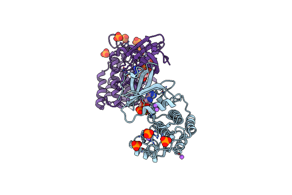
Structure Of Ttbk1 Kinase Domain In Complex With Compound 18
Organism: Homo sapiens
Method: X-RAY DIFFRACTION Resolution:2.15 Å Release Date: 2021-05-19 Classification: TRANSFERASE/INHIBITOR Ligands: VSY |
Organism: Homo sapiens
Method: X-RAY DIFFRACTION
Release Date: 2021-05-19
Ligands: VSY
 |
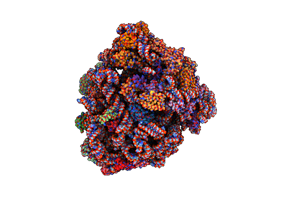
Adp Bound Ttbk2 Kinase Domain
Organism: Homo sapiens
Method: X-RAY DIFFRACTION Resolution:1.50 Å Release Date: 2020-06-03 Classification: TRANSFERASE Ligands: ADP, PO4, NA, MG, GOL |
Organism: Homo sapiens
Method: X-RAY DIFFRACTION
Release Date: 2020-06-03
Ligands: ADP, PO4, NA, MG, GOL
 |
70S Ribosome Bound To Hiv Frameshifting Stem-Loop (Fss) And P/E Trna (Rotated Conformation)
Organism: Escherichia coli, Human immunodeficiency virus 1
Method: ELECTRON MICROSCOPY Release Date: 2020-06-03 Classification: RIBOSOME Ligands: MG |
Organism: Escherichia coli, Human immunodeficiency virus 1
Method: ELECTRON MICROSCOPY
Release Date: 2020-06-03
Ligands: MG
 |
70S Ribosome Bound To Hiv Frameshifting Stem-Loop (Fss) And P-Site Trna (Non-Rotated Conformation, Structure I)
Organism: Human immunodeficiency virus 1, Escherichia coli
Method: ELECTRON MICROSCOPY Release Date: 2020-06-03 Classification: RIBOSOME Ligands: MG |
Organism: Human immunodeficiency virus 1, Escherichia coli
Method: ELECTRON MICROSCOPY
Release Date: 2020-06-03
Ligands: MG