

Search Count: 40
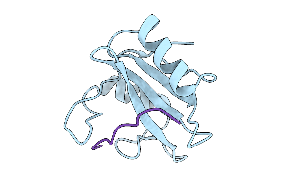 |
Structure Of The N-Sh2 Domain Of Shp2 In Complex With The Phosphoy627-Gab1 (613-651) Peptide
Organism: Homo sapiens
Method: X-RAY DIFFRACTION Resolution:2.08 Å Release Date: 2025-12-31 Classification: SIGNALING PROTEIN |
Organism: Homo sapiens
Method: X-RAY DIFFRACTION
Release Date: 2025-12-31
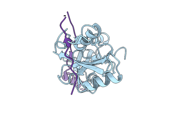 |
Micro-Ed Structure Of The Nsh2-Csh2 Tandem Domain Of Shp2 In Complex With The Bis-Phosphorylated Py627-Py659-Gab1 (613-694) Peptide
Organism: Homo sapiens
Method: ELECTRON CRYSTALLOGRAPHY Resolution:3.20 Å Release Date: 2025-12-31 Classification: SIGNALING PROTEIN |
Organism: Homo sapiens
Method: ELECTRON CRYSTALLOGRAPHY
Release Date: 2025-12-31
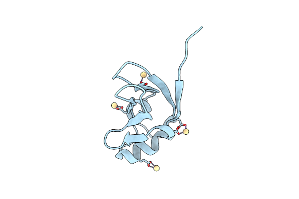 |
Crystal Structure Of S.Cerevisiae Kti11P
Organism: Saccharomyces cerevisiae s288c
Method: X-RAY DIFFRACTION Resolution:2.40 Å Release Date: 2016-07-20 Classification: METAL BINDING PROTEIN Ligands: CD |
Organism: Saccharomyces cerevisiae s288c
Method: X-RAY DIFFRACTION
Release Date: 2016-07-20
Ligands: CD
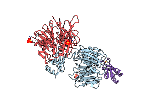 |
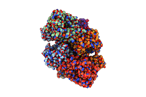
Crystal Structure Of The Kti11 Kti13 Heterodimer Spacegroup P64
Organism: Saccharomyces cerevisiae
Method: X-RAY DIFFRACTION Resolution:2.90 Å Release Date: 2015-01-14 Classification: TRANSLATION Ligands: FE, SO4 |
Organism: Saccharomyces cerevisiae
Method: X-RAY DIFFRACTION
Release Date: 2015-01-14
Ligands: FE, SO4
 |
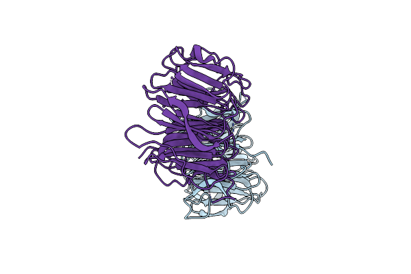
Crystal Structure Of The Kti11 Kti13 Heterodimer Spacegroup P65
Organism: Saccharomyces cerevisiae
Method: X-RAY DIFFRACTION Resolution:3.00 Å Release Date: 2015-01-14 Classification: TRANSLATION Ligands: FE, SO4 |
Organism: Saccharomyces cerevisiae
Method: X-RAY DIFFRACTION
Release Date: 2015-01-14
Ligands: FE, SO4
 |
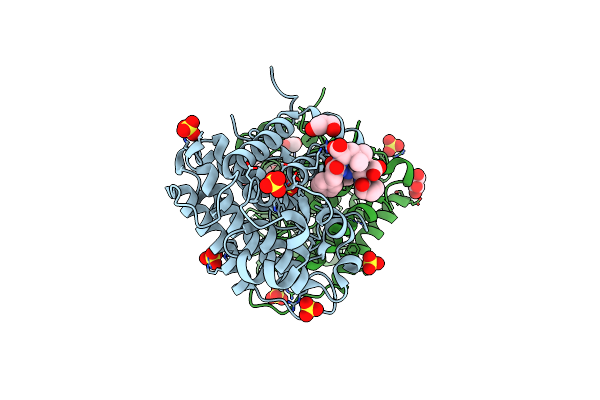
Crystal Structure Of Kti13/Ats1
Organism: Saccharomyces cerevisiae
Method: X-RAY DIFFRACTION Resolution:2.40 Å Release Date: 2015-01-14 Classification: TRANSLATION |
Organism: Saccharomyces cerevisiae
Method: X-RAY DIFFRACTION
Release Date: 2015-01-14
 |
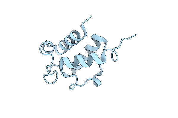
Human Ptpa In Complex With Peptide
Organism: Homo sapiens
Method: X-RAY DIFFRACTION Resolution:1.80 Å Release Date: 2014-07-23 Classification: HYDROLASE ACTIVATOR Ligands: SO4, GOL |
Organism: Homo sapiens
Method: X-RAY DIFFRACTION
Release Date: 2014-07-23
Ligands: SO4, GOL
 |
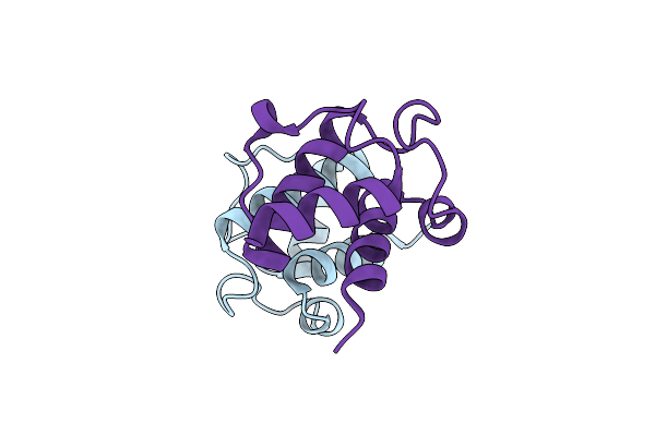
High Resolution Crystal Structure Of Bacillus Subtilis Dltc S36A
Organism: Bacillus subtilis
Method: X-RAY DIFFRACTION Resolution:1.01 Å Release Date: 2014-06-11 Classification: LIGASE |
Organism: Bacillus subtilis
Method: X-RAY DIFFRACTION
Release Date: 2014-06-11
 |
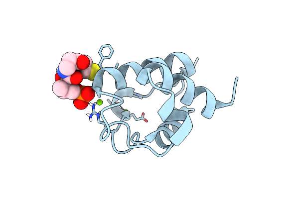
Crystal Structure Of Bacillus Subtilis Dltc
Organism: Bacillus subtilis
Method: X-RAY DIFFRACTION Resolution:2.20 Å Release Date: 2014-06-11 Classification: LIGASE |
Organism: Bacillus subtilis
Method: X-RAY DIFFRACTION
Release Date: 2014-06-11
 |
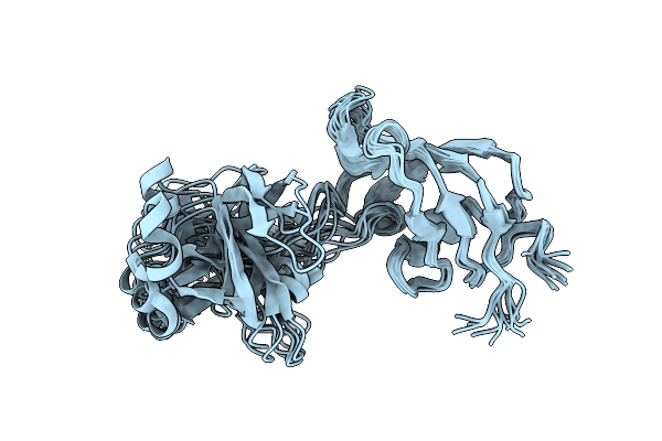
High Resolution Crystal Structure Of Bacillus Subtilis Dltc
Organism: Bacillus subtilis
Method: X-RAY DIFFRACTION Resolution:1.80 Å Release Date: 2014-06-11 Classification: LIGASE Ligands: PNS, MG |
Organism: Bacillus subtilis
Method: X-RAY DIFFRACTION
Release Date: 2014-06-11
Ligands: PNS, MG
 |
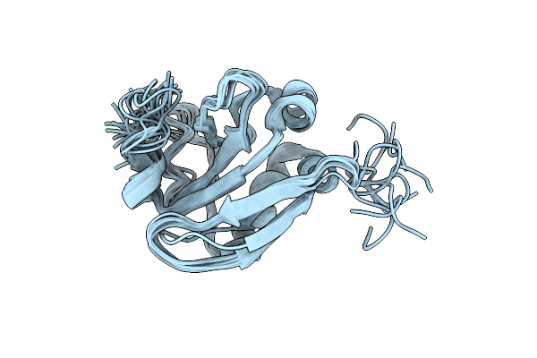
Nmr Solution Structure Of The Two Domain Ppiase Slpa From Escherichia Coli
|
Organism: Escherichia coli
Method: SOLUTION NMR
Release Date: 2013-12-18
 |
Solution Structure Of The Psiaa4 Oligomerization Domain Reveals Interaction Modes For Transcription Factors In Early Auxin Response
|
Organism: Pisum sativum
Method: SOLUTION NMR
Release Date: 2013-12-11
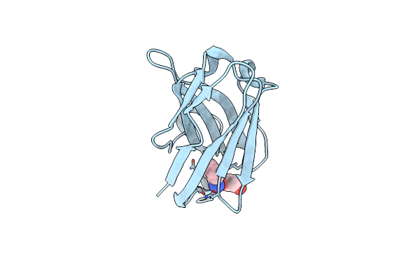 |
Crystal Structure Of The Oligomer-Specific Kw1 Antibody Fragment
Organism: Camelidae
Method: X-RAY DIFFRACTION Resolution:1.30 Å Release Date: 2012-07-11 Classification: IMMUNE SYSTEM Ligands: BEN, EDO |
Organism: Camelidae
Method: X-RAY DIFFRACTION
Release Date: 2012-07-11
Ligands: BEN, EDO
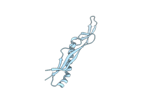 |
Crystal Structure Of The Pilus Binding Domain Of The Filamentous Phage Ike
Organism: Enterobacteria phage ike
Method: X-RAY DIFFRACTION Resolution:1.61 Å Release Date: 2012-05-30 Classification: VIRAL PROTEIN |
Organism: Enterobacteria phage ike
Method: X-RAY DIFFRACTION
Release Date: 2012-05-30
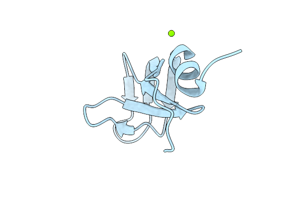 |
Crystal Structure Of The Tola Binding Domain From The Filamentous Phage Ike
Organism: Enterobacteria phage ike
Method: X-RAY DIFFRACTION Resolution:1.80 Å Release Date: 2012-05-30 Classification: VIRAL PROTEIN Ligands: MG |
Organism: Enterobacteria phage ike
Method: X-RAY DIFFRACTION
Release Date: 2012-05-30
Ligands: MG
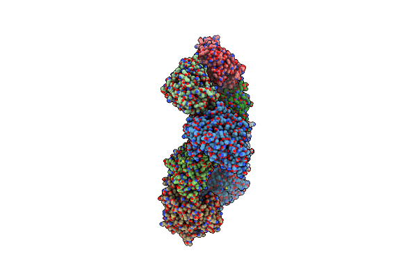 |
Crystal Structure Of E. Coli Oppa Complexed With Endogenous Ligands
Organism: Escherichia coli
Method: X-RAY DIFFRACTION Resolution:2.00 Å Release Date: 2011-10-12 Classification: PROTEIN TRANSPORT |
Organism: Escherichia coli
Method: X-RAY DIFFRACTION
Release Date: 2011-10-12
 |
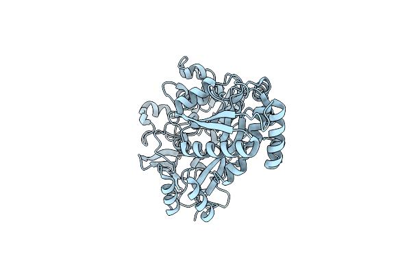
Crystal Structure Of E. Coli Oppa Complexed With The Tripeptide Kge
Organism: Escherichia coli
Method: X-RAY DIFFRACTION Resolution:2.00 Å Release Date: 2011-10-12 Classification: PROTEIN TRANSPORT |
Organism: Escherichia coli
Method: X-RAY DIFFRACTION
Release Date: 2011-10-12
 |
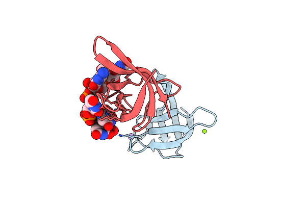
Crystal Structure Of E. Coli Oppa In An Open Conformation
Organism: Escherichia coli
Method: X-RAY DIFFRACTION Resolution:1.98 Å Release Date: 2011-10-12 Classification: PROTEIN TRANSPORT |
Organism: Escherichia coli
Method: X-RAY DIFFRACTION
Release Date: 2011-10-12
 |
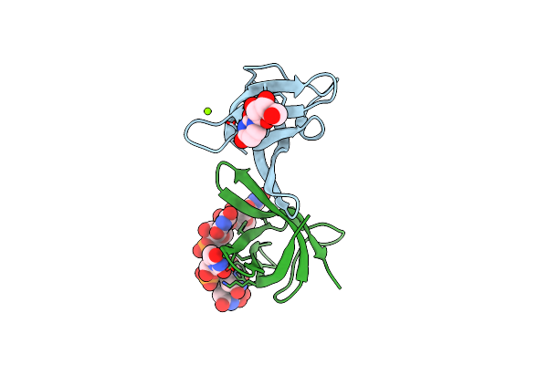
Crystal Structure Of Bs-Cspb In Complex With R(Gucuuua)
Organism: Bacillus subtilis
Method: X-RAY DIFFRACTION Resolution:1.38 Å Release Date: 2011-09-21 Classification: GENE REGULATION/RNA |
Organism: Bacillus subtilis
Method: X-RAY DIFFRACTION
Release Date: 2011-09-21
 |
Crystal Structure Of Bs-Cspb In Complex With Ru6
Organism: Bacillus subtilis
Method: X-RAY DIFFRACTION Resolution:1.68 Å Release Date: 2011-09-21 Classification: GENE REGULATION/RNA Ligands: MG |
Organism: Bacillus subtilis
Method: X-RAY DIFFRACTION
Release Date: 2011-09-21
Ligands: MG