

Search Count: 18
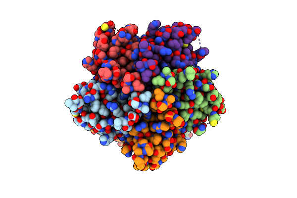 |
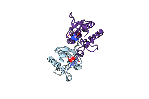
Structure Of The Disease-Causing Mutant P20S Of Human Kctd1
Organism: Homo sapiens
Method: X-RAY DIFFRACTION Resolution:2.60 Å Release Date: 2025-04-23 Classification: SIGNALING PROTEIN Ligands: PO4, K |
Organism: Homo sapiens
Method: X-RAY DIFFRACTION
Release Date: 2025-04-23
Ligands: PO4, K
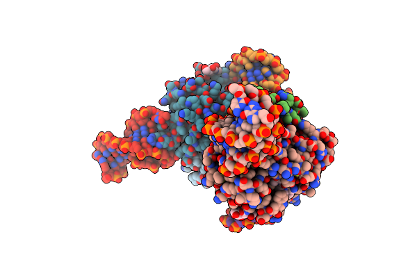 |
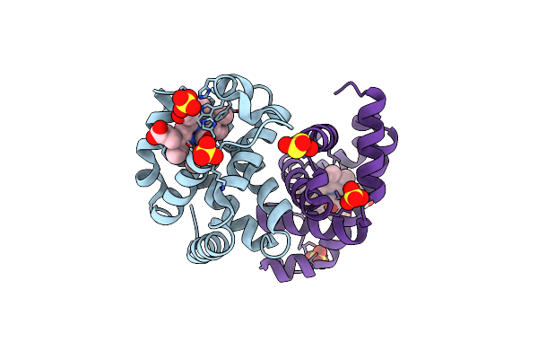
X-Ray Structure Of The Complex Between Human Alpha Thrombin And Two Duplex/Quadruplex Aptamers: Nu172 And Hd22_27Mer
Organism: Synthetic construct, Homo sapiens
Method: X-RAY DIFFRACTION Resolution:3.10 Å Release Date: 2021-04-28 Classification: HYDROLASE Ligands: 0G6, NAG, NA |
Organism: Synthetic construct, Homo sapiens
Method: X-RAY DIFFRACTION
Release Date: 2021-04-28
Ligands: 0G6, NAG, NA
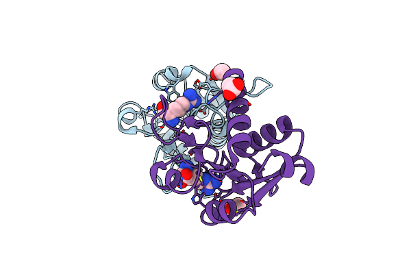 |
Crystal Structure Of The Phe57Trp Mutant Of The Arginine-Bound Form Of Domain 1 From Tmargbp
Organism: Thermotoga maritima, Thermotoga maritima (strain atcc 43589 / msb8 / dsm 3109 / jcm 10099)
Method: X-RAY DIFFRACTION Resolution:1.79 Å Release Date: 2020-11-04 Classification: TRANSPORT PROTEIN Ligands: ARG, ACT |
Organism: Thermotoga maritima, Thermotoga maritima (strain atcc 43589 / msb8 / dsm 3109 / jcm 10099)
Method: X-RAY DIFFRACTION
Release Date: 2020-11-04
Ligands: ARG, ACT
 |
Crystal Structure Of The Cisplatin Beta-Lactoglobulin Adduct Formed After 18 H Of Soaking
Organism: Bos taurus
Method: X-RAY DIFFRACTION Resolution:2.00 Å Release Date: 2020-09-30 Classification: TRANSPORT PROTEIN Ligands: NH3, PT, SO4 |
Organism: Bos taurus
Method: X-RAY DIFFRACTION
Release Date: 2020-09-30
Ligands: NH3, PT, SO4
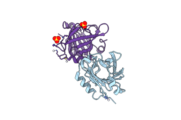 |
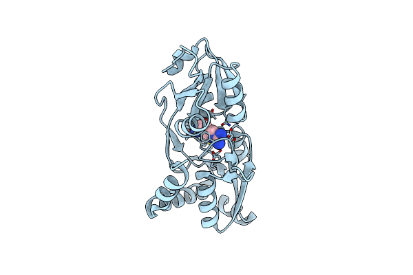
Crystal Structure Of The Cisplatin Beta-Lactoglobulin Adduct Formed After 72 H Of Soaking
Organism: Bos taurus
Method: X-RAY DIFFRACTION Resolution:2.01 Å Release Date: 2020-09-30 Classification: TRANSPORT PROTEIN Ligands: NH3, PT, SO4 |
Organism: Bos taurus
Method: X-RAY DIFFRACTION
Release Date: 2020-09-30
Ligands: NH3, PT, SO4
 |
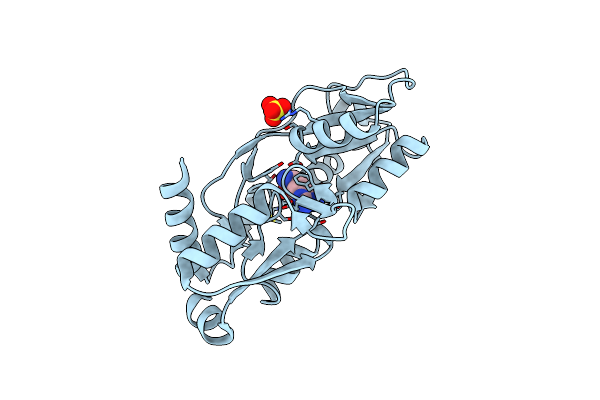
Crystal Structure Of Tmargbp Domain 1 In Complex With The Guanidinium Ion
Organism: Thermotoga maritima msb8
Method: X-RAY DIFFRACTION Resolution:1.70 Å Release Date: 2020-07-15 Classification: TRANSPORT PROTEIN Ligands: GAI, EDO |
Organism: Thermotoga maritima msb8
Method: X-RAY DIFFRACTION
Release Date: 2020-07-15
Ligands: GAI, EDO
 |
Crystal Structure Of Monocarboxylated Hemoglobin From The Sub-Antarctic Fish Eleginops Maclovinus
Organism: Eleginops maclovinus
Method: X-RAY DIFFRACTION Resolution:1.49 Å Release Date: 2019-12-25 Classification: OXYGEN TRANSPORT Ligands: CMO, HEM, SO4, DTN |
Organism: Eleginops maclovinus
Method: X-RAY DIFFRACTION
Release Date: 2019-12-25
Ligands: CMO, HEM, SO4, DTN
 |
Crystal Structure Of The P235Gk Mutant Of Argbp From T. Maritima
Organism: Thermotoga maritima (strain atcc 43589 / msb8 / dsm 3109 / jcm 10099)
Method: X-RAY DIFFRACTION Resolution:1.60 Å Release Date: 2019-11-20 Classification: TRANSPORT PROTEIN Ligands: ARG |
Organism: Thermotoga maritima (strain atcc 43589 / msb8 / dsm 3109 / jcm 10099)
Method: X-RAY DIFFRACTION
Release Date: 2019-11-20
Ligands: ARG
 |
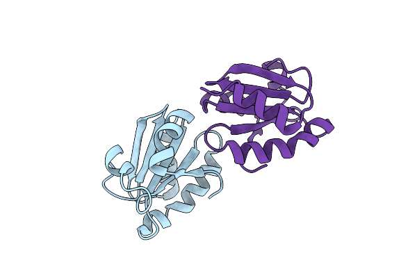
Gly52Ala Mutant Of Arginine-Bound Argbp From T. Maritima
Organism: Thermotoga maritima (strain atcc 43589 / msb8 / dsm 3109 / jcm 10099)
Method: X-RAY DIFFRACTION Resolution:1.64 Å Release Date: 2019-05-08 Classification: TRANSPORT PROTEIN Ligands: ARG, SO4 |
Organism: Thermotoga maritima (strain atcc 43589 / msb8 / dsm 3109 / jcm 10099)
Method: X-RAY DIFFRACTION
Release Date: 2019-05-08
Ligands: ARG, SO4
 |
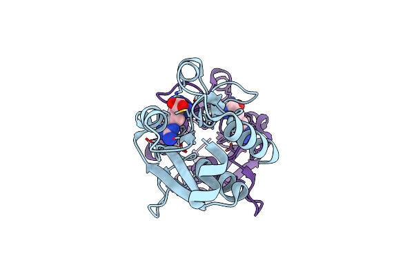
Crystal Structure Of A Chimeric Variant Of Thioredoxin From Escherichia Coli
Organism: Escherichia coli, Sulfolobus solfataricus
Method: X-RAY DIFFRACTION Resolution:2.99 Å Release Date: 2019-02-06 Classification: OXIDOREDUCTASE |
Organism: Escherichia coli, Sulfolobus solfataricus
Method: X-RAY DIFFRACTION
Release Date: 2019-02-06
 |
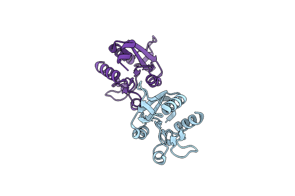
Crystal Structure Of The Arginine-Bound Form Of Domain 1 From Tmargbp
Organism: Thermotoga maritima (strain atcc 43589 / msb8 / dsm 3109 / jcm 10099)
Method: X-RAY DIFFRACTION Resolution:1.75 Å Release Date: 2018-08-15 Classification: TRANSPORT PROTEIN Ligands: ARG |
Organism: Thermotoga maritima (strain atcc 43589 / msb8 / dsm 3109 / jcm 10099)
Method: X-RAY DIFFRACTION
Release Date: 2018-08-15
Ligands: ARG
 |
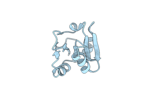
Crystal Structure Of The Ligand-Free Form Of Domain 1 From Tmargbp
Organism: Thermotoga maritima msb8
Method: X-RAY DIFFRACTION Resolution:1.75 Å Release Date: 2018-08-15 Classification: TRANSPORT PROTEIN |
Organism: Thermotoga maritima msb8
Method: X-RAY DIFFRACTION
Release Date: 2018-08-15
 |
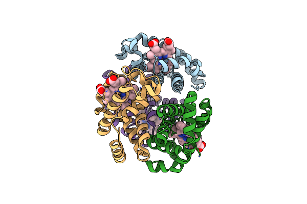
Crystal Structure Of Domain 2 From Tmargbp
Organism: Thermotoga maritima
Method: X-RAY DIFFRACTION Resolution:1.73 Å Release Date: 2018-08-15 Classification: TRANSPORT PROTEIN |
Organism: Thermotoga maritima
Method: X-RAY DIFFRACTION
Release Date: 2018-08-15
 |
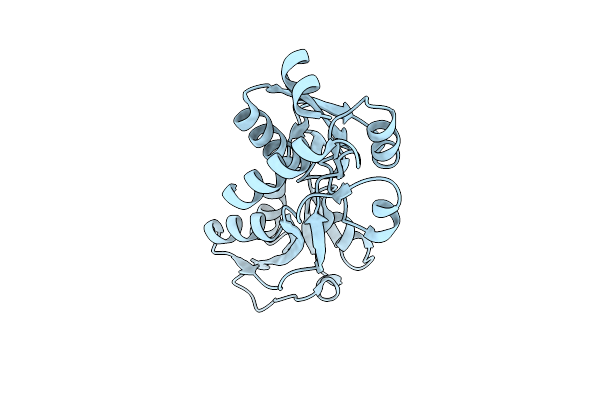
The X-Ray Structure Of Ferric-Beta4 Human Hemoglobin
Organism: Homo sapiens
Method: X-RAY DIFFRACTION Resolution:2.10 Å Release Date: 2018-06-13 Classification: OXYGEN TRANSPORT Ligands: HEM |
Organism: Homo sapiens
Method: X-RAY DIFFRACTION
Release Date: 2018-06-13
Ligands: HEM
 |
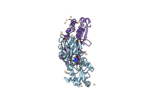
Structure Of The Ligand-Free Form Of Truncated Argbp (Residues 20-233) From T. Maritima
Organism: Thermotoga maritima (strain atcc 43589 / msb8 / dsm 3109 / jcm 10099)
Method: X-RAY DIFFRACTION Resolution:1.03 Å Release Date: 2018-06-13 Classification: TRANSPORT PROTEIN |
 |
Structure Of The Arginine-Bound Form Of Truncated (Residues 20-233) Argbp From T. Maritima
Organism: Thermotoga maritima
Method: X-RAY DIFFRACTION Resolution:2.69 Å Release Date: 2018-06-13 Classification: TRANSPORT PROTEIN Ligands: ARG, CD |
Organism: Thermotoga maritima
Method: X-RAY DIFFRACTION
Release Date: 2018-06-13
Ligands: ARG, CD
 |
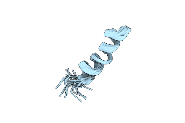
Cullin3 - Btb Interface: A Novel Target For Stapled Peptides
Organism: Homo sapiens
Method: SOLUTION NMR Release Date: 2015-04-22 Classification: PROTEIN BINDING |
Organism: Homo sapiens
Method: SOLUTION NMR
Release Date: 2015-04-22
 |
Cullin3 - Btb Interface: A Novel Target For Stapled Peptides
|
Organism: Human
Method: SOLUTION NMR
Release Date: 2015-04-22