

Search Count: 43
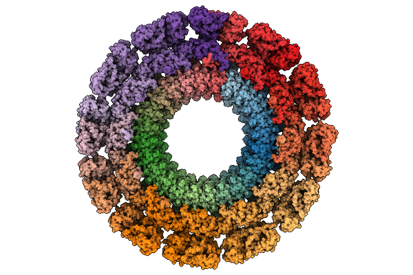 |
Cryo-Em Map Of The In Vitro Reconstituted Razr:Gp77 Complex With Alphafold-Predicted Models Fitted Into The Density.
Organism: Escherichia phage secphi27, Escherichia coli
Method: ELECTRON MICROSCOPY Release Date: 2025-12-24 Classification: RNA BINDING PROTEIN Ligands: ZN |
Organism: Escherichia phage secphi27, Escherichia coli
Method: ELECTRON MICROSCOPY
Release Date: 2025-12-24
Ligands: ZN
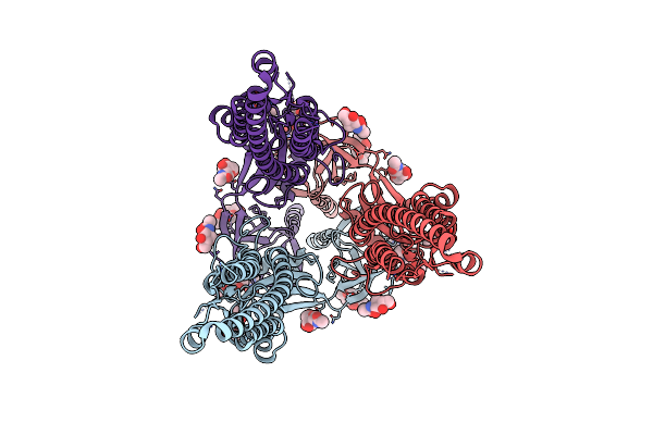 |
Structure Of The Sars-Cov-2 Spike Glycoprotein S2 Subunit
Organism: Severe acute respiratory syndrome coronavirus 2
Method: ELECTRON MICROSCOPY Release Date: 2022-11-30 Classification: VIRAL PROTEIN Ligands: NAG |
Organism: Severe acute respiratory syndrome coronavirus 2
Method: ELECTRON MICROSCOPY
Release Date: 2022-11-30
Ligands: NAG
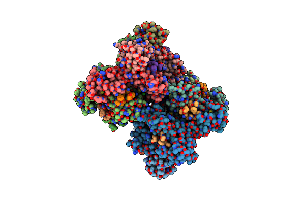 |
Hexamer Hiv-1 Ca In Complex With Cpsf6 Peptide And Ip6 Ligand
Organism: Human immunodeficiency virus type 1 group m subtype b (isolate bh10), Human immunodeficiency virus 1
Method: X-RAY DIFFRACTION Resolution:2.81 Å Release Date: 2022-09-07 Classification: VIRAL PROTEIN Ligands: IHP, CL |
Organism: Human immunodeficiency virus type 1 group m subtype b (isolate bh10), Human immunodeficiency virus 1
Method: X-RAY DIFFRACTION
Release Date: 2022-09-07
Ligands: IHP, CL
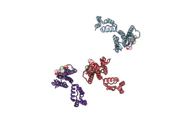 |
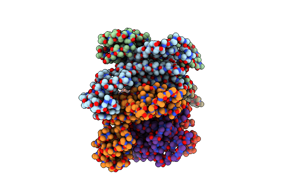
Co-Crystal Structure Of Gs-6207 Bound To Hiv-1 Capsid Hexamer
Organism: Human immunodeficiency virus 1
Method: X-RAY DIFFRACTION Resolution:2.22 Å Release Date: 2020-10-21 Classification: VIRAL PROTEIN Ligands: QNG, IOD, CL |
Organism: Human immunodeficiency virus 1
Method: X-RAY DIFFRACTION
Release Date: 2020-10-21
Ligands: QNG, IOD, CL
 |
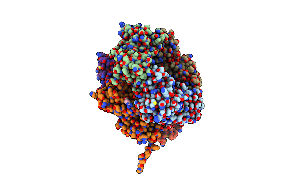
Hexamer Of Helical Hiv Capsid By Rastr Method
Organism: Human immunodeficiency virus 1
Method: ELECTRON MICROSCOPY Release Date: 2020-10-21 Classification: VIRAL PROTEIN |
Organism: Human immunodeficiency virus 1
Method: ELECTRON MICROSCOPY
Release Date: 2020-10-21
 |
Crystal Structure Of The Complex Of Proliferating Cell Nuclear Antigen From Leishmania Donovani With Arginine At 3.19 A Resolution.
Organism: Leishmania donovani
Method: X-RAY DIFFRACTION Resolution:3.19 Å Release Date: 2019-05-29 Classification: DNA BINDING PROTEIN Ligands: ARG |
Organism: Leishmania donovani
Method: X-RAY DIFFRACTION
Release Date: 2019-05-29
Ligands: ARG
 |
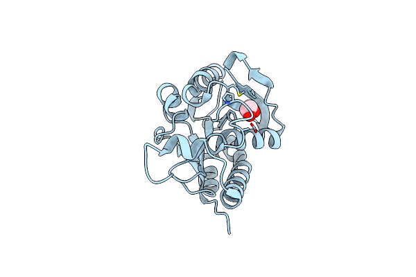
Crystal Structure Of The Complex Of Phosphopantetheine Adenylyltransferasefrom Acinetobacter Baumannii With Ascorbic Acid (Vitamin-C) At 2.0A Resolution
Organism: Acinetobacter baumannii
Method: X-RAY DIFFRACTION Resolution:2.00 Å Release Date: 2019-03-27 Classification: TRANSFERASE Ligands: ASC, SO4, NA |
Organism: Acinetobacter baumannii
Method: X-RAY DIFFRACTION
Release Date: 2019-03-27
Ligands: ASC, SO4, NA
 |
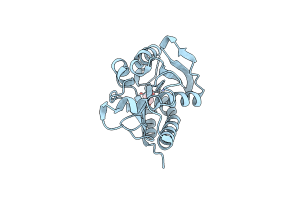
Crystal Structure Of Peptidyl-Trna Hydrolase From Acinetobacter Baumannii At 0.97 A Resolution With Disordered Five N-Terminal Residues
Organism: Acinetobacter baumannii (strain atcc 19606 / dsm 30007 / cip 70.34 / jcm 6841 / nbrc 109757 / ncimb 12457 / nctc 12156 / 81)
Method: X-RAY DIFFRACTION Resolution:0.97 Å Release Date: 2019-03-13 Classification: HYDROLASE Ligands: CL, EDO |
 |
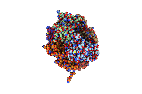
Crystal Structure Of Peptidyl-Trna Hydrolase From Acinetobacter Baumannii At 0.95 A Resolution
Organism: Acinetobacter baumannii (strain atcc 19606 / dsm 30007 / cip 70.34 / jcm 6841 / nbrc 109757 / ncimb 12457 / nctc 12156 / 81)
Method: X-RAY DIFFRACTION Resolution:0.95 Å Release Date: 2019-02-06 Classification: HYDROLASE Ligands: EDO |
 |
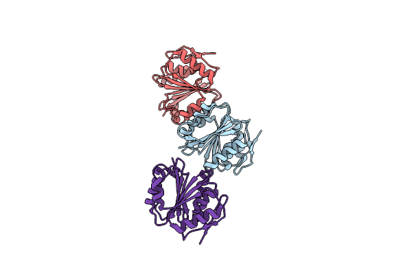
Crystal Structure Of Proliferating Cell Nuclear Antigen From Leishmania Donovani With An Unexplained Density Near Residues Pro229,Pr0267 And Ala285
Organism: Leishmania donovani
Method: X-RAY DIFFRACTION Resolution:3.10 Å Release Date: 2019-01-23 Classification: DNA BINDING PROTEIN Ligands: PEG, GOL |
Organism: Leishmania donovani
Method: X-RAY DIFFRACTION
Release Date: 2019-01-23
Ligands: PEG, GOL
 |
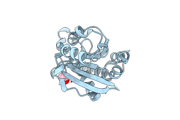
Crystal Structure Of A New Form Of Rsmd-Like Rna Methyl Transferase From Mycobacterium Tuberculosis Determined At 1.74 A Resolution
Organism: Mycobacterium tuberculosis
Method: X-RAY DIFFRACTION Resolution:1.74 Å Release Date: 2018-09-26 Classification: TRANSFERASE |
Organism: Mycobacterium tuberculosis
Method: X-RAY DIFFRACTION
Release Date: 2018-09-26
 |
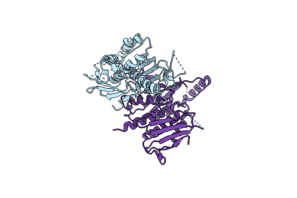
Crystal Structure Of Peg-1500 Crystallized Peptidyl-Trna Hydrolase From Acinetobacter Baumannii At 1.5 A Resolution
Organism: Acinetobacter baumannii (strain atcc 19606 / dsm 30007 / cip 70.34 / jcm 6841 / nbrc 109757 / ncimb 12457 / nctc 12156 / 81)
Method: X-RAY DIFFRACTION Resolution:1.57 Å Release Date: 2018-06-20 Classification: HYDROLASE Ligands: EDO |
 |
Crystal Structure Of Chorismate Synthase From Pseudomonas Aeruginosa
Organism: Pseudomonas aeruginosa (strain lesb58)
Method: X-RAY DIFFRACTION Resolution:2.79 Å Release Date: 2018-02-28 Classification: LYASE |
Organism: Pseudomonas aeruginosa (strain lesb58)
Method: X-RAY DIFFRACTION
Release Date: 2018-02-28
 |
Crystal Structure Of Sulfated Dehydroquinate Dehydratase From Acinetobacter Baumannii At 2.65 A Resolution
Organism: Acinetobacter baumannii
Method: X-RAY DIFFRACTION Resolution:2.65 Å Release Date: 2017-11-22 Classification: LYASE Ligands: SO4, GOL |
Organism: Acinetobacter baumannii
Method: X-RAY DIFFRACTION
Release Date: 2017-11-22
Ligands: SO4, GOL
 |
The Crystal Structure Of Phosphopantetheine Adenylyltransferase From Acinetobacter Baumannii With Coenzyme A At 2.88 A Resolution
Organism: Acinetobacter baumannii
Method: X-RAY DIFFRACTION Resolution:2.88 Å Release Date: 2017-11-22 Classification: TRANSFERASE Ligands: COA, SO4 |
Organism: Acinetobacter baumannii
Method: X-RAY DIFFRACTION
Release Date: 2017-11-22
Ligands: COA, SO4
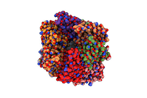 |
Crystal Structure Of Dodecameric Dehydroquinate Dehydratase From Acinetobacter Baumannii At 1.9A Resolution
Organism: Acinetobacter baumannii (strain atcc 17978 / cip 53.77 / lmg 1025 / ncdc kc755 / 5377)
Method: X-RAY DIFFRACTION Resolution:1.90 Å Release Date: 2017-11-01 Classification: LYASE Ligands: SO4, GOL, TRS |
Organism: Acinetobacter baumannii (strain atcc 17978 / cip 53.77 / lmg 1025 / ncdc kc755 / 5377)
Method: X-RAY DIFFRACTION
Release Date: 2017-11-01
Ligands: SO4, GOL, TRS
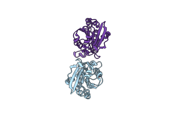 |
The Crystal Structure Of Inactive Dimeric Peptidyl-Trna Hydrolase From Acinetobacter Baumannii At 1.79 A Resolution
Organism: Acinetobacter baumannii (strain atcc 19606 / dsm 30007 / cip 70.34 / jcm 6841 / nbrc 109757 / ncimb 12457 / nctc 12156 / 81)
Method: X-RAY DIFFRACTION Resolution:1.79 Å Release Date: 2017-11-01 Classification: HYDROLASE |
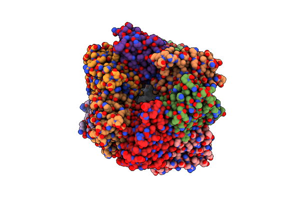 |
Crystal Structure Of Dehydroquinate Dehydratase With Tris Induced Oligomerisation At 1.907 Angstrom Resolution
Organism: Acinetobacter baumannii (strain atcc 17978 / cip 53.77 / lmg 1025 / ncdc kc755 / 5377)
Method: X-RAY DIFFRACTION Resolution:1.91 Å Release Date: 2017-10-25 Classification: LYASE Ligands: SO4, TRS |
Organism: Acinetobacter baumannii (strain atcc 17978 / cip 53.77 / lmg 1025 / ncdc kc755 / 5377)
Method: X-RAY DIFFRACTION
Release Date: 2017-10-25
Ligands: SO4, TRS
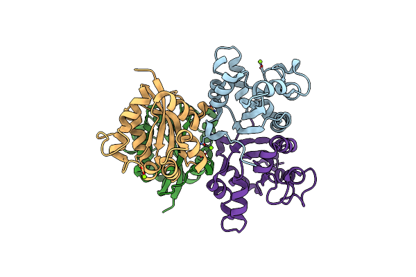 |
Crystal Structure Of Tetrameric Nucleoside Diphosphate Kinase At 1.98 A Resolution From Acinetobacter Baumannii
Organism: Acinetobacter baumannii
Method: X-RAY DIFFRACTION Resolution:1.98 Å Release Date: 2017-10-25 Classification: TRANSFERASE Ligands: MG |
Organism: Acinetobacter baumannii
Method: X-RAY DIFFRACTION
Release Date: 2017-10-25
Ligands: MG
 |
Crystal Structure Of The Complex Of Phosphopantetheine Adenylyltransferase From Acinetobacter Baumannii With Coenzyme A At 2.0 A Resolution
Organism: Acinetobacter baumannii (strain acicu)
Method: X-RAY DIFFRACTION Resolution:2.03 Å Release Date: 2017-10-11 Classification: TRANSFERASE Ligands: COA, SO4 |
Organism: Acinetobacter baumannii (strain acicu)
Method: X-RAY DIFFRACTION
Release Date: 2017-10-11
Ligands: COA, SO4