

Search Count: 14
 |
Crystal Structure Of Enl Yeats In Complex With Histone H3 Methacrylated At K18
Organism: Homo sapiens
Method: X-RAY DIFFRACTION Resolution:2.60 Å Release Date: 2025-04-09 Classification: TRANSCRIPTION Ligands: MES |
Organism: Homo sapiens
Method: X-RAY DIFFRACTION
Release Date: 2025-04-09
Ligands: MES
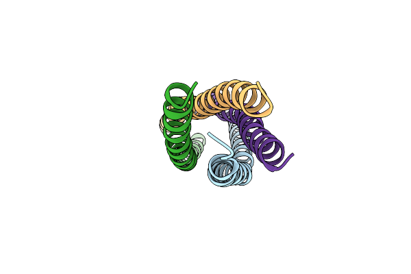 |
Crystal Structure Of Coiled Coil Domain Of Golm1 (Golgi Membrane Protein 1)
Organism: Homo sapiens
Method: X-RAY DIFFRACTION Resolution:2.28 Å Release Date: 2025-02-12 Classification: ONCOPROTEIN |
Organism: Homo sapiens
Method: X-RAY DIFFRACTION
Release Date: 2025-02-12
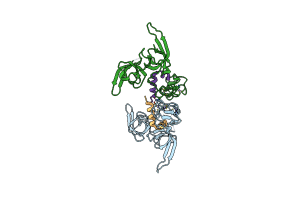 |
Crystal Structure Of Ttd-Phd Domain Of Uhrf1 In Complex With Mstella Peptide (Residues 85-119)
Organism: Homo sapiens, Mus musculus
Method: X-RAY DIFFRACTION Resolution:3.20 Å Release Date: 2024-11-06 Classification: GENE REGULATION/INHIBITOR Ligands: ZN |
Organism: Homo sapiens, Mus musculus
Method: X-RAY DIFFRACTION
Release Date: 2024-11-06
Ligands: ZN
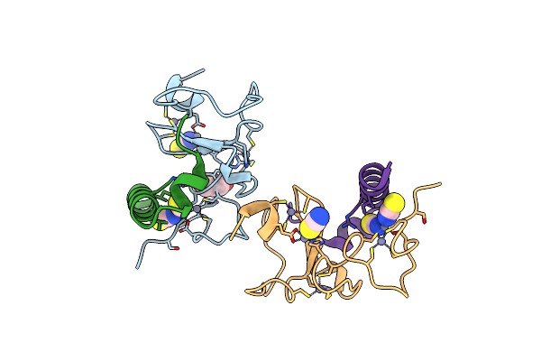 |
Crystal Structure Of Phd Domain Of Uhrf1 In Complex With Mstella Peptide (Residues 85-119)
Organism: Homo sapiens, Mus musculus
Method: X-RAY DIFFRACTION Resolution:1.60 Å Release Date: 2024-11-06 Classification: GENE REGULATION Ligands: SCN, GOL, ZN |
Organism: Homo sapiens, Mus musculus
Method: X-RAY DIFFRACTION
Release Date: 2024-11-06
Ligands: SCN, GOL, ZN
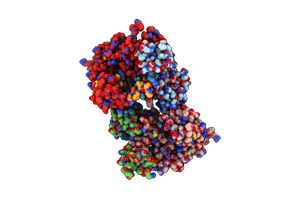 |
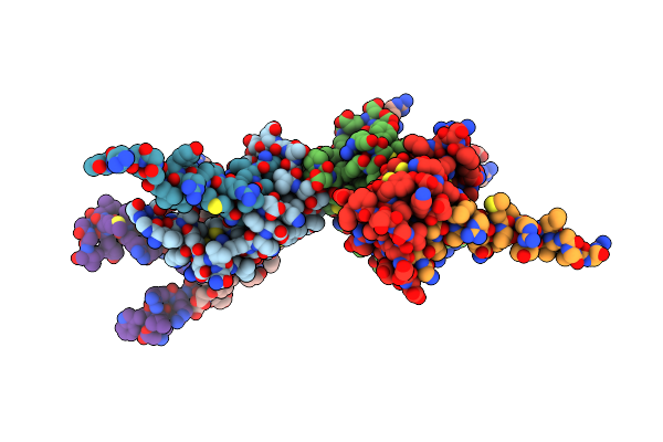
Crystal Structure Of Ttd-Phd Domain Of Uhrf1 In Complex With Hstella Peptide (Residues 75-121)
Organism: Homo sapiens
Method: X-RAY DIFFRACTION Resolution:2.25 Å Release Date: 2024-11-06 Classification: GENE REGULATION Ligands: ZN, GOL, ACY |
Organism: Homo sapiens
Method: X-RAY DIFFRACTION
Release Date: 2024-11-06
Ligands: ZN, GOL, ACY
 |
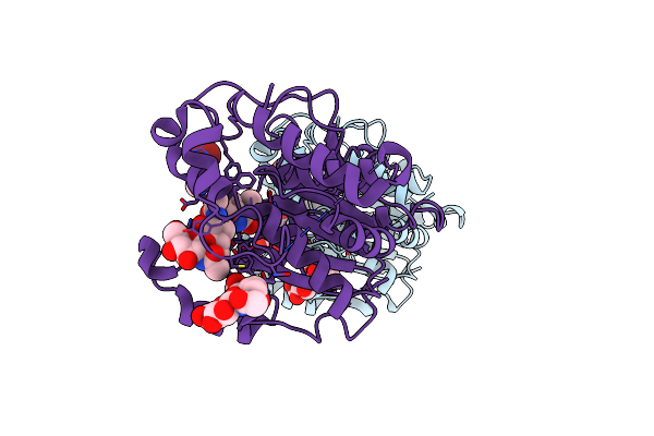
Crystal Structure Of Phd Domain Of Uhrf1 In Complex With Hstella Peptide (Residues 75-121)
Organism: Homo sapiens
Method: X-RAY DIFFRACTION Resolution:2.05 Å Release Date: 2024-11-06 Classification: GENE REGULATION Ligands: ZN |
Organism: Homo sapiens
Method: X-RAY DIFFRACTION
Release Date: 2024-11-06
Ligands: ZN
 |
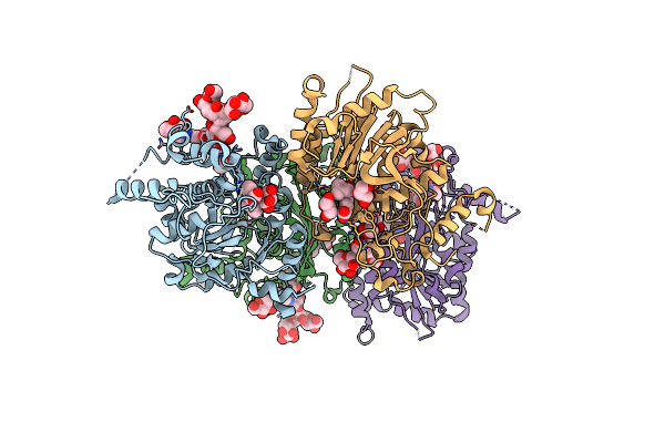
Structure Of Human Beta 1,3-N-Acetylglucosaminyltransferase 2 With Compound 7J
Organism: Homo sapiens
Method: X-RAY DIFFRACTION Resolution:2.32 Å Release Date: 2023-12-06 Classification: TRANSFERASE Ligands: X18, EDO |
Organism: Homo sapiens
Method: X-RAY DIFFRACTION
Release Date: 2023-12-06
Ligands: X18, EDO
 |
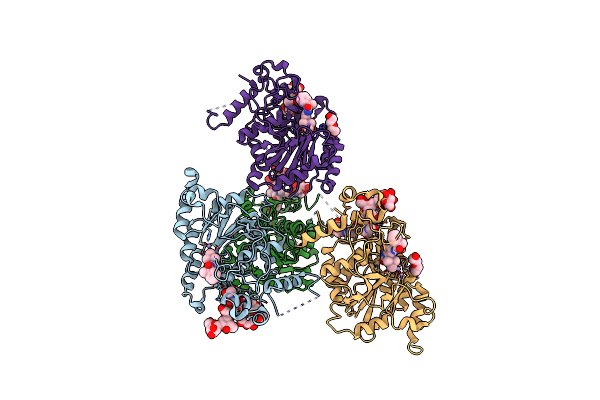
Structure Of Human Beta 1,3-N-Acetylglucosaminyltransferase 2 With Compound 1
Organism: Homo sapiens
Method: X-RAY DIFFRACTION Resolution:2.70 Å Release Date: 2023-12-06 Classification: TRANSFERASE Ligands: NAG, FKX, GOL, CL |
Organism: Homo sapiens
Method: X-RAY DIFFRACTION
Release Date: 2023-12-06
Ligands: NAG, FKX, GOL, CL
 |
Structure Of Human Beta 1,3-N-Acetylglucosaminyltransferase 2 With Compound 8A
Organism: Homo sapiens
Method: X-RAY DIFFRACTION Resolution:2.20 Å Release Date: 2023-12-06 Classification: TRANSFERASE Ligands: MN, HI8, NAG, PEG |
Organism: Homo sapiens
Method: X-RAY DIFFRACTION
Release Date: 2023-12-06
Ligands: MN, HI8, NAG, PEG
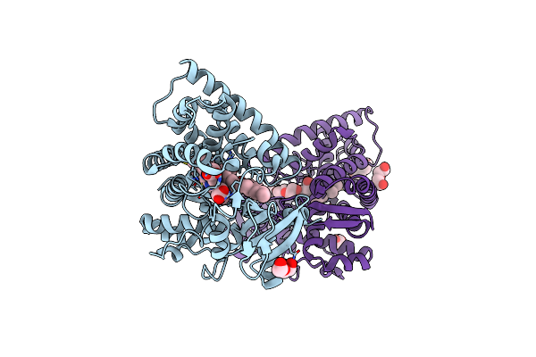 |
Crystal Structure Of P450Bsbeta-L78G/Q85F/F173S/G290I Variant In Complex With Palmitoleic Acid
Organism: Bacillus subtilis
Method: X-RAY DIFFRACTION Resolution:2.35 Å Release Date: 2023-11-08 Classification: HYDROLASE Ligands: HEM, PAM, PEG |
Organism: Bacillus subtilis
Method: X-RAY DIFFRACTION
Release Date: 2023-11-08
Ligands: HEM, PAM, PEG
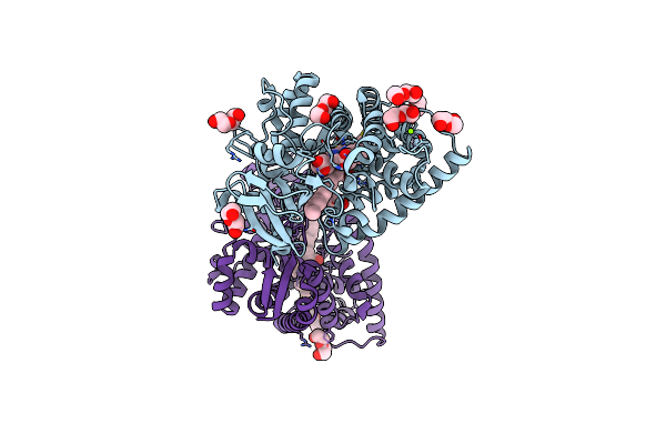 |
Crystal Structure Of P450Bsbeta-L78I/Q85H/G290I Variant In Complex With Palmitic Acid.
Organism: Bacillus subtilis
Method: X-RAY DIFFRACTION Resolution:2.00 Å Release Date: 2022-12-21 Classification: OXIDOREDUCTASE Ligands: HEM, PAM, PEG, MG |
Organism: Bacillus subtilis
Method: X-RAY DIFFRACTION
Release Date: 2022-12-21
Ligands: HEM, PAM, PEG, MG
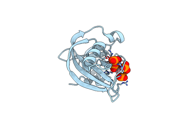 |
Crystal Structure Of Human Diphosphoinositol Polyphosphate Phosphohydrolase 1 In Complex With 1-Ip7
Organism: Homo sapiens
Method: X-RAY DIFFRACTION Resolution:1.20 Å Release Date: 2020-04-29 Classification: HYDROLASE Ligands: O81, MG, CL |
Organism: Homo sapiens
Method: X-RAY DIFFRACTION
Release Date: 2020-04-29
Ligands: O81, MG, CL
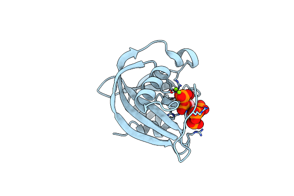 |
Crystal Structure Of Human Diphosphoinositol Polyphosphate Phosphohydrolase 1 In Complex With 5-Ip7
Organism: Homo sapiens
Method: X-RAY DIFFRACTION Resolution:1.30 Å Release Date: 2020-04-29 Classification: HYDROLASE Ligands: I7P, MG, CL |
Organism: Homo sapiens
Method: X-RAY DIFFRACTION
Release Date: 2020-04-29
Ligands: I7P, MG, CL
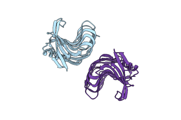 |
Crystal Structure Of Endo-Beta-1,4-Xylanase From The Alkaliphilic Bacillus Sp. Sn5
Organism: Bacillus sp
Method: X-RAY DIFFRACTION Resolution:1.49 Å Release Date: 2014-02-12 Classification: HYDROLASE Ligands: SO4 |
Organism: Bacillus sp
Method: X-RAY DIFFRACTION
Release Date: 2014-02-12
Ligands: SO4