

Search Count: 54
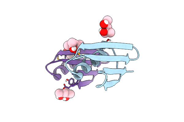 |
Selenomethionine Mutant (V29Sem) Of Protein Gb1 Examined By X-Ray Diffraction
Organism: Streptococcus sp. group g
Method: X-RAY DIFFRACTION Resolution:1.20 Å Release Date: 2019-07-10 Classification: IMMUNE SYSTEM Ligands: MPD |
Organism: Streptococcus sp. group g
Method: X-RAY DIFFRACTION
Release Date: 2019-07-10
Ligands: MPD
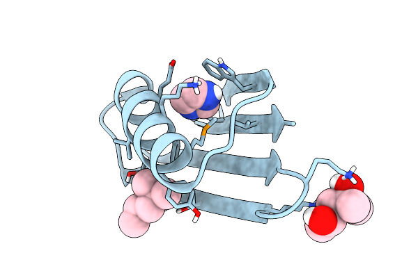 |
Selenomethionine Mutant (A34Sem) Of Protein Gb1 Examined By X-Ray Diffraction
Organism: Streptococcus sp. group g
Method: X-RAY DIFFRACTION Resolution:1.10 Å Release Date: 2019-07-10 Classification: IMMUNE SYSTEM Ligands: MPD, IMD |
Organism: Streptococcus sp. group g
Method: X-RAY DIFFRACTION
Release Date: 2019-07-10
Ligands: MPD, IMD
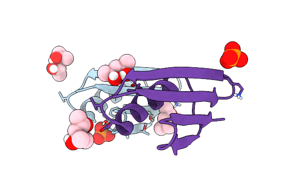 |
Selenomethionine Variant (V29Sem) Of Protein Gb1
Organism: Streptococcus sp. group g
Method: X-RAY DIFFRACTION Resolution:1.20 Å Release Date: 2019-07-10 Classification: IMMUNE SYSTEM Ligands: MPD, PO4 |
Organism: Streptococcus sp. group g
Method: X-RAY DIFFRACTION
Release Date: 2019-07-10
Ligands: MPD, PO4
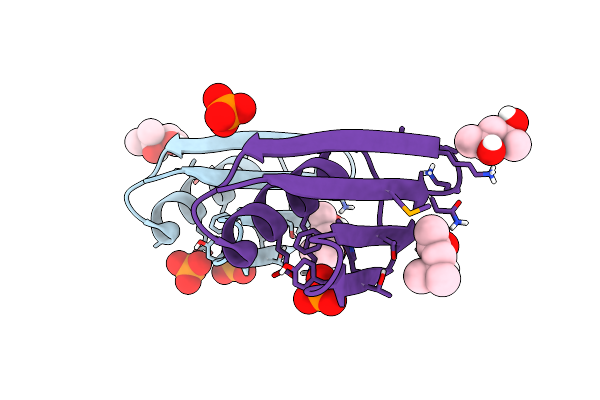 |
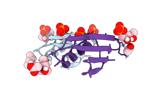
Selenomethionine Mutant (I6Sem) Of Protein Gb1 Examined By X-Ray Diffraction
Organism: Streptococcus sp. group g
Method: X-RAY DIFFRACTION Resolution:1.12 Å Release Date: 2019-07-10 Classification: IMMUNE SYSTEM Ligands: MRD, MPD, PO4 |
Organism: Streptococcus sp. group g
Method: X-RAY DIFFRACTION
Release Date: 2019-07-10
Ligands: MRD, MPD, PO4
 |
77Se-Nmr Probes The Protein Environment Of Selenomethionine
Organism: Streptococcus sp. group g
Method: X-RAY DIFFRACTION Resolution:1.20 Å Release Date: 2019-07-10 Classification: IMMUNE SYSTEM Ligands: MPD, PO4, MRD, ACT |
Organism: Streptococcus sp. group g
Method: X-RAY DIFFRACTION
Release Date: 2019-07-10
Ligands: MPD, PO4, MRD, ACT
 |
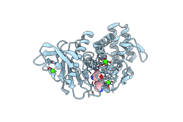
Conformational Sampling Differences Across The Arrhenius Plot Biphasic Break Point At Ambient Temperature In The Enzyme Thermolysin
Organism: Bacillus thermoproteolyticus
Method: X-RAY DIFFRACTION Resolution:2.09 Å Release Date: 2017-08-30 Classification: HYDROLASE Ligands: CA, ZN, VAL, LYS |
Organism: Bacillus thermoproteolyticus
Method: X-RAY DIFFRACTION
Release Date: 2017-08-30
Ligands: CA, ZN, VAL, LYS
 |
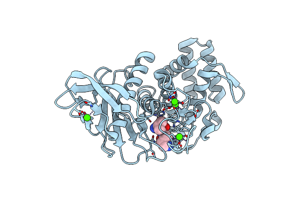
Conformational Sampling Differences Across The Arrhenius Plot Biphasic Break Point At Ambient Temperature In The Enzyme Thermolysin
Organism: Bacillus thermoproteolyticus
Method: X-RAY DIFFRACTION Resolution:2.10 Å Release Date: 2017-08-30 Classification: HYDROLASE Ligands: CA, ZN, VAL, LYS |
Organism: Bacillus thermoproteolyticus
Method: X-RAY DIFFRACTION
Release Date: 2017-08-30
Ligands: CA, ZN, VAL, LYS
 |
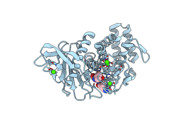
Conformational Sampling Differences Across The Arrhenius Plot Biphasic Break Point At Ambient Temperature In The Enzyme Thermolysin
Organism: Bacillus thermoproteolyticus
Method: X-RAY DIFFRACTION Resolution:2.10 Å Release Date: 2017-08-30 Classification: HYDROLASE Ligands: CA, ZN, VAL, LYS, PO4 |
Organism: Bacillus thermoproteolyticus
Method: X-RAY DIFFRACTION
Release Date: 2017-08-30
Ligands: CA, ZN, VAL, LYS, PO4
 |
Conformational Sampling Differences Across The Arrhenius Plot Biphasic Break Point At Ambient Temperature In The Enzyme Thermolysin
Organism: Bacillus thermoproteolyticus
Method: X-RAY DIFFRACTION Resolution:2.04 Å Release Date: 2017-08-30 Classification: HYDROLASE Ligands: CA, ZN, VAL, LYS |
Organism: Bacillus thermoproteolyticus
Method: X-RAY DIFFRACTION
Release Date: 2017-08-30
Ligands: CA, ZN, VAL, LYS
 |
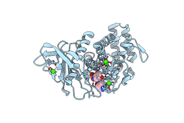
Conformational Sampling Differences Across The Arrhenius Plot Biphasic Break Point At Ambient Temperature In The Enzyme Thermolysin
Organism: Bacillus thermoproteolyticus
Method: X-RAY DIFFRACTION Resolution:2.09 Å Release Date: 2017-08-30 Classification: HYDROLASE Ligands: CA, ZN, VAL, LYS, PO4 |
Organism: Bacillus thermoproteolyticus
Method: X-RAY DIFFRACTION
Release Date: 2017-08-30
Ligands: CA, ZN, VAL, LYS, PO4
 |
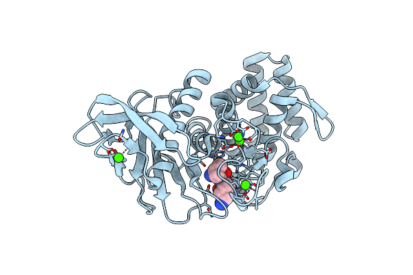
Conformational Sampling Differences Across The Arrhenius Plot Biphasic Break Point At Ambient Temperature In The Enzyme Thermolysin
Organism: Bacillus thermoproteolyticus
Method: X-RAY DIFFRACTION Resolution:2.30 Å Release Date: 2017-08-30 Classification: HYDROLASE Ligands: CA, ZN, VAL, LYS, PO4 |
Organism: Bacillus thermoproteolyticus
Method: X-RAY DIFFRACTION
Release Date: 2017-08-30
Ligands: CA, ZN, VAL, LYS, PO4
 |
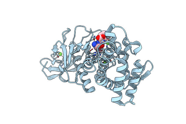
Conformational Sampling Differences Across The Arrhenius Plot Biphasic Break Point At Ambient Temperature In The Enzyme Thermolysin
Organism: Bacillus thermoproteolyticus
Method: X-RAY DIFFRACTION Resolution:2.30 Å Release Date: 2017-08-30 Classification: HYDROLASE Ligands: CA, ZN, VAL, LYS |
Organism: Bacillus thermoproteolyticus
Method: X-RAY DIFFRACTION
Release Date: 2017-08-30
Ligands: CA, ZN, VAL, LYS
 |
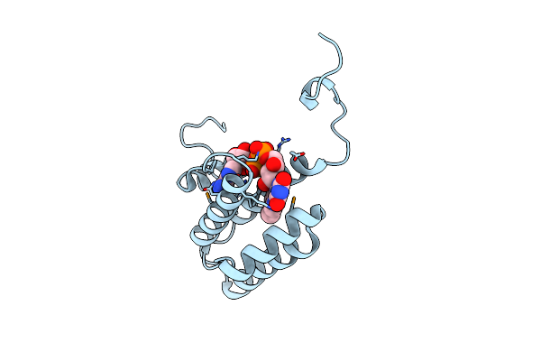
Conformational Sampling Differences Across The Arrhenius Plot Biphasic Break Point At Ambient Temperature In The Enzyme Thermolysin
Organism: Bacillus thermoproteolyticus
Method: X-RAY DIFFRACTION Resolution:2.03 Å Release Date: 2017-08-30 Classification: HYDROLASE Ligands: CA, ZN, VAL, LYS |
Organism: Bacillus thermoproteolyticus
Method: X-RAY DIFFRACTION
Release Date: 2017-08-30
Ligands: CA, ZN, VAL, LYS
 |
Conformational Sampling Differences Across The Arrhenius Plot Biphasic Break Point At Ambient Temperature In The Enzyme Thermolysin
Organism: Bacillus thermoproteolyticus
Method: X-RAY DIFFRACTION Resolution:2.00 Å Release Date: 2017-08-30 Classification: HYDROLASE Ligands: CA, ZN, VAL, LYS |
Organism: Bacillus thermoproteolyticus
Method: X-RAY DIFFRACTION
Release Date: 2017-08-30
Ligands: CA, ZN, VAL, LYS
 |
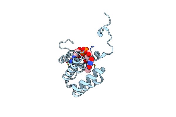
Selenium Substituted Human Augmenter Of Liver Regeneration
Organism: Homo sapiens
Method: X-RAY DIFFRACTION Resolution:1.50 Å Release Date: 2012-10-17 Classification: OXIDOREDUCTASE Ligands: FAD |
Organism: Homo sapiens
Method: X-RAY DIFFRACTION
Release Date: 2012-10-17
Ligands: FAD
 |
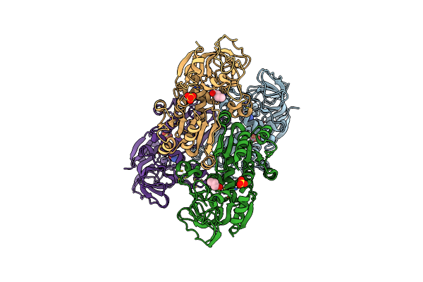
Mutation Of Sfalr
Organism: Homo sapiens
Method: X-RAY DIFFRACTION Resolution:1.61 Å Release Date: 2012-08-29 Classification: FLAVOPROTEIN Ligands: FAD |
Organism: Homo sapiens
Method: X-RAY DIFFRACTION
Release Date: 2012-08-29
Ligands: FAD
 |
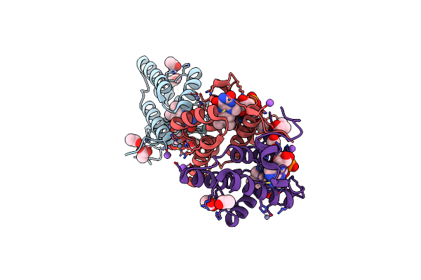
Crystal Structure Of Mutant Of Ht- Alcohol Dehydrogenase With Substrate Analogue Butyramide
Organism: Geobacillus stearothermophilus
Method: X-RAY DIFFRACTION Resolution:2.90 Å Release Date: 2011-11-09 Classification: OXIDOREDUCTASE Ligands: ZN, BMD, SO4 |
Organism: Geobacillus stearothermophilus
Method: X-RAY DIFFRACTION
Release Date: 2011-11-09
Ligands: ZN, BMD, SO4
 |
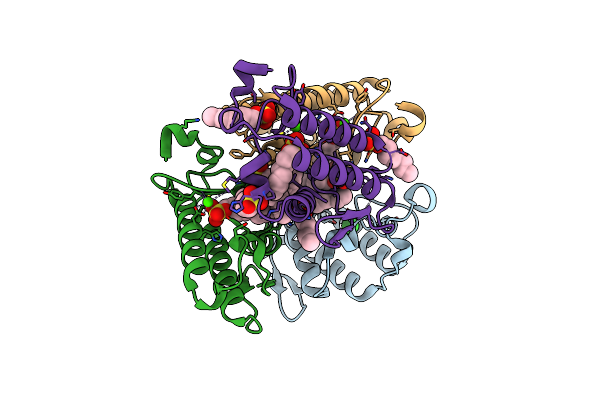
Crystal Structure Of Human Augmenter Of Liver Regeneration (Alr)
Organism: Homo sapiens
Method: X-RAY DIFFRACTION Resolution:1.85 Å Release Date: 2010-07-21 Classification: FLAVOPROTEIN Ligands: FAD, ZN, NA, ACT |
Organism: Homo sapiens
Method: X-RAY DIFFRACTION
Release Date: 2010-07-21
Ligands: FAD, ZN, NA, ACT
 |
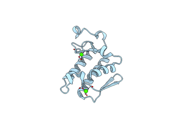
Crystal Structure Of Complex Of Phospholipase A2 With Octyl Sulfates
Organism: Sus scrofa
Method: X-RAY DIFFRACTION Resolution:2.70 Å Release Date: 2010-03-16 Classification: HYDROLASE Ligands: CA, CL, OSF, NA |
Organism: Sus scrofa
Method: X-RAY DIFFRACTION
Release Date: 2010-03-16
Ligands: CA, CL, OSF, NA
 |
Crystal Structure Of Phospholipase A2 1B Crystallized In The Presence Of Octyl Sulfate
Organism: Sus scrofa
Method: X-RAY DIFFRACTION Resolution:2.30 Å Release Date: 2010-03-16 Classification: HYDROLASE Ligands: CA, CL |
Organism: Sus scrofa
Method: X-RAY DIFFRACTION
Release Date: 2010-03-16
Ligands: CA, CL