

Search Count: 22
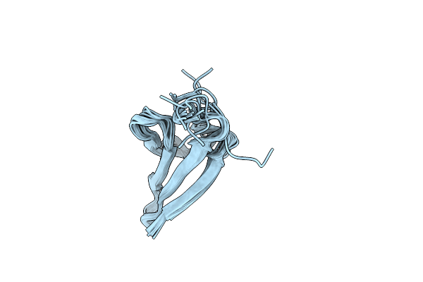 |
Nmr Structure Of Phosphorylated 4E-Bp2
Organism: Homo sapiens
Method: SOLUTION NMR Release Date: 2015-01-07 Classification: Translation,protein Binding |
Organism: Homo sapiens
Method: SOLUTION NMR
Release Date: 2015-01-07
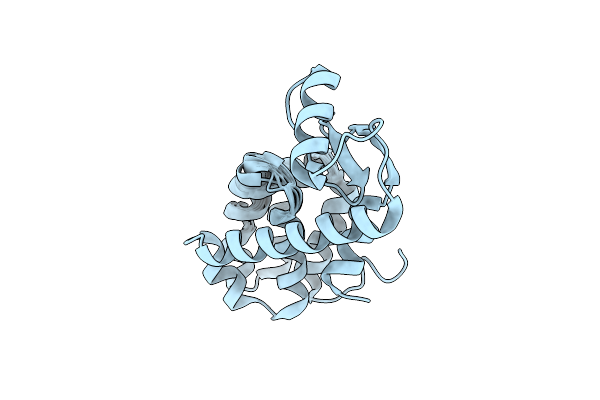 |
Solution Structure Of A Minor And Transiently Formed State Of A T4 Lysozyme Mutant
Organism: Enterobacteria phage t4
Method: SOLUTION NMR Release Date: 2011-08-17 Classification: HYDROLASE |
Organism: Enterobacteria phage t4
Method: SOLUTION NMR
Release Date: 2011-08-17
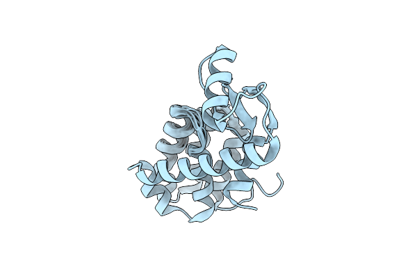 |
Solution Structure Of A Minor And Transiently Formed State Of A T4 Lysozyme Mutant
Organism: Enterobacteria phage t4
Method: SOLUTION NMR Release Date: 2011-08-17 Classification: HYDROLASE |
Organism: Enterobacteria phage t4
Method: SOLUTION NMR
Release Date: 2011-08-17
 |
Structure Of Human Thrombin With Residues 145-150 Of Murine Thrombin.
Organism: Homo sapiens
Method: X-RAY DIFFRACTION Resolution:1.75 Å Release Date: 2011-05-11 Classification: HYDROLASE Ligands: NAG, NA |
Organism: Homo sapiens
Method: X-RAY DIFFRACTION
Release Date: 2011-05-11
Ligands: NAG, NA
 |
Crystal Structure Of Human Thrombin Mutant N143P In E:Na+ Form
Organism: Homo sapiens
Method: X-RAY DIFFRACTION Resolution:1.60 Å Release Date: 2009-10-20 Classification: HYDROLASE |
Organism: Homo sapiens
Method: X-RAY DIFFRACTION
Release Date: 2009-10-20
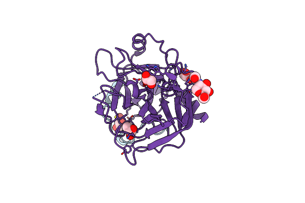 |
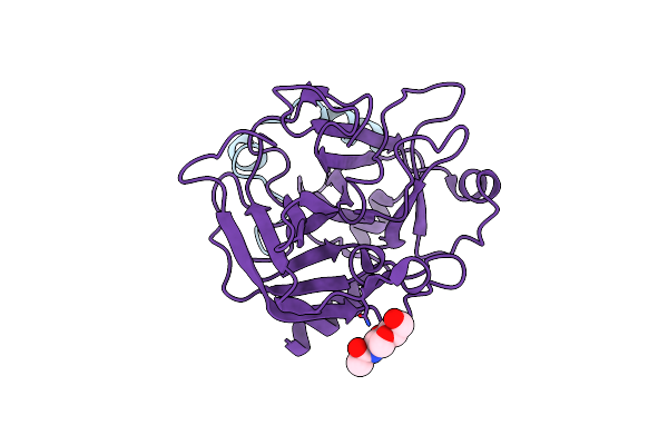
Crystal Structure Of Human Thrombin Mutant N143P In E* Form
Organism: Homo sapiens
Method: X-RAY DIFFRACTION Resolution:2.40 Å Release Date: 2009-10-20 Classification: HYDROLASE Ligands: NAG, GOL |
Organism: Homo sapiens
Method: X-RAY DIFFRACTION
Release Date: 2009-10-20
Ligands: NAG, GOL
 |
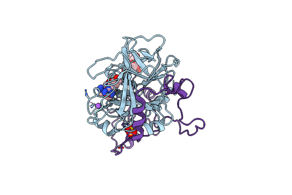
Structure Of Thrombin Mutant Delta(146-149E) In The Free Form
Organism: Homo sapiens
Method: X-RAY DIFFRACTION Resolution:1.55 Å Release Date: 2009-06-02 Classification: HYDROLASE Ligands: NAG |
Organism: Homo sapiens
Method: X-RAY DIFFRACTION
Release Date: 2009-06-02
Ligands: NAG
 |
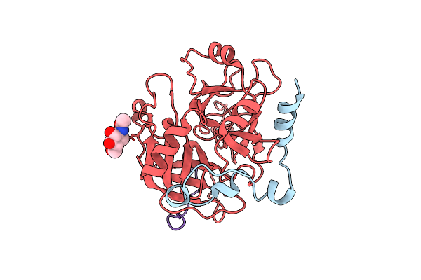
Crystal Structure Of Human Meizothrombin Desf1
Organism: Homo sapiens
Method: X-RAY DIFFRACTION Resolution:2.10 Å Release Date: 2008-11-18 Classification: HYDROLASE Ligands: SO4, DFK, NA |
Organism: Homo sapiens
Method: X-RAY DIFFRACTION
Release Date: 2008-11-18
Ligands: SO4, DFK, NA
 |
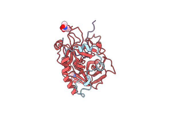
Crystal Structure Of Murine Thrombin In Complex With The Extracellular Fragment Of Murine Par3
Organism: Mus musculus
Method: X-RAY DIFFRACTION Resolution:2.00 Å Release Date: 2007-07-10 Classification: HYDROLASE Ligands: NAG |
Organism: Mus musculus
Method: X-RAY DIFFRACTION
Release Date: 2007-07-10
Ligands: NAG
 |
Crystal Structure Of Murine Thrombin In Complex With The Extracellular Fragment Of Murine Par4
Organism: Mus musculus
Method: X-RAY DIFFRACTION Resolution:3.50 Å Release Date: 2007-07-10 Classification: HYDROLASE Ligands: NAG |
Organism: Mus musculus
Method: X-RAY DIFFRACTION
Release Date: 2007-07-10
Ligands: NAG
 |
Crystal Structure Of The Slow Form Of Thrombin In A Self-Inhibited Conformation
Organism: Homo sapiens
Method: X-RAY DIFFRACTION Resolution:1.87 Å Release Date: 2006-09-12 Classification: HYDROLASE Ligands: EPE |
Organism: Homo sapiens
Method: X-RAY DIFFRACTION
Release Date: 2006-09-12
Ligands: EPE
 |
Solution Structure Of Hstx3P
|
Method: SOLUTION NMR
Release Date: 2005-11-01
 |
Lebetin Peptides, A New Class Of Potent Aggregation Inhibitors
Organism: Macrovipera lebetina
Method: SOLUTION NMR Release Date: 2005-05-03 Classification: BLOOD CLOTTING |
Organism: Macrovipera lebetina
Method: SOLUTION NMR
Release Date: 2005-05-03
 |
Solution Structure Of Pi1-3P
|
Organism: Pandinus imperator
Method: SOLUTION NMR
Release Date: 2005-04-19
 |
Cobatoxin 1 From Centruroides Noxius Scorpion Venom: Chemical Synthesis, 3-D Structure In Solution, Pharmacology And Docking On K+ Channels
|
Method: SOLUTION NMR
Release Date: 2004-03-09
 |
Solution Structure Of The Viscotoxin B
|
Organism: Viscum album
Method: SOLUTION NMR
Release Date: 2003-11-11
 |
Solution Structure Of The Viscotoxin A2
|
Organism: Viscum album
Method: SOLUTION NMR
Release Date: 2003-06-24
 |
Nmr Solution Structure Of The Chemosensory Protein Csp2 From Moth Mamestra Brassicae
Organism: Mamestra brassicae
Method: SOLUTION NMR Release Date: 2002-12-04 Classification: LIPID TRANSPORT |
Organism: Mamestra brassicae
Method: SOLUTION NMR
Release Date: 2002-12-04
 |
Carbohydrate Binding Module Cbm6 From Xylanase U Clostridium Thermocellum
Organism: Clostridium thermocellum
Method: X-RAY DIFFRACTION Resolution:2.00 Å Release Date: 2001-11-28 Classification: XYLANASE Ligands: SO4, NA, CA |
Organism: Clostridium thermocellum
Method: X-RAY DIFFRACTION
Release Date: 2001-11-28
Ligands: SO4, NA, CA
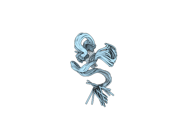 |
Solution Structure Of Ptu-1, A Toxin From The Assassin Bugs Peirates Turpis That Blocks The Voltage Sensitive Calcium Channel N-Type
|
Organism: Peirates turpis
Method: SOLUTION NMR
Release Date: 2001-11-21