

Search Count: 67
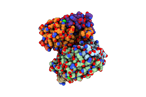 |
The 1.8 Angstrom Crystal Structure Of The Periplasmic Divalent Cation Tolerance Protein Cuta From Pyrococcus Horikoshii Ot3
Organism: Pyrococcus horikoshii
Method: X-RAY DIFFRACTION Resolution:1.80 Å Release Date: 2014-01-01 Classification: METAL BINDING PROTEIN |
Organism: Pyrococcus horikoshii
Method: X-RAY DIFFRACTION
Release Date: 2014-01-01
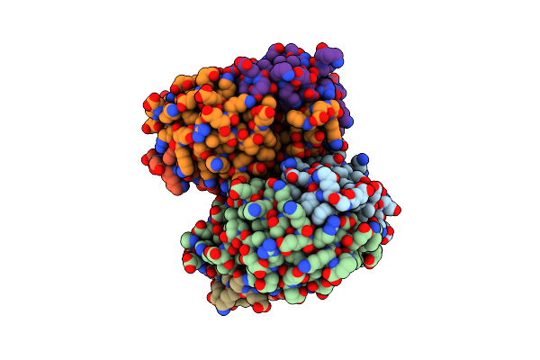 |
The 2.0 Angstrom Crystal Structure Of Pyrococcus Horikoshii Cuta1 Complexed With Na+
Organism: Pyrococcus horikoshii
Method: X-RAY DIFFRACTION Resolution:2.00 Å Release Date: 2014-01-01 Classification: METAL BINDING PROTEIN |
Organism: Pyrococcus horikoshii
Method: X-RAY DIFFRACTION
Release Date: 2014-01-01
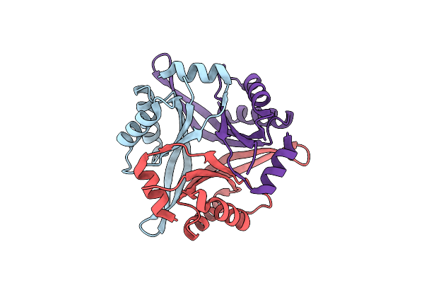 |
Crystal Structure Analysis Of The Mutant Cuta1 (S11V/E61V) From E. Coli
Organism: Escherichia coli
Method: X-RAY DIFFRACTION Resolution:2.30 Å Release Date: 2010-08-11 Classification: UNKNOWN FUNCTION |
Organism: Escherichia coli
Method: X-RAY DIFFRACTION
Release Date: 2010-08-11
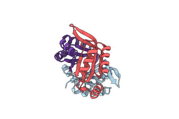 |
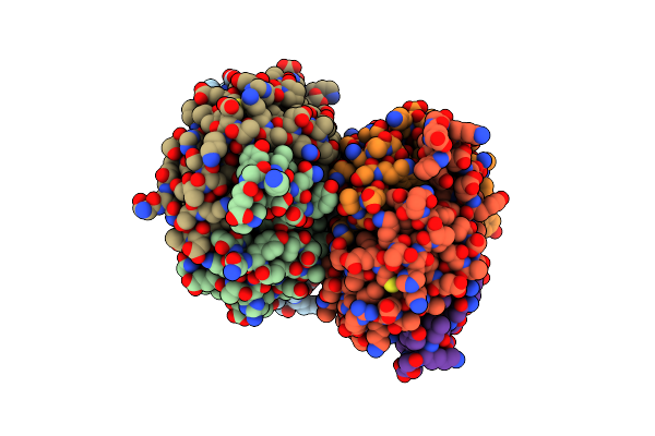
Crystal Structure Analysis Of The Mutant Cuta1 (E61V) From E. Coli
Organism: Escherichia coli
Method: X-RAY DIFFRACTION Resolution:2.30 Å Release Date: 2010-08-11 Classification: UNKNOWN FUNCTION |
Organism: Escherichia coli
Method: X-RAY DIFFRACTION
Release Date: 2010-08-11
 |
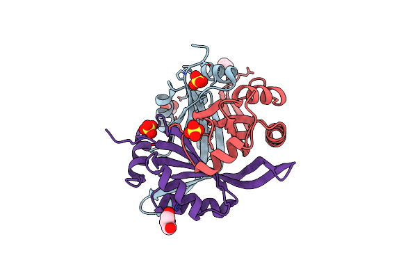
Remarkable Improvement Of The Heat Stability Of Cuta1 From E.Coli By Rational Protein Designing
Organism: Escherichia coli
Method: X-RAY DIFFRACTION Resolution:2.40 Å Release Date: 2010-08-11 Classification: UNKNOWN FUNCTION |
Organism: Escherichia coli
Method: X-RAY DIFFRACTION
Release Date: 2010-08-11
 |
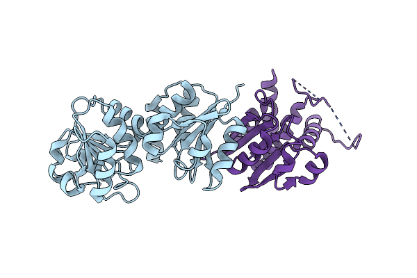
Crystal Structure Of Cuta1 From Oryza Sativa
Organism: Oryza sativa subsp. japonica
Method: X-RAY DIFFRACTION Resolution:3.02 Å Release Date: 2009-05-26 Classification: UNKNOWN FUNCTION Ligands: GOL, SO4 |
Organism: Oryza sativa subsp. japonica
Method: X-RAY DIFFRACTION
Release Date: 2009-05-26
Ligands: GOL, SO4
 |
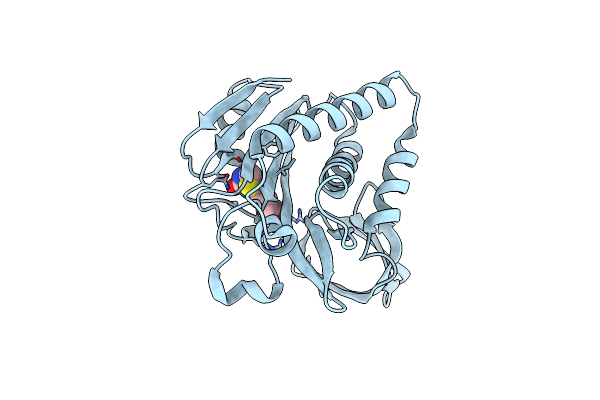
Crystal Structure Of Shikimate 5-Dehydrogenase (Aroe) From Geobacillus Kaustophilus
Organism: Geobacillus kaustophilus
Method: X-RAY DIFFRACTION Resolution:2.25 Å Release Date: 2008-03-18 Classification: OXIDOREDUCTASE Ligands: CL |
Organism: Geobacillus kaustophilus
Method: X-RAY DIFFRACTION
Release Date: 2008-03-18
Ligands: CL
 |
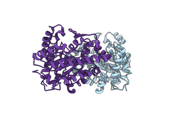
Crystal Structure Of Biotin Protein Ligase From Methanococcus Jannaschii
Organism: Methanocaldococcus jannaschii
Method: X-RAY DIFFRACTION Resolution:2.00 Å Release Date: 2008-03-18 Classification: LIGASE Ligands: BTN |
Organism: Methanocaldococcus jannaschii
Method: X-RAY DIFFRACTION
Release Date: 2008-03-18
Ligands: BTN
 |
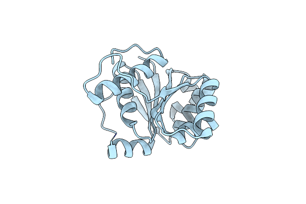
Crystal Structure Of Uroporphyrinogen Decarboxylase From Aquifex Aeolicus
Organism: Aquifex aeolicus
Method: X-RAY DIFFRACTION Resolution:1.90 Å Release Date: 2008-03-18 Classification: LYASE Ligands: ACT |
Organism: Aquifex aeolicus
Method: X-RAY DIFFRACTION
Release Date: 2008-03-18
Ligands: ACT
 |
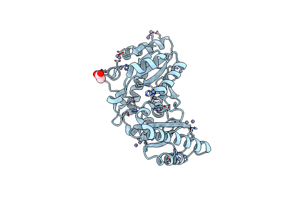
Crystal Structure Of Phenylacrylic Acid Decarboxylase From Aquifex Aeolicus
Organism: Aquifex aeolicus
Method: X-RAY DIFFRACTION Resolution:2.15 Å Release Date: 2008-03-18 Classification: LYASE |
Organism: Aquifex aeolicus
Method: X-RAY DIFFRACTION
Release Date: 2008-03-18
 |
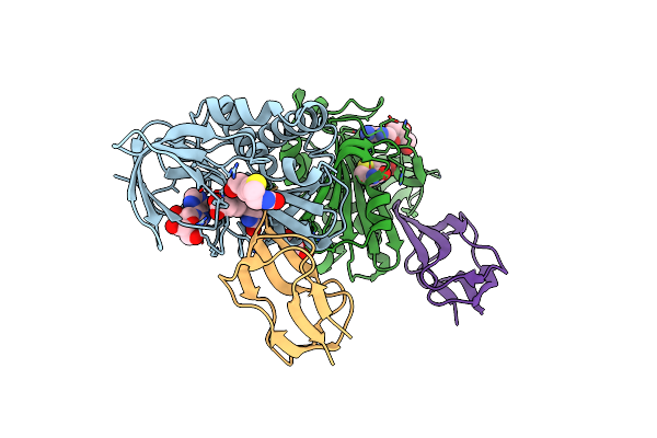
Crystal Structure Of Pantoate--Beta-Alanine Ligase (Panc) From Thermotoga Maritima
Organism: Thermotoga maritima
Method: X-RAY DIFFRACTION Resolution:2.40 Å Release Date: 2008-03-18 Classification: LIGASE Ligands: ZN, ACT |
Organism: Thermotoga maritima
Method: X-RAY DIFFRACTION
Release Date: 2008-03-18
Ligands: ZN, ACT
 |
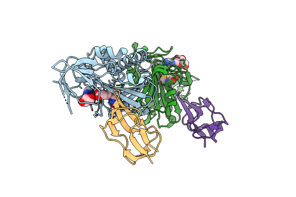
Crystal Structure Of The Biotin Protein Ligase (Mutations R48A And K111A) And Biotin Carboxyl Carrier Protein Complex From Pyrococcus Horikoshii Ot3
Organism: Pyrococcus horikoshii
Method: X-RAY DIFFRACTION Resolution:2.00 Å Release Date: 2008-03-18 Classification: LIGASE Ligands: BTN, ADN, GOL |
Organism: Pyrococcus horikoshii
Method: X-RAY DIFFRACTION
Release Date: 2008-03-18
Ligands: BTN, ADN, GOL
 |
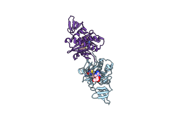
Crystal Structure Of The Biotin Protein Ligase (Mutation R48A) And Biotin Carboxyl Carrier Protein Complex From Pyrococcus Horikoshii Ot3
Organism: Pyrococcus horikoshii
Method: X-RAY DIFFRACTION Resolution:2.71 Å Release Date: 2008-03-18 Classification: LIGASE Ligands: BTN, ADN |
Organism: Pyrococcus horikoshii
Method: X-RAY DIFFRACTION
Release Date: 2008-03-18
Ligands: BTN, ADN
 |
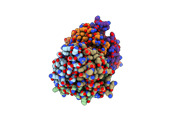
Crystal Structure Of Biotin Protein Ligase From Pyrococcus Horikoshii Complexed With Adenosine And Biotin, Mutations R48A And K111A
Organism: Pyrococcus horikoshii
Method: X-RAY DIFFRACTION Resolution:1.50 Å Release Date: 2008-02-05 Classification: LIGASE Ligands: ADN, BTN |
Organism: Pyrococcus horikoshii
Method: X-RAY DIFFRACTION
Release Date: 2008-02-05
Ligands: ADN, BTN
 |
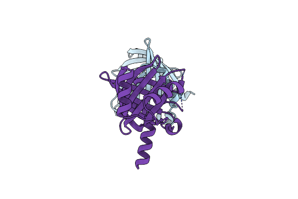
Crystal Structure Of Putative Cuta1 From Homo Sapiens At 2.05A Resolution
Organism: Homo sapiens
Method: X-RAY DIFFRACTION Resolution:2.05 Å Release Date: 2008-01-22 Classification: STRUCTURAL GENOMICS, UNKNOWN FUNCTION |
Organism: Homo sapiens
Method: X-RAY DIFFRACTION
Release Date: 2008-01-22
 |
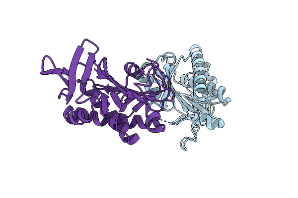
Structure Of Ph1819 Protein From Pyrococcus Horikoshii Ot3
Organism: Pyrococcus horikoshii
Method: X-RAY DIFFRACTION Resolution:1.65 Å Release Date: 2007-08-21 Classification: Structural Genomics, Unknown Function |
Organism: Pyrococcus horikoshii
Method: X-RAY DIFFRACTION
Release Date: 2007-08-21
 |
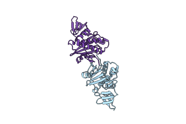
Crystal Structure Of Biotin Protein Ligase From Aquifex Aeolicus
Organism: Aquifex aeolicus
Method: X-RAY DIFFRACTION Resolution:1.95 Å Release Date: 2007-08-14 Classification: LIGASE |
Organism: Aquifex aeolicus
Method: X-RAY DIFFRACTION
Release Date: 2007-08-14
 |
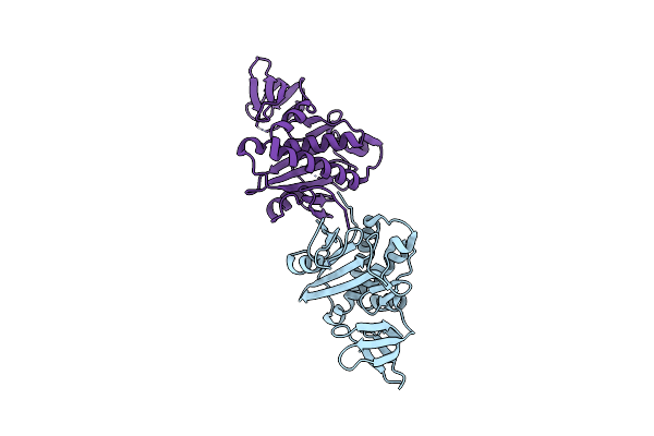
Crystal Structure Of Biotin Protein Ligase From Pyrococcus Horikoshii, Mutations R48A And K111A
Organism: Pyrococcus horikoshii
Method: X-RAY DIFFRACTION Resolution:1.50 Å Release Date: 2007-06-26 Classification: LIGASE |
Organism: Pyrococcus horikoshii
Method: X-RAY DIFFRACTION
Release Date: 2007-06-26
 |
Crystal Structure Of Biotin Protein Ligase From Pyrococcus Horikoshii Ot3, Mutation D104A
Organism: Pyrococcus horikoshii
Method: X-RAY DIFFRACTION Resolution:1.65 Å Release Date: 2007-06-26 Classification: LIGASE |
Organism: Pyrococcus horikoshii
Method: X-RAY DIFFRACTION
Release Date: 2007-06-26
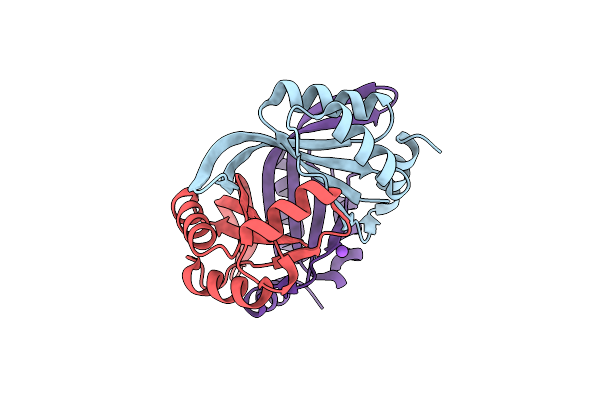 |
Crystal Structure Of Cuta1 From Pyrococcus Horikoshii Ot3, Mutation D60A
Organism: Pyrococcus horikoshii
Method: X-RAY DIFFRACTION Resolution:2.00 Å Release Date: 2007-06-26 Classification: STRUCTURAL GENOMICS, UNKNOWN FUNCTION |
Organism: Pyrococcus horikoshii
Method: X-RAY DIFFRACTION
Release Date: 2007-06-26