

Search Count: 18
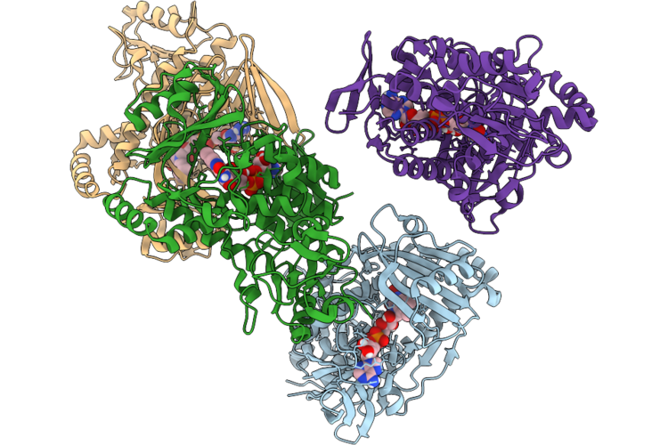 |
Structure Of The Diels-Alderase Chle3 In Complex With Cofactor Fad
Organism: Streptomyces antibioticus
Method: X-RAY DIFFRACTION Resolution:1.84 Å Release Date: 2026-06-24 Classification: OXIDOREDUCTASE Ligands: FAD |
Organism: Streptomyces antibioticus
Method: X-RAY DIFFRACTION
Release Date: 2026-06-24
Ligands: FAD
 |
Structure Of The Diels Alderase Tedj, In Complex With Cofactor Fad
Organism: Streptomyces
Method: X-RAY DIFFRACTION Resolution:1.59 Å Release Date: 2024-11-13 Classification: OXIDOREDUCTASE Ligands: FAD |
Organism: Streptomyces
Method: X-RAY DIFFRACTION
Release Date: 2024-11-13
Ligands: FAD
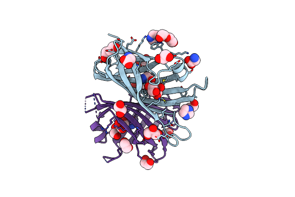 |
Cyc15 Diels Alderase
Organism: Streptomyces sp. nl15-2k
Method: X-RAY DIFFRACTION Resolution:1.66 Å Release Date: 2023-06-21 Classification: LIGASE Ligands: IMD, PEG, EDO, GLY, SER, GOL |
Organism: Streptomyces sp. nl15-2k
Method: X-RAY DIFFRACTION
Release Date: 2023-06-21
Ligands: IMD, PEG, EDO, GLY, SER, GOL
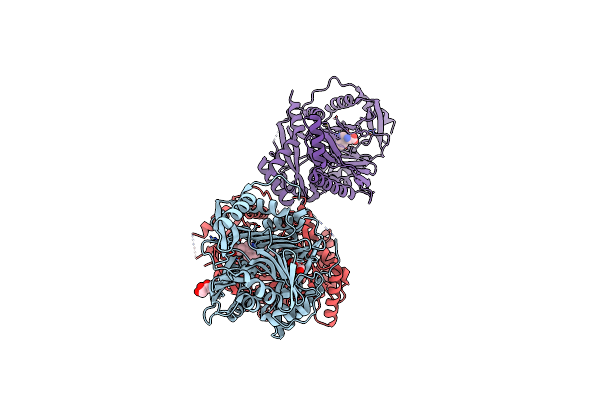 |
Structure Of Aminodeoxychorismate Synthase Component 1 (Pabb) From Bacillus Subtilis Spizizenii.
Organism: Bacillus subtilis
Method: X-RAY DIFFRACTION Resolution:2.14 Å Release Date: 2023-01-25 Classification: TRANSFERASE Ligands: GOL, TRP |
Organism: Bacillus subtilis
Method: X-RAY DIFFRACTION
Release Date: 2023-01-25
Ligands: GOL, TRP
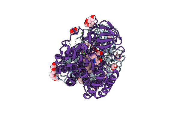 |
Cytochrome P450 Enzyme Abyv
Organism: Micromonospora maris (strain dsm 45365 / jcm 31040 / nbrc 109089 / nrrl b-24793 / ab-18-032)
Method: X-RAY DIFFRACTION Resolution:2.01 Å Release Date: 2022-11-30 Classification: OXIDOREDUCTASE Ligands: HEM, PGE, GOL, PEG, P6G, P33, CL, MG |
Organism: Micromonospora maris (strain dsm 45365 / jcm 31040 / nbrc 109089 / nrrl b-24793 / ab-18-032)
Method: X-RAY DIFFRACTION
Release Date: 2022-11-30
Ligands: HEM, PGE, GOL, PEG, P6G, P33, CL, MG
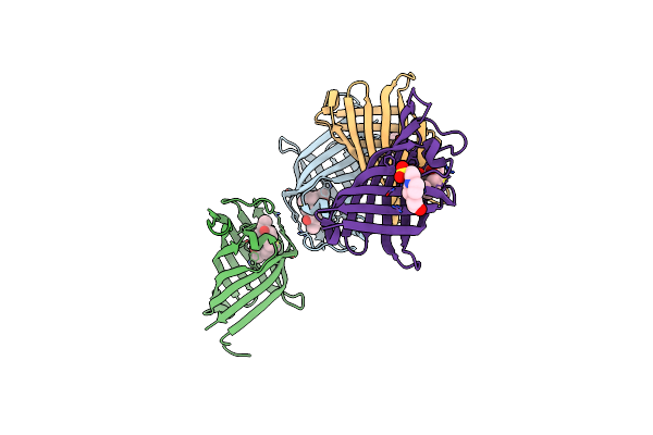 |
Structure Of The Diels Alderase Enzyme Abyu, From Micromonospora Maris, Co-Crystallised With A Non Transformable Substrate Analogue
Organism: Micromonospora maris ab-18-032
Method: X-RAY DIFFRACTION Resolution:1.95 Å Release Date: 2022-11-16 Classification: LIGASE Ligands: 8IF, EPE |
Organism: Micromonospora maris ab-18-032
Method: X-RAY DIFFRACTION
Release Date: 2022-11-16
Ligands: 8IF, EPE
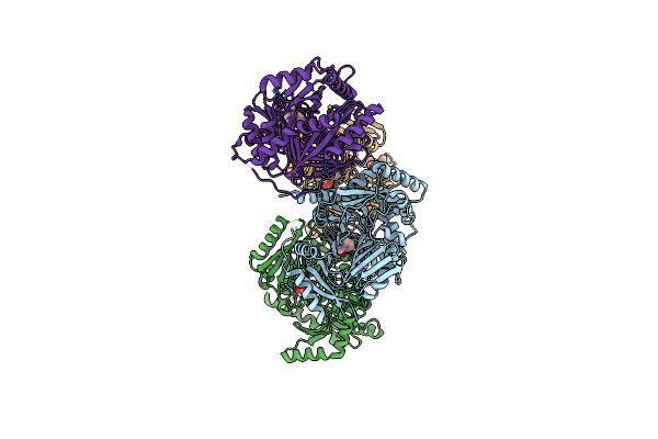 |
Bacillus Subtilis Pabb
Organism: Bacillus subtilis (strain 168)
Method: X-RAY DIFFRACTION Resolution:1.73 Å Release Date: 2022-09-07 Classification: TRANSFERASE Ligands: MG, TRP |
Organism: Bacillus subtilis (strain 168)
Method: X-RAY DIFFRACTION
Release Date: 2022-09-07
Ligands: MG, TRP
 |
Streptococcal Surface Adhesin - Cshb Nr2
Organism: Streptococcus gordonii (strain challis / atcc 35105 / bcrc 15272 / ch1 / dl1 / v288)
Method: X-RAY DIFFRACTION Resolution:1.40 Å Release Date: 2021-05-26 Classification: CELL ADHESION |
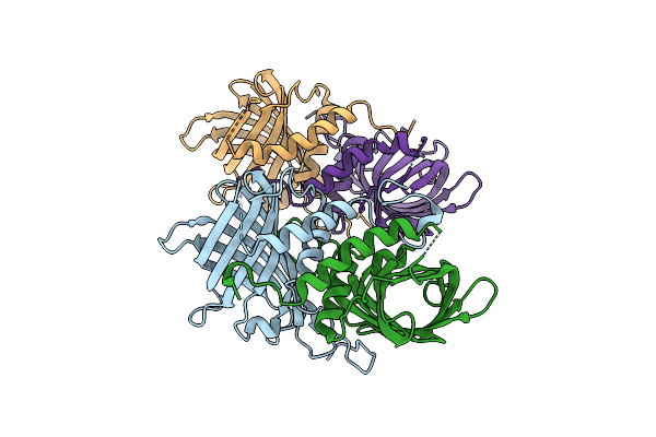 |
Crystal Structure Of The Diels Alderase Abmu From Streptomyces Koyangensis
Organism: Streptomyces koyangensis
Method: X-RAY DIFFRACTION Resolution:2.05 Å Release Date: 2021-04-21 Classification: LIGASE Ligands: BR |
Organism: Streptomyces koyangensis
Method: X-RAY DIFFRACTION
Release Date: 2021-04-21
Ligands: BR
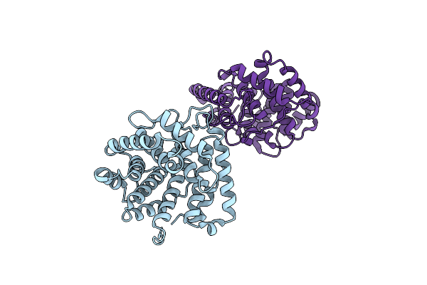 |
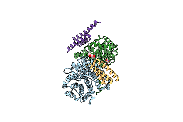
Crystal Structure Of Disulphide-Linked Human C3D Dimer
Organism: Homo sapiens
Method: X-RAY DIFFRACTION Resolution:2.00 Å Release Date: 2020-11-18 Classification: IMMUNE SYSTEM Ligands: CL |
Organism: Homo sapiens
Method: X-RAY DIFFRACTION
Release Date: 2020-11-18
Ligands: CL
 |
Crystal Structure Of Disulphide-Linked Human C3D Dimer In Complex With Staphylococcus Aureus Complement Subversion Protein Sbi-Iv
Organism: Homo sapiens, Staphylococcus aureus subsp. aureus mu50
Method: X-RAY DIFFRACTION Resolution:2.40 Å Release Date: 2020-11-18 Classification: IMMUNE SYSTEM Ligands: PEG, EDO |
Organism: Homo sapiens, Staphylococcus aureus subsp. aureus mu50
Method: X-RAY DIFFRACTION
Release Date: 2020-11-18
Ligands: PEG, EDO
 |
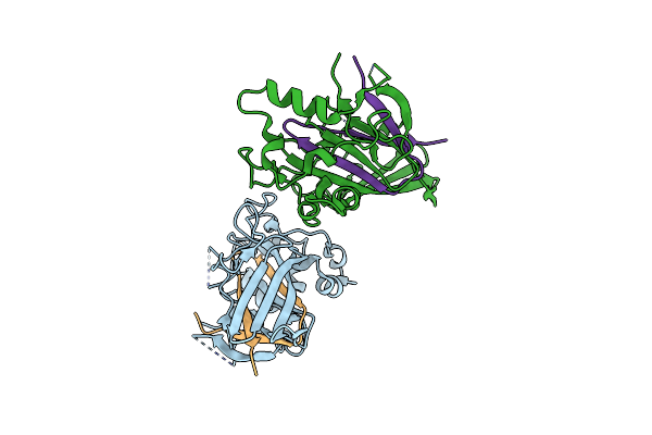
Nmr Structure Of Repeat Domain 13 Of The Fibrillar Adhesin Csha From Streptococcus Gordonii.
Organism: Streptococcus gordonii (strain challis / atcc 35105 / bcrc 15272 / ch1 / dl1 / v288)
Method: SOLUTION NMR Release Date: 2020-04-08 Classification: CELL ADHESION |
 |
Streptococcal Surface Adhesin - Csha Nr2
Organism: Streptococcus gordonii (strain challis / atcc 35105 / bcrc 15272 / ch1 / dl1 / v288)
Method: X-RAY DIFFRACTION Resolution:2.66 Å Release Date: 2016-12-14 Classification: CELL ADHESION |
 |
Structure And Mechanism Of A Dehydratase/Decarboxylase Enzyme Couple Involved In Polyketide Beta-Branching
Organism: Bacillus subtilis
Method: X-RAY DIFFRACTION Resolution:2.10 Å Release Date: 2015-05-06 Classification: LYASE Ligands: EDO, GOL |
Organism: Bacillus subtilis
Method: X-RAY DIFFRACTION
Release Date: 2015-05-06
Ligands: EDO, GOL
 |
Structure And Mechanism Of A Dehydratase/Decarboxylase Enzyme Couple Involved In Polyketide Beta-Branching
Organism: Bacillus subtilis
Method: X-RAY DIFFRACTION Resolution:1.93 Å Release Date: 2015-05-06 Classification: LYASE Ligands: GOL, NA, EPE |
Organism: Bacillus subtilis
Method: X-RAY DIFFRACTION
Release Date: 2015-05-06
Ligands: GOL, NA, EPE
 |
Structure And Mechanism Of A Dehydratase/Decarboxylase Enzyme Couple Involved In Polyketide Beta-Branching
Organism: Bacillus subtilis
Method: X-RAY DIFFRACTION Resolution:2.10 Å Release Date: 2015-05-06 Classification: LYASE Ligands: GOL |
Organism: Bacillus subtilis
Method: X-RAY DIFFRACTION
Release Date: 2015-05-06
Ligands: GOL
 |
Structure And Mechanism Of A Dehydratase/Decarboxylase Enzyme Couple Involved In Polyketide Beta-Branching
Organism: Bacillus subtilis
Method: X-RAY DIFFRACTION Resolution:2.17 Å Release Date: 2015-05-06 Classification: LYASE Ligands: EDO, NA |
Organism: Bacillus subtilis
Method: X-RAY DIFFRACTION
Release Date: 2015-05-06
Ligands: EDO, NA
 |
Structure And Mechanism Of A Dehydratase/Decarboxylase Enzyme Couple Involved In Polyketide Beta-Branching
Organism: Bacillus subtilis
Method: X-RAY DIFFRACTION Resolution:1.75 Å Release Date: 2015-05-06 Classification: LYASE Ligands: GOL, PO4 |
Organism: Bacillus subtilis
Method: X-RAY DIFFRACTION
Release Date: 2015-05-06
Ligands: GOL, PO4