
Search Count: 75
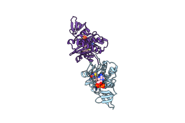 |
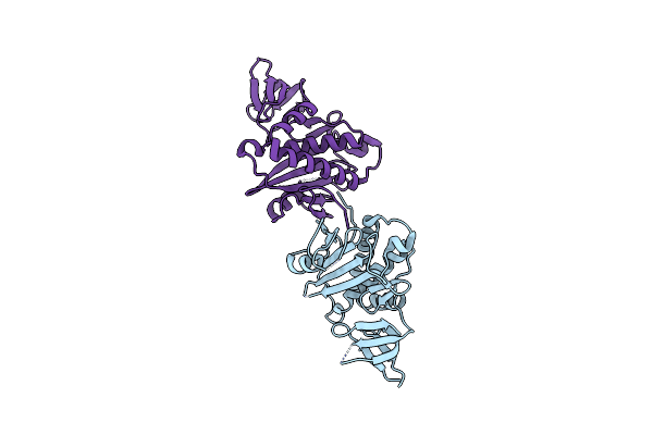
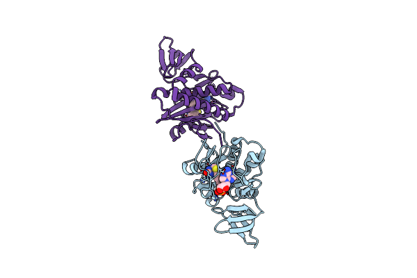
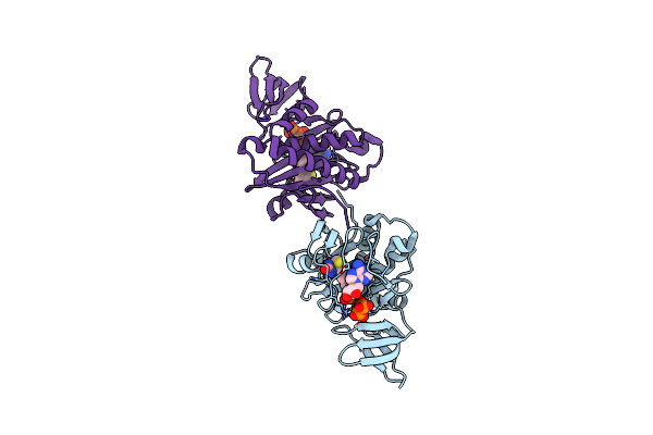
Crystal Structure Of Biotin Protein Ligase From Pyrococcus Horikoshii Complexed With Atp And Biotin, Mutation D104A
Organism: Pyrococcus horikoshii
Method: X-RAY DIFFRACTION Resolution:1.60 Å Release Date: 2007-03-01 Classification: LIGASE Ligands: ATP, BTN |
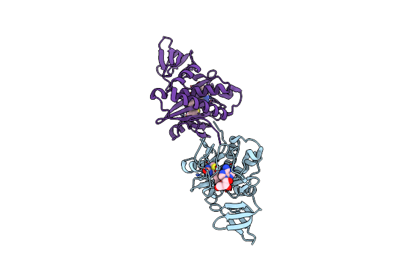 |
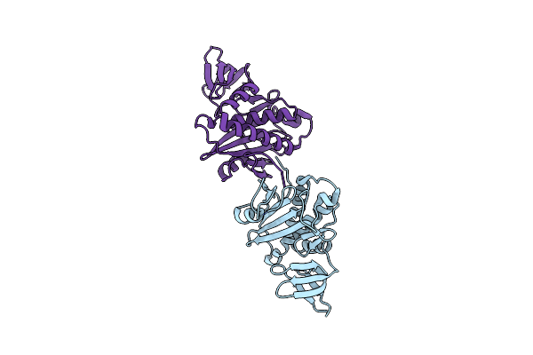
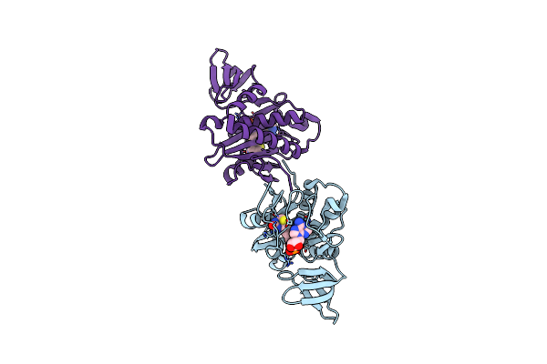
Crystal Structure Of Biotin Protein Ligase From Pyrococcus Horikoshii Complexed With Adenosine And Biotin, Mutations R48A And K111A
Organism: Pyrococcus horikoshii
Method: X-RAY DIFFRACTION Resolution:1.50 Å Release Date: 2008-02-05 Classification: LIGASE Ligands: ADN, BTN |
 |
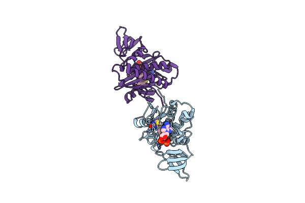
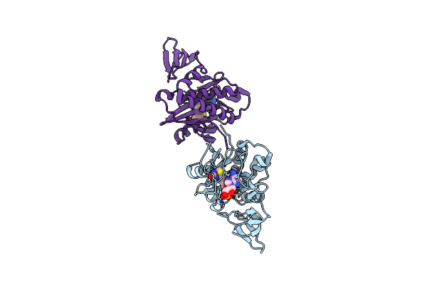
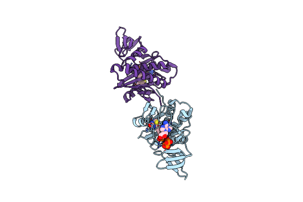
Crystal Structure Of Biotin Protein Ligase From Pyrococcus Horikoshii Ot3 Complexed With Atp And Biotin
Organism: Pyrococcus horikoshii
Method: X-RAY DIFFRACTION Resolution:1.50 Å Release Date: 2007-01-13 Classification: LIGASE Ligands: ATP, BTN |
 |
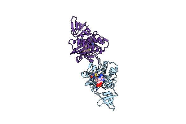
The Crystal Structure Of The Orthorhombic Form Of Biotin Protein Ligase From Pyrococcus Horikoshii Ot3 In Complex With Biotin And Adp
Organism: Pyrococcus horikoshii
Method: X-RAY DIFFRACTION Resolution:1.95 Å Release Date: 2007-01-12 Classification: LIGASE Ligands: ADP, BTN |
 |
Crystal Structure Of Biotin Protein Ligase From Pyrococcus Horikoshii Ot3 In Complex With Adp And Biotin
Organism: Pyrococcus horikoshii
Method: X-RAY DIFFRACTION Resolution:1.60 Å Release Date: 2006-08-08 Classification: LIGASE Ligands: ADP, BTN |
 |
Mechanistic Implications And Family Relationships From The Structure Of Dethiobiotin Synthetase
Organism: Escherichia coli
Method: X-RAY DIFFRACTION Resolution:1.80 Å Release Date: 1995-04-20 Classification: BIOTIN BIOSYNTHESIS Ligands: SO4 |
 |
Organism: Methanocaldococcus jannaschii
Method: X-RAY DIFFRACTION Resolution:2.00 Å Release Date: 2008-03-18 Classification: LIGASE Ligands: BTN |
 |
Dethiobiotin Synthetase From Escherichia Coli, Complex With Substrates Atp And Diaminopelargonic Acid
Organism: Escherichia coli
Method: X-RAY DIFFRACTION Resolution:1.80 Å Release Date: 1999-05-11 Classification: BIOTIN BIOSYNTHESIS Ligands: MG, DNN, ATP |
 |
Organism: Aquifex aeolicus
Method: X-RAY DIFFRACTION Resolution:1.95 Å Release Date: 2007-08-14 Classification: LIGASE |
 |
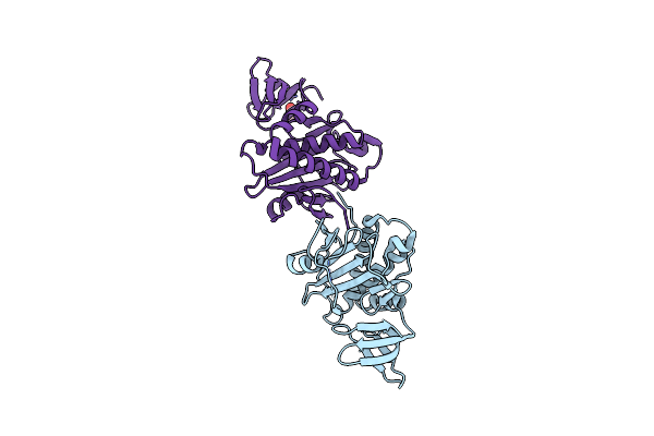
Crystal Structure Of Biotin Protein Ligase From Pyrococcus Horikoshii, Mutation R48A
Organism: Pyrococcus horikoshii
Method: X-RAY DIFFRACTION Resolution:1.45 Å Release Date: 2007-03-27 Classification: LIGASE |
 |
Crystal Structure Of Biotin Protein Ligase From Pyrococcus Horikoshii Ot3, Mutation D104A
Organism: Pyrococcus horikoshii
Method: X-RAY DIFFRACTION Resolution:1.65 Å Release Date: 2007-06-26 Classification: LIGASE |
 |
Crystal Structure Of Biotin Protein Ligase From Pyrococcus Horikoshii, Mutations R48A And K111A
Organism: Pyrococcus horikoshii
Method: X-RAY DIFFRACTION Resolution:1.50 Å Release Date: 2007-06-26 Classification: LIGASE |
 |
Crystal Structure Of Biotin Protein Ligase From Pyrococcus Horikoshii Complexed With Biotinyl-5'-Amp, Mutation R48A
Organism: Pyrococcus horikoshii
Method: X-RAY DIFFRACTION Resolution:1.28 Å Release Date: 2007-03-01 Classification: LIGASE Ligands: BT5 |
 |
Crystal Structure Of Biotin Protein Ligase From Pyrococcus Horikoshii Complexed With Biotinyl-5'-Amp, Mutation D104A
Organism: Pyrococcus horikoshii
Method: X-RAY DIFFRACTION Resolution:1.84 Å Release Date: 2007-03-26 Classification: LIGASE Ligands: BT5 |
 |
Crystal Structure Of Biotin Protein Ligase From Pyrococcus Horikoshii Complexed With The Reaction Product Analog Biotinol-5'-Amp, Mutations R48A And K111A
Organism: Pyrococcus horikoshii
Method: X-RAY DIFFRACTION Resolution:1.75 Å Release Date: 2007-06-05 Classification: LIGASE Ligands: BTX |
 |
Crystal Structure Of Biotin Protein Ligase From Pyrococcus Horikoshii Ot3, K111A Mutation
Organism: Pyrococcus horikoshii
Method: X-RAY DIFFRACTION Resolution:1.90 Å Release Date: 2007-01-13 Classification: LIGASE Ligands: ACY |
 |
Crystal Structure Of Biotin Protein Ligase From Pyrococcus Horikoshii Ot3 In Complex With Biotinyl-5'-Amp, K111A Mutation
Organism: Pyrococcus horikoshii
Method: X-RAY DIFFRACTION Resolution:1.85 Å Release Date: 2006-10-06 Classification: LIGASE Ligands: BT5 |
 |
Crystal Structure Of Biotin Protein Ligase From Pyrococcus Horikoshii Ot3 In Complex With Biotinyl-5'-Amp, K111G Mutation
Organism: Pyrococcus horikoshii
Method: X-RAY DIFFRACTION Resolution:1.85 Å Release Date: 2006-08-16 Classification: LIGASE Ligands: BT5 |
 |
Crystal Structure Of Biotin Protein Ligase From Pyrococcus Horikoshii Ot3 In Complex With Biotinyl-5'-Amp, Pyrophosphate And Mg(2+)
Organism: Pyrococcus horikoshii
Method: X-RAY DIFFRACTION Resolution:2.00 Å Release Date: 2006-10-11 Classification: LIGASE Ligands: MG, POP, BT5 |
 |
Crystal Structure Of Biotin Protein Ligase From Pyrococcus Horikoshii Ot3 In Complex With Biotinyl-5'-Amp, Pyrophosphate And Mn(2+)
Organism: Pyrococcus horikoshii
Method: X-RAY DIFFRACTION Resolution:2.20 Å Release Date: 2007-01-12 Classification: LIGASE Ligands: MN, POP, BT5 |