

Search Count: 349
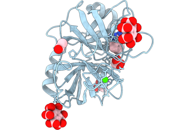 |
Structure Of Bovine Trypsin Complexed With Mellitic Acid At Room Temperature
Organism: Bos taurus
Method: X-RAY DIFFRACTION Resolution:1.30 Å Release Date: 2026-05-27 Classification: HYDROLASE Ligands: BEN, MLI, ACT, BHC, CA |
Organism: Bos taurus
Method: X-RAY DIFFRACTION
Release Date: 2026-05-27
Ligands: BEN, MLI, ACT, BHC, CA
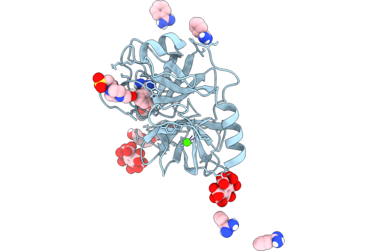 |
The Structure Of Bovine Trypsin Complexed With Mellitic Acid At 173 Degrees
Organism: Bos taurus
Method: X-RAY DIFFRACTION Resolution:1.50 Å Release Date: 2026-05-27 Classification: HYDROLASE Ligands: EPE, BHC, PG4, CA, BEN, ACT |
Organism: Bos taurus
Method: X-RAY DIFFRACTION
Release Date: 2026-05-27
Ligands: EPE, BHC, PG4, CA, BEN, ACT
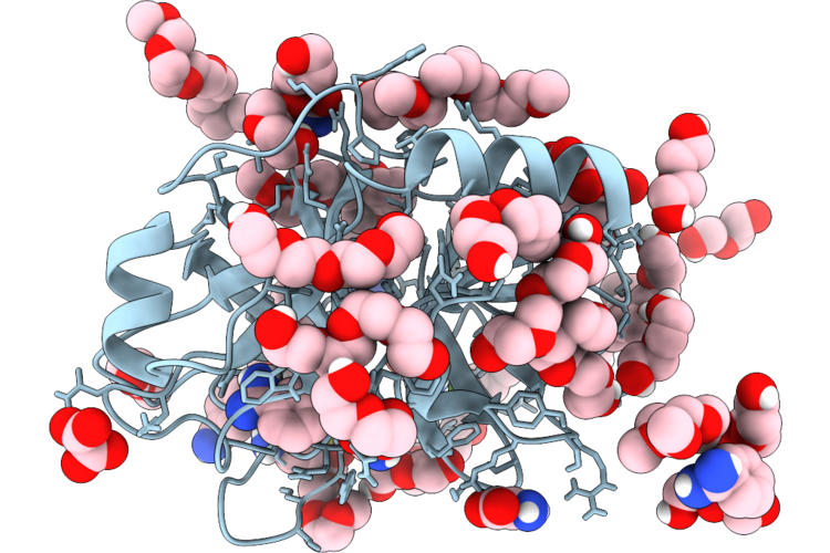 |
The Structure Of Porcine Trypsin In Complex With Crystallization Additives I
Organism: Sus scrofa
Method: X-RAY DIFFRACTION Resolution:1.28 Å Release Date: 2026-05-27 Classification: HYDROLASE Ligands: CA, BEN, PG5, PEG, PG4, OXL, A1CHW, OXM, PG6 |
Organism: Sus scrofa
Method: X-RAY DIFFRACTION
Release Date: 2026-05-27
Ligands: CA, BEN, PG5, PEG, PG4, OXL, A1CHW, OXM, PG6
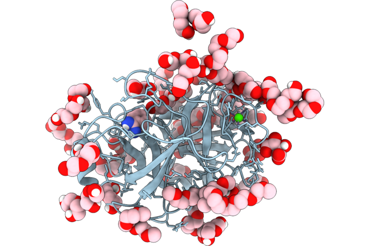 |
Porcine Trypsin Grown From Peg And Complexed With Crystallization Additives Ii
Organism: Sus scrofa
Method: X-RAY DIFFRACTION Resolution:1.27 Å Release Date: 2026-05-27 Classification: HYDROLASE Ligands: CA, TLA, PEG, PG4, PG6, PG5, BEN |
Organism: Sus scrofa
Method: X-RAY DIFFRACTION
Release Date: 2026-05-27
Ligands: CA, TLA, PEG, PG4, PG6, PG5, BEN
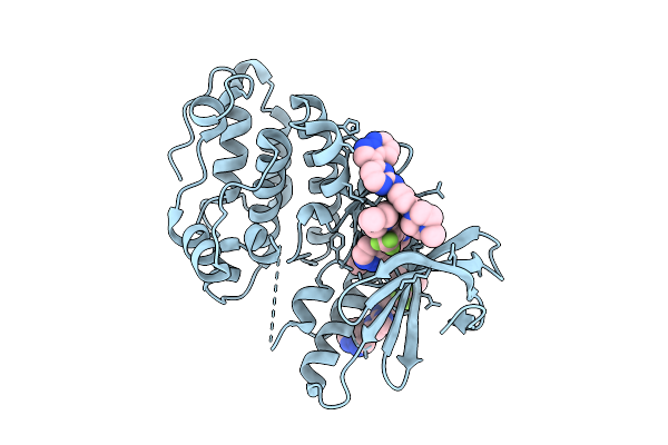 |
Staphylococcus Aureus Serine/Threonine Kinase Stk1 With Inhibitor Gw779439X
Organism: Staphylococcus aureus subsp. aureus n315
Method: X-RAY DIFFRACTION Resolution:2.07 Å Release Date: 2026-03-11 Classification: TRANSFERASE/INHIBITOR Ligands: A1CYJ, BEN, DMS |
Organism: Staphylococcus aureus subsp. aureus n315
Method: X-RAY DIFFRACTION
Release Date: 2026-03-11
Ligands: A1CYJ, BEN, DMS
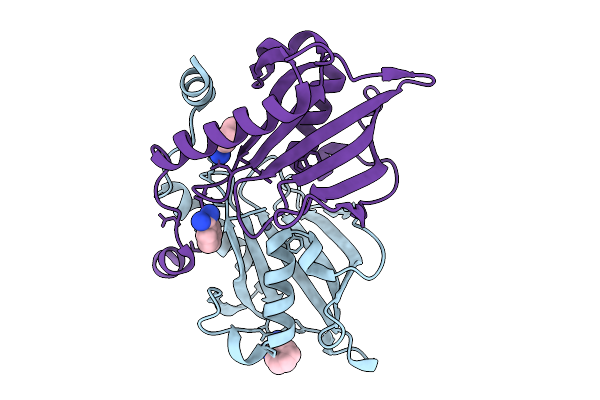 |
Crystal Structure Of Adp-Ribosylated Endolytic Muramidase Tdem From Pseudomonas Aeruginosa
Organism: Pseudomonas aeruginosa (strain atcc 15692 / dsm 22644 / cip 104116 / jcm 14847 / lmg 12228 / 1c / prs 101 / pao1)
Method: X-RAY DIFFRACTION Resolution:2.00 Å Release Date: 2026-01-21 Classification: DE NOVO PROTEIN Ligands: BEN, ZN |
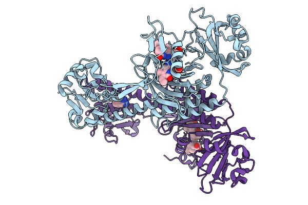 |
Photoactivation In Bacteriophytochromes, Reference (Dark) Structure For The 3 Ps Time Point
Organism: Stigmatella aurantiaca
Method: X-RAY DIFFRACTION Resolution:2.30 Å Release Date: 2025-10-08 Classification: SIGNALING PROTEIN Ligands: 3Q8, BEN |
Organism: Stigmatella aurantiaca
Method: X-RAY DIFFRACTION
Release Date: 2025-10-08
Ligands: 3Q8, BEN
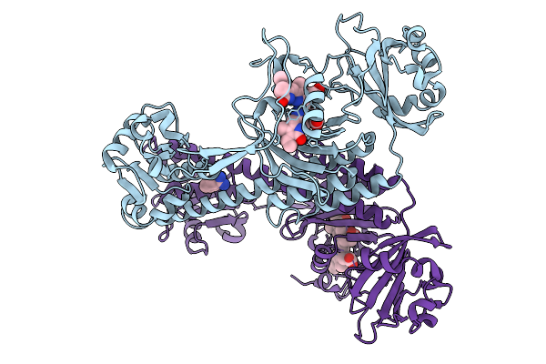 |
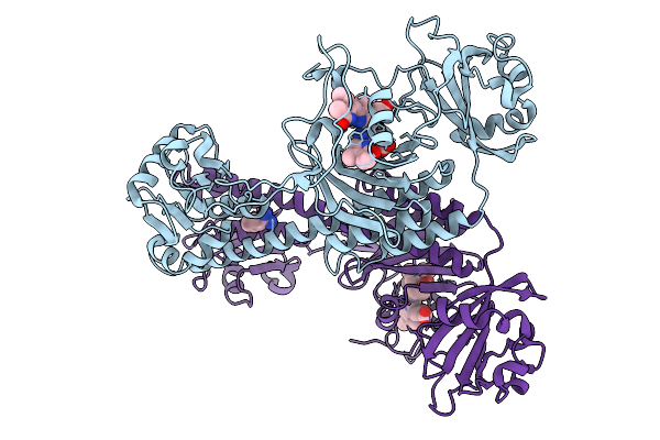
Photoactivation In Bacteriophytochromes, Reference (Dark) Structure For The 100 Ps Time Point
Organism: Stigmatella aurantiaca
Method: X-RAY DIFFRACTION Resolution:1.93 Å Release Date: 2025-10-08 Classification: SIGNALING PROTEIN Ligands: EL5, BEN |
Organism: Stigmatella aurantiaca
Method: X-RAY DIFFRACTION
Release Date: 2025-10-08
Ligands: EL5, BEN
 |
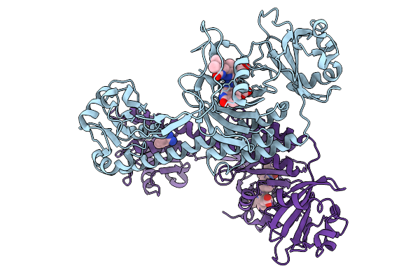
Photoactivation In Bacteriophytochromes; 3Ps Tr-Sfx Structure
Organism: Stigmatella aurantiaca
Method: X-RAY DIFFRACTION Resolution:2.30 Å Release Date: 2025-10-08 Classification: SIGNALING PROTEIN Ligands: BLA, BEN |
Organism: Stigmatella aurantiaca
Method: X-RAY DIFFRACTION
Release Date: 2025-10-08
Ligands: BLA, BEN
 |
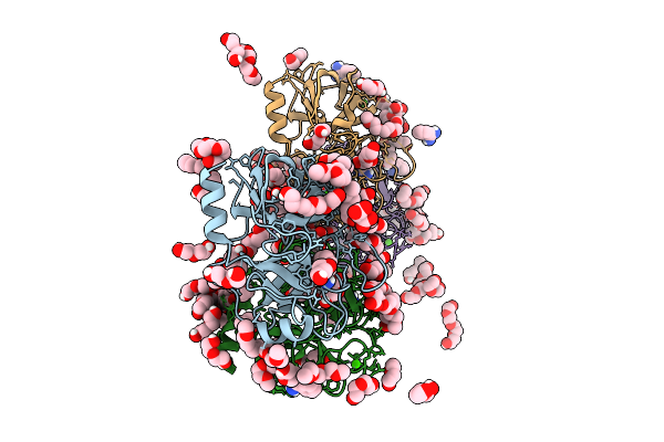
Photoactivation In Bacteriophytochrome, 100 Ps Structure
Organism: Stigmatella aurantiaca
Method: X-RAY DIFFRACTION Resolution:2.30 Å Release Date: 2025-10-08 Classification: SIGNALING PROTEIN Ligands: BLA, BEN |
Organism: Stigmatella aurantiaca
Method: X-RAY DIFFRACTION
Release Date: 2025-10-08
Ligands: BLA, BEN
 |
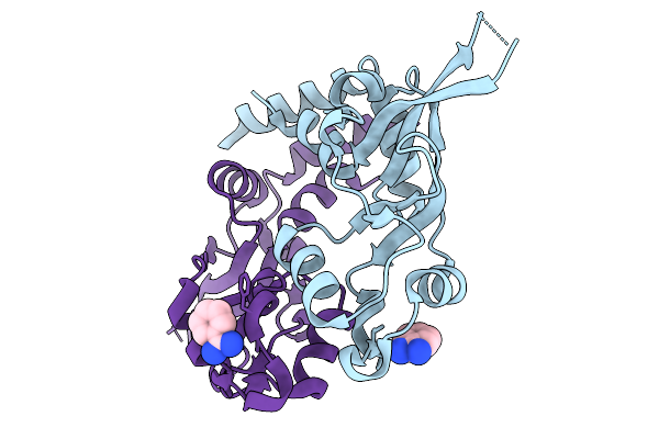
Structure Of Porcine Trypsin Crystals Grown From Peg And Complexed With Crystallization Additives Iv
Organism: Sus scrofa
Method: X-RAY DIFFRACTION Resolution:1.18 Å Release Date: 2025-09-24 Classification: PEPTIDE BINDING PROTEIN Ligands: BEN, PG5, PG6, PEG, CA, MLI, PG4, PMA, EPE, CL |
Organism: Sus scrofa
Method: X-RAY DIFFRACTION
Release Date: 2025-09-24
Ligands: BEN, PG5, PG6, PEG, CA, MLI, PG4, PMA, EPE, CL
 |
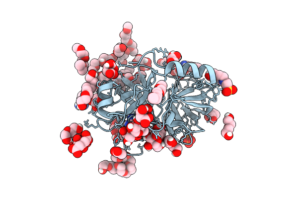
Structure Of The Cbs Pair Domain From Zebrafish Magnesium Transporter Cnnm2
Organism: Danio rerio
Method: X-RAY DIFFRACTION Resolution:3.00 Å Release Date: 2025-09-24 Classification: TRANSPORT PROTEIN Ligands: BEN |
Organism: Danio rerio
Method: X-RAY DIFFRACTION
Release Date: 2025-09-24
Ligands: BEN
 |
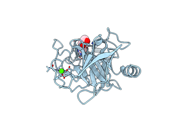
Structure Of Porcine Trypsin Crystals Grown From Peg Complexed With Crystallization Additives Iii
Organism: Sus scrofa
Method: X-RAY DIFFRACTION Resolution:1.41 Å Release Date: 2025-09-17 Classification: PEPTIDE BINDING PROTEIN Ligands: CA, BEN, FLC, PG4, PEG, GOL, PG5, MLT, PG6, EPE, ACT, PO4, TAR |
 |
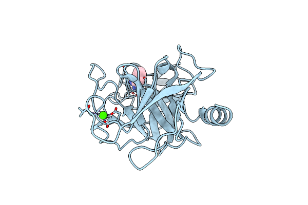
Crystal Structure Of Trypsin At 100 Kelvin With Benzamidine
Organism: Bos taurus
Method: X-RAY DIFFRACTION Resolution:1.50 Å Release Date: 2025-03-12 Classification: HYDROLASE Ligands: CA, BEN, GOL |
Organism: Bos taurus
Method: X-RAY DIFFRACTION
Release Date: 2025-03-12
Ligands: CA, BEN, GOL
 |
Crystal Structure Of Trypsin At 100 Kelvin With Benzamidine (Duplicate)
Organism: Bos taurus
Method: X-RAY DIFFRACTION Resolution:1.50 Å Release Date: 2025-03-12 Classification: HYDROLASE Ligands: CA, BEN |
Organism: Bos taurus
Method: X-RAY DIFFRACTION
Release Date: 2025-03-12
Ligands: CA, BEN
 |
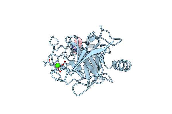
Crystal Structure Of Trypsin At 100 Kelvin With Benzamidine (Triplicate)
Organism: Bos taurus
Method: X-RAY DIFFRACTION Resolution:1.50 Å Release Date: 2025-03-12 Classification: HYDROLASE Ligands: CA, BEN |
Organism: Bos taurus
Method: X-RAY DIFFRACTION
Release Date: 2025-03-12
Ligands: CA, BEN
 |
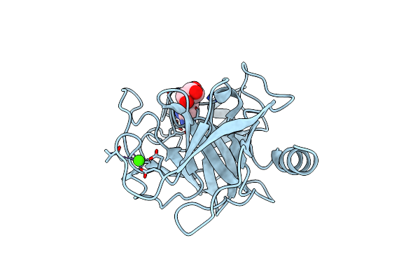
Crystal Structure Of Trypsin At 100 Kelvin With Benzamidine (Quadruplicate)
Organism: Bos taurus
Method: X-RAY DIFFRACTION Resolution:1.50 Å Release Date: 2025-03-12 Classification: HYDROLASE Ligands: CA, BEN |
Organism: Bos taurus
Method: X-RAY DIFFRACTION
Release Date: 2025-03-12
Ligands: CA, BEN
 |
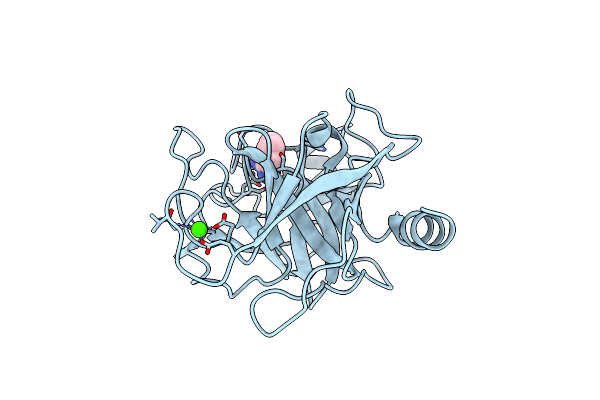
Crystal Structure Of Trypsin At 125 Kelvin With Benzamidine
Organism: Bos taurus
Method: X-RAY DIFFRACTION Resolution:1.50 Å Release Date: 2025-03-12 Classification: HYDROLASE Ligands: CA, BEN, GOL |
Organism: Bos taurus
Method: X-RAY DIFFRACTION
Release Date: 2025-03-12
Ligands: CA, BEN, GOL
 |
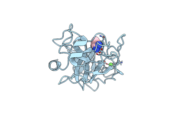
Crystal Structure Of Trypsin At 125 Kelvin With Benzamidine (Duplicate)
Organism: Bos taurus
Method: X-RAY DIFFRACTION Resolution:1.50 Å Release Date: 2025-03-12 Classification: HYDROLASE Ligands: CA, BEN |
Organism: Bos taurus
Method: X-RAY DIFFRACTION
Release Date: 2025-03-12
Ligands: CA, BEN
 |
Crystal Structure Of Trypsin At 125 Kelvin With Benzamidine (Triplicate)
Organism: Bos taurus
Method: X-RAY DIFFRACTION Resolution:1.50 Å Release Date: 2025-03-12 Classification: HYDROLASE Ligands: CA, BEN |
Organism: Bos taurus
Method: X-RAY DIFFRACTION
Release Date: 2025-03-12
Ligands: CA, BEN