

Search Count: 52
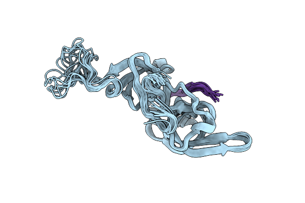 |
Nmr Structure Of Uhrf1 Tandem Tudor Domains In A Complex With Histone H3 Peptide
Organism: Homo sapiens
Method: SOLUTION NMR Release Date: 2011-04-13 Classification: DNA BINDING PROTEIN/GENE REGULATION |
Organism: Homo sapiens
Method: SOLUTION NMR
Release Date: 2011-04-13
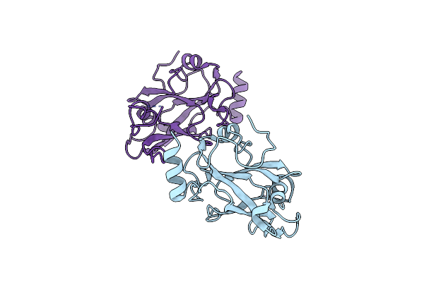 |
Crystal Structure Of The Sra Domain Of E3 Ubiquitin-Protein Ligase Uhrf2
Organism: Homo sapiens
Method: X-RAY DIFFRACTION Resolution:2.30 Å Release Date: 2010-10-13 Classification: LIGASE |
Organism: Homo sapiens
Method: X-RAY DIFFRACTION
Release Date: 2010-10-13
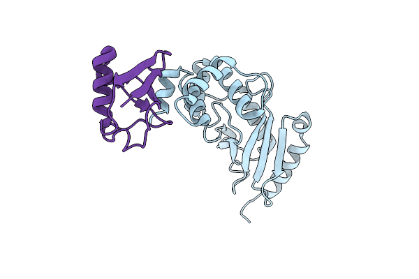 |
The Crystal Structure Of E2-25K And Ubiquitin Complex
Organism: Homo sapiens
Method: X-RAY DIFFRACTION Resolution:2.80 Å Release Date: 2010-09-08 Classification: LIGASE/SIGNALING PROTEIN |
Organism: Homo sapiens
Method: X-RAY DIFFRACTION
Release Date: 2010-09-08
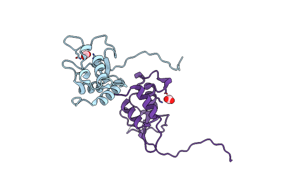 |
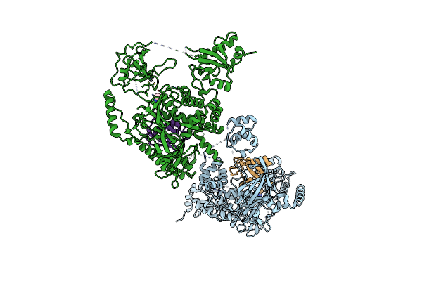
Oligomeric Structure Of The Dusp Domain Of Human Usp15
Organism: Homo sapiens
Method: X-RAY DIFFRACTION Resolution:2.15 Å Release Date: 2010-03-23 Classification: HYDROLASE Ligands: FMT, ACY |
Organism: Homo sapiens
Method: X-RAY DIFFRACTION
Release Date: 2010-03-23
Ligands: FMT, ACY
 |
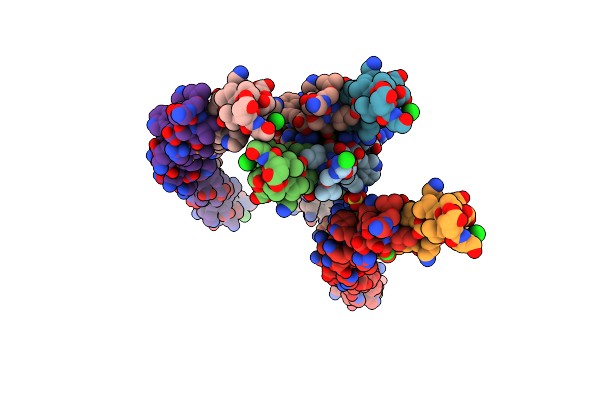
Covalent Ubiquitin-Usp5 Complex
Organism: Homo sapiens
Method: X-RAY DIFFRACTION Resolution:2.80 Å Release Date: 2009-12-29 Classification: HYDROLASE Ligands: ZN, NEH, CL |
Organism: Homo sapiens
Method: X-RAY DIFFRACTION
Release Date: 2009-12-29
Ligands: ZN, NEH, CL
 |
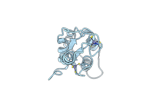
Coiled-Coil Domain Of Tumor Susceptibility Gene 101
Organism: Homo sapiens
Method: X-RAY DIFFRACTION Resolution:2.50 Å Release Date: 2009-11-03 Classification: HYDROLASE Ligands: SO4, CL |
Organism: Homo sapiens
Method: X-RAY DIFFRACTION
Release Date: 2009-11-03
Ligands: SO4, CL
 |
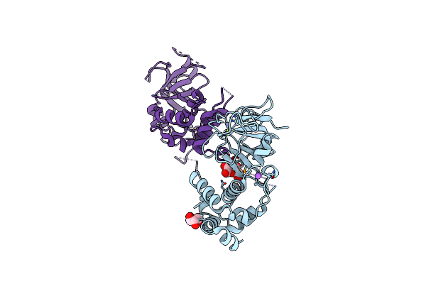
Crystal Structure Of The Ring Domain Of The E3 Ubiquitin-Protein Ligase Uhrf1
Organism: Homo sapiens
Method: X-RAY DIFFRACTION Resolution:1.75 Å Release Date: 2009-01-20 Classification: LIGASE Ligands: ZN |
Organism: Homo sapiens
Method: X-RAY DIFFRACTION
Release Date: 2009-01-20
Ligands: ZN
 |
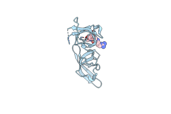
Structure Of The Replication Foci-Targeting Sequence Of Human Dna Cytosine Methyltransferase Dnmt1
Organism: Homo sapiens
Method: X-RAY DIFFRACTION Resolution:2.31 Å Release Date: 2008-11-25 Classification: TRANSFERASE Ligands: ZN, BGC, GOL, NA |
Organism: Homo sapiens
Method: X-RAY DIFFRACTION
Release Date: 2008-11-25
Ligands: ZN, BGC, GOL, NA
 |
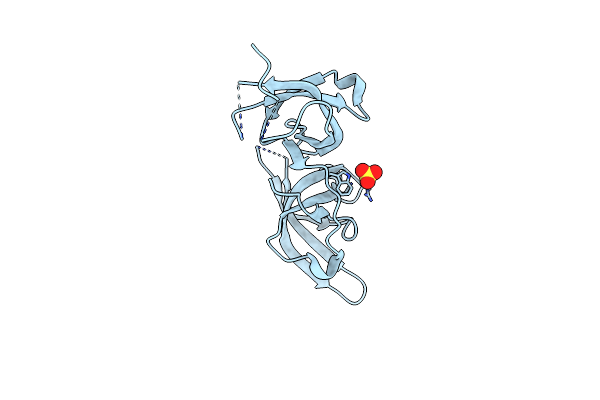
Crystal Structure Of The Tandem Tudor Domains Of The E3 Ubiquitin-Protein Ligase Uhrf1 In Complex With Trimethylated Histone H3-K9 Peptide
Organism: Homo sapiens
Method: X-RAY DIFFRACTION Resolution:2.40 Å Release Date: 2008-09-16 Classification: LIGASE |
Organism: Homo sapiens
Method: X-RAY DIFFRACTION
Release Date: 2008-09-16
 |
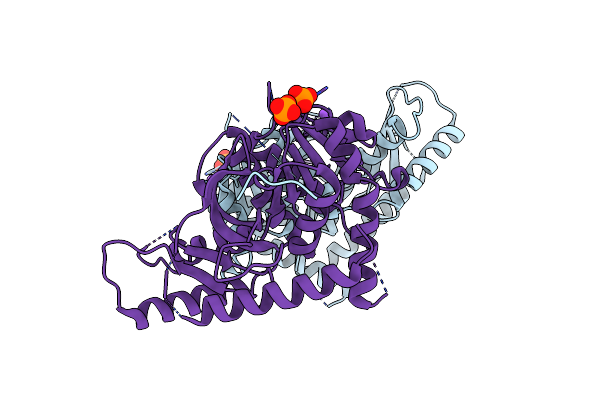
Crystal Structure Of The Tandem Tudor Domains Of The E3 Ubiquitin-Protein Ligase Uhrf1
Organism: Homo sapiens
Method: X-RAY DIFFRACTION Resolution:2.40 Å Release Date: 2008-09-16 Classification: LIGASE Ligands: SO4 |
Organism: Homo sapiens
Method: X-RAY DIFFRACTION
Release Date: 2008-09-16
Ligands: SO4
 |
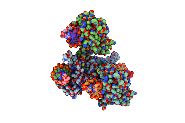
Hexagonal Structure Of The Ldh Domain Of Human Ubiquitin-Conjugating Enzyme E2-Like Isoform A
Organism: Homo sapiens
Method: X-RAY DIFFRACTION Resolution:2.10 Å Release Date: 2008-07-15 Classification: STRUCTURAL GENOMICS, UNKNOWN FUNCTION |
Organism: Homo sapiens
Method: X-RAY DIFFRACTION
Release Date: 2008-07-15
 |
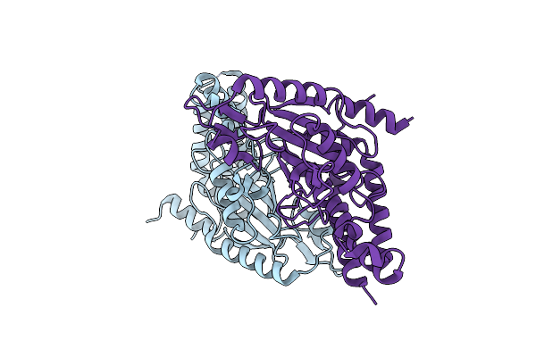
The Set And Ring Associated (Sra) Domain Of Uhrf1 Bound To Methylated Dna
Organism: Homo sapiens
Method: X-RAY DIFFRACTION Resolution:2.20 Å Release Date: 2008-04-29 Classification: LIGASE |
Organism: Homo sapiens
Method: X-RAY DIFFRACTION
Release Date: 2008-04-29
 |
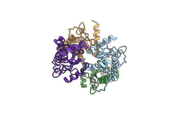
Crystal Structure Of The Ubc Domain Of Baculoviral Iap Repeat-Containing Protein 6
Organism: Homo sapiens
Method: X-RAY DIFFRACTION Resolution:2.01 Å Release Date: 2008-04-01 Classification: LIGASE |
Organism: Homo sapiens
Method: X-RAY DIFFRACTION
Release Date: 2008-04-01
 |
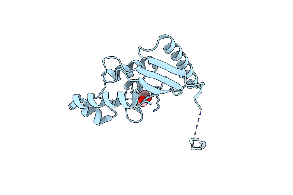
Ubiquitin-Conjugating Enzyme E2Q
Organism: Homo sapiens
Method: X-RAY DIFFRACTION Resolution:2.56 Å Release Date: 2008-03-18 Classification: LIGASE |
Organism: Homo sapiens
Method: X-RAY DIFFRACTION
Release Date: 2008-03-18
 |
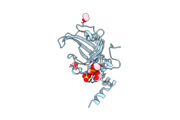
Crystal Structure Of Human Ubiquitin-Conjugating Enzyme E2 E1
Organism: Homo sapiens
Method: X-RAY DIFFRACTION Resolution:1.60 Å Release Date: 2008-02-26 Classification: LIGASE Ligands: GOL |
Organism: Homo sapiens
Method: X-RAY DIFFRACTION
Release Date: 2008-02-26
Ligands: GOL
 |
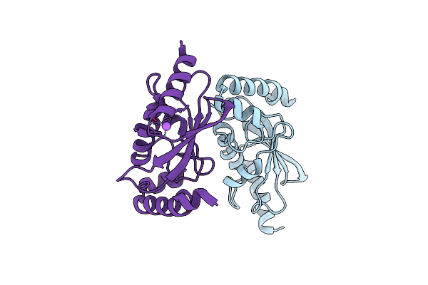
Crystal Structure Of The Sra Domain Of E3 Ubiquitin-Protein Ligase Uhrf1
Organism: Homo sapiens
Method: X-RAY DIFFRACTION Resolution:1.70 Å Release Date: 2007-12-18 Classification: LIGASE Ligands: SO4, EDO, UNL |
Organism: Homo sapiens
Method: X-RAY DIFFRACTION
Release Date: 2007-12-18
Ligands: SO4, EDO, UNL
 |
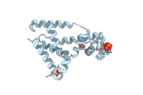
Human Ubiquitin-Conjugating Enzyme E2 H
Organism: Homo sapiens
Method: X-RAY DIFFRACTION Resolution:2.10 Å Release Date: 2007-10-09 Classification: LIGASE |
Organism: Homo sapiens
Method: X-RAY DIFFRACTION
Release Date: 2007-10-09
 |
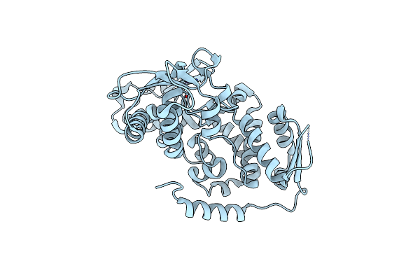
Crystal Structure Of Putative Transcriptional Regulator Sco4942 From Streptomyces Coelicolor
Organism: Streptomyces coelicolor a3(2)
Method: X-RAY DIFFRACTION Resolution:2.80 Å Release Date: 2007-06-19 Classification: TRANSCRIPTION Ligands: SO4 |
Organism: Streptomyces coelicolor a3(2)
Method: X-RAY DIFFRACTION
Release Date: 2007-06-19
Ligands: SO4
 |
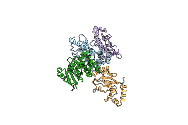
Catalytic Domain Of The Human Nedd4-Like E3 Ligase
Organism: Homo sapiens
Method: X-RAY DIFFRACTION Resolution:2.20 Å Release Date: 2007-04-17 Classification: LIGASE |
Organism: Homo sapiens
Method: X-RAY DIFFRACTION
Release Date: 2007-04-17
 |
Ubiquitin-Conjugating Enzyme E2-25 Kda Complexed With Sumo-1-Conjugating Enzyme Ubc9
Organism: Homo sapiens
Method: X-RAY DIFFRACTION Resolution:2.60 Å Release Date: 2007-01-16 Classification: LIGASE |
Organism: Homo sapiens
Method: X-RAY DIFFRACTION
Release Date: 2007-01-16