

Search Count: 30
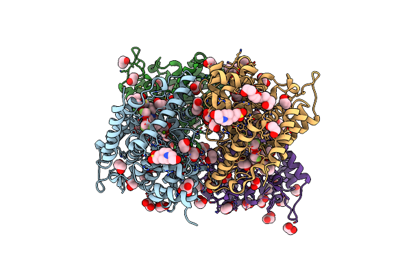 |
Crystal Structure Of Human Phosphodiesterase 4D2 Catalytic Domain With Inhibitor Npd-1439
Organism: Homo sapiens
Method: X-RAY DIFFRACTION Resolution:1.88 Å Release Date: 2019-04-24 Classification: HYDROLASE Ligands: ZN, MG, EDO, PEG, D68, EPE, NI, PG4 |
Organism: Homo sapiens
Method: X-RAY DIFFRACTION
Release Date: 2019-04-24
Ligands: ZN, MG, EDO, PEG, D68, EPE, NI, PG4
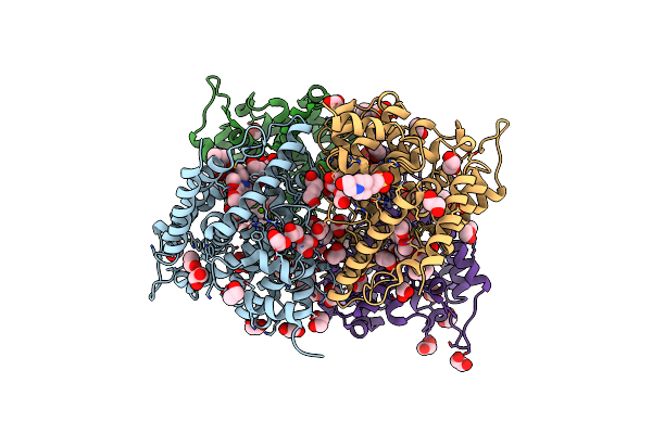 |
Crystal Structure Of Human Phosphodiesterase 4D2 Catalytic Domain With Inhibitor Npd-226
Organism: Homo sapiens
Method: X-RAY DIFFRACTION Resolution:1.90 Å Release Date: 2019-04-10 Classification: HYDROLASE Ligands: ZN, MG, EDO, PEG, D5T, EPE, PG4 |
Organism: Homo sapiens
Method: X-RAY DIFFRACTION
Release Date: 2019-04-10
Ligands: ZN, MG, EDO, PEG, D5T, EPE, PG4
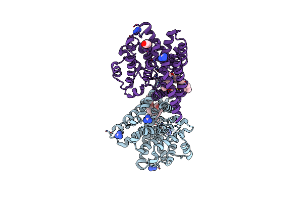 |
Crystal Structure Of T. Brucei Pde-B1 Catalytic Domain With Inhibitor Npd-226
Organism: Trypanosoma brucei
Method: X-RAY DIFFRACTION Resolution:2.20 Å Release Date: 2019-04-10 Classification: HYDROLASE Ligands: GAI, D5T, ZN, MG, GOL |
Organism: Trypanosoma brucei
Method: X-RAY DIFFRACTION
Release Date: 2019-04-10
Ligands: GAI, D5T, ZN, MG, GOL
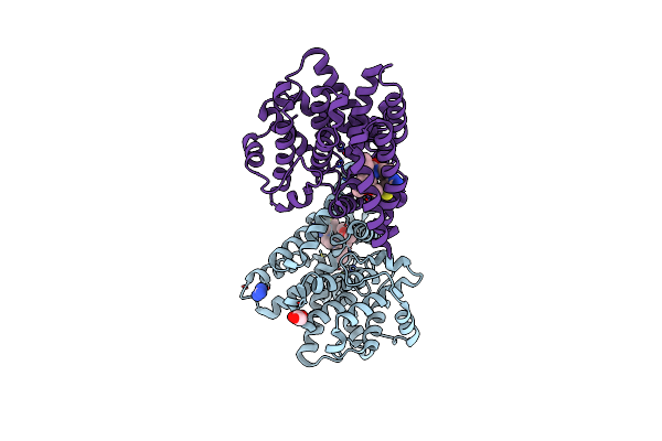 |
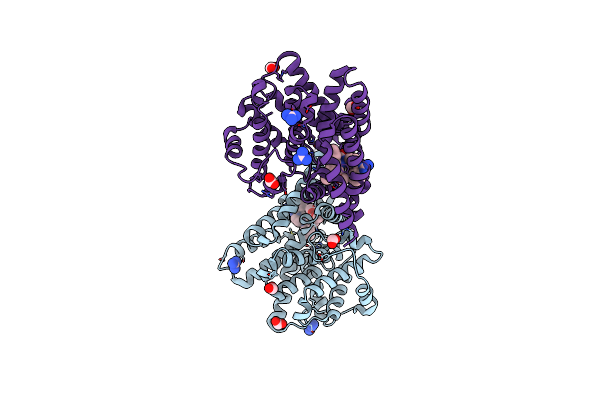
Crystal Structure Of T. Brucei Pde-B1 Catalytic Domain With Inhibitor Npd-356
Organism: Trypanosoma brucei
Method: X-RAY DIFFRACTION Resolution:1.96 Å Release Date: 2019-04-10 Classification: HYDROLASE Ligands: D62, ZN, MG, GAI, GOL |
Organism: Trypanosoma brucei
Method: X-RAY DIFFRACTION
Release Date: 2019-04-10
Ligands: D62, ZN, MG, GAI, GOL
 |
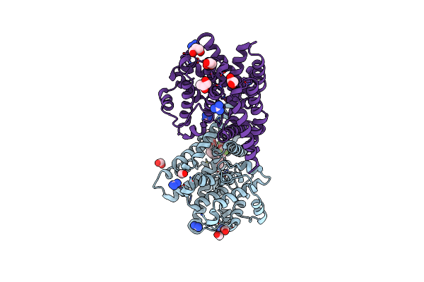
Crystal Structure Of T. Brucei Pde-B1 Catalytic Domain With Inhibitor Npd-1086
Organism: Trypanosoma brucei
Method: X-RAY DIFFRACTION Resolution:2.31 Å Release Date: 2019-04-10 Classification: HYDROLASE Ligands: ZN, MG, D5Z, FMT, GAI |
Organism: Trypanosoma brucei
Method: X-RAY DIFFRACTION
Release Date: 2019-04-10
Ligands: ZN, MG, D5Z, FMT, GAI
 |
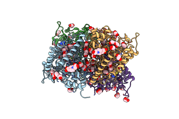
Crystal Structure Of T. Brucei Pde-B1 Catalytic Domain With Inhibitor Npd-1439
Organism: Trypanosoma brucei
Method: X-RAY DIFFRACTION Resolution:1.62 Å Release Date: 2019-04-10 Classification: HYDROLASE Ligands: GAI, D68, ZN, MG, FMT, GOL |
Organism: Trypanosoma brucei
Method: X-RAY DIFFRACTION
Release Date: 2019-04-10
Ligands: GAI, D68, ZN, MG, FMT, GOL
 |
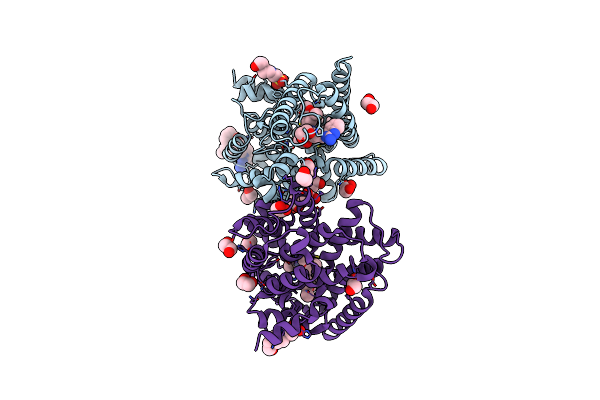
Crystal Structure Of Human Phosphodiesterase 4D2 Catalytic Domain With Inhibitor Npd-356
Organism: Homo sapiens
Method: X-RAY DIFFRACTION Resolution:2.00 Å Release Date: 2019-04-10 Classification: HYDROLASE Ligands: ZN, MG, EDO, PEG, D62, EPE, PG4 |
Organism: Homo sapiens
Method: X-RAY DIFFRACTION
Release Date: 2019-04-10
Ligands: ZN, MG, EDO, PEG, D62, EPE, PG4
 |
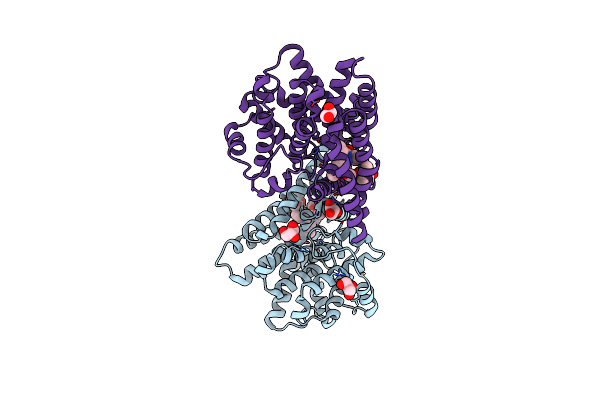
Crystal Structure Of Human Phosphodiesterase 4D2 Catalytic Domain With Inhibitor Npd-1086
Organism: Homo sapiens
Method: X-RAY DIFFRACTION Resolution:1.93 Å Release Date: 2019-04-10 Classification: HYDROLASE Ligands: EDO, D5Z, MG, ZN, EPE |
Organism: Homo sapiens
Method: X-RAY DIFFRACTION
Release Date: 2019-04-10
Ligands: EDO, D5Z, MG, ZN, EPE
 |
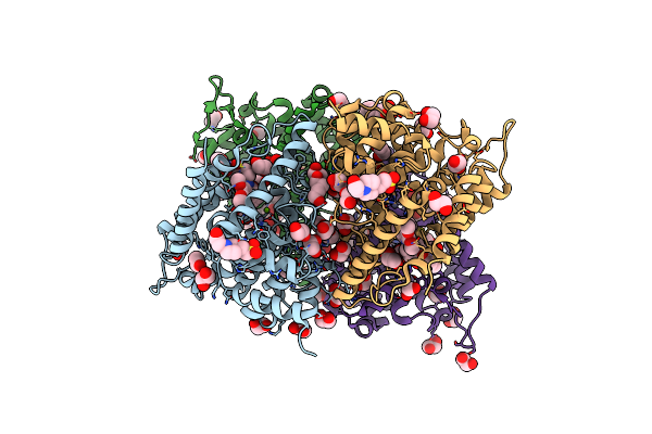
Crystal Structure Of T. Brucei Pde-B1 Catalytic Domain With Inhibitor Npd-007
Organism: Trypanosoma brucei
Method: X-RAY DIFFRACTION Resolution:2.48 Å Release Date: 2019-03-20 Classification: HYDROLASE Ligands: MG, ZN, GOL, E8H |
Organism: Trypanosoma brucei
Method: X-RAY DIFFRACTION
Release Date: 2019-03-20
Ligands: MG, ZN, GOL, E8H
 |
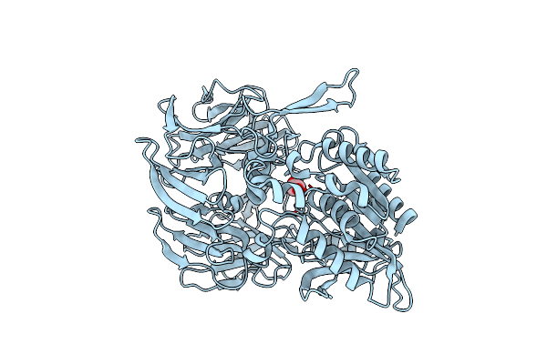
Crystal Structure Of Human Phosphodiesterase 4D2 Catalytic Domain With Inhibitor Npd-007
Organism: Homo sapiens
Method: X-RAY DIFFRACTION Resolution:1.78 Å Release Date: 2019-03-20 Classification: HYDROLASE Ligands: ZN, MG, EDO, PEG, EPE, E8H, PG4 |
Organism: Homo sapiens
Method: X-RAY DIFFRACTION
Release Date: 2019-03-20
Ligands: ZN, MG, EDO, PEG, EPE, E8H, PG4
 |
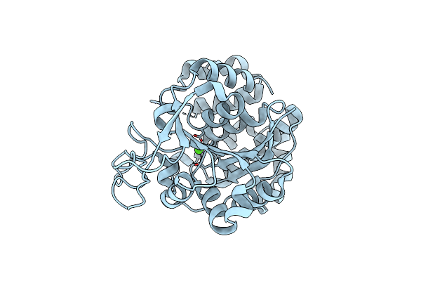
Crystal Structure Of Porphyromonas Gingivalis Dipeptidyl Peptidase 4
Organism: Porphyromonas gingivalis
Method: X-RAY DIFFRACTION Resolution:2.20 Å Release Date: 2017-09-06 Classification: HYDROLASE Ligands: GOL |
Organism: Porphyromonas gingivalis
Method: X-RAY DIFFRACTION
Release Date: 2017-09-06
Ligands: GOL
 |
Crystal Structure Of The Trypanosoma Brucei Inosine-Adenosine-Guanosine Nucleoside Hydrolase
Organism: Trypanosoma brucei brucei
Method: X-RAY DIFFRACTION Resolution:1.60 Å Release Date: 2013-08-07 Classification: HYDROLASE Ligands: CA |
Organism: Trypanosoma brucei brucei
Method: X-RAY DIFFRACTION
Release Date: 2013-08-07
Ligands: CA
 |
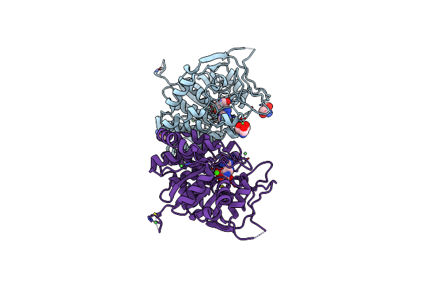
Crystal Structure Of The Trypanosoma Brucei Inosine-Adenosine-Guanosine Nucleoside Hydrolase In Complex With A Trypanocidal Compound
Organism: Trypanosoma brucei brucei
Method: X-RAY DIFFRACTION Resolution:1.28 Å Release Date: 2013-08-07 Classification: HYDROLASE/HYDROLASE INHIBITOR Ligands: AGV, CA, NI, TRS |
Organism: Trypanosoma brucei brucei
Method: X-RAY DIFFRACTION
Release Date: 2013-08-07
Ligands: AGV, CA, NI, TRS
 |
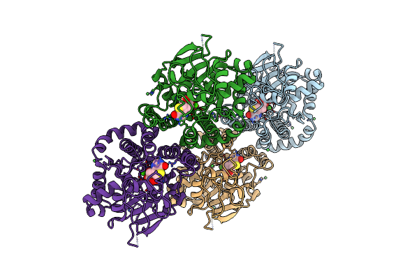
Crystal Structure Of The Trypanosoma Brucei Inosine-Adenosine-Guanosine Nucleoside Hydrolase In Complex With Immucillin A
Organism: Trypanosoma brucei brucei
Method: X-RAY DIFFRACTION Resolution:2.05 Å Release Date: 2013-08-07 Classification: HYDROLASE/HYDROLASE INHIBITOR Ligands: CA, UA2, NI, TRS |
Organism: Trypanosoma brucei brucei
Method: X-RAY DIFFRACTION
Release Date: 2013-08-07
Ligands: CA, UA2, NI, TRS
 |
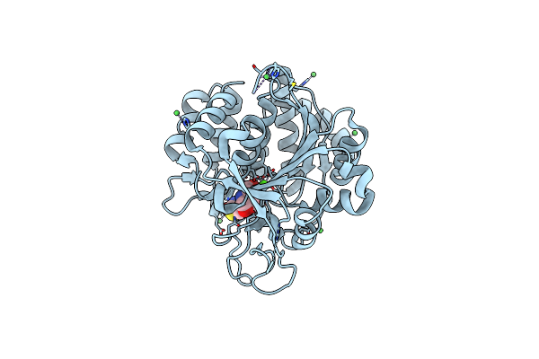
Crystal Structure Of The Trypanosoma Brucei Inosine-Adenosine-Guanosine Nucleoside Hydrolase In Complex With Compound Uamc-00312
Organism: Trypanosoma brucei brucei
Method: X-RAY DIFFRACTION Resolution:2.18 Å Release Date: 2013-08-07 Classification: HYDROLASE/HYDROLASE INHIBITOR Ligands: CA, MBY, NI |
Organism: Trypanosoma brucei brucei
Method: X-RAY DIFFRACTION
Release Date: 2013-08-07
Ligands: CA, MBY, NI
 |
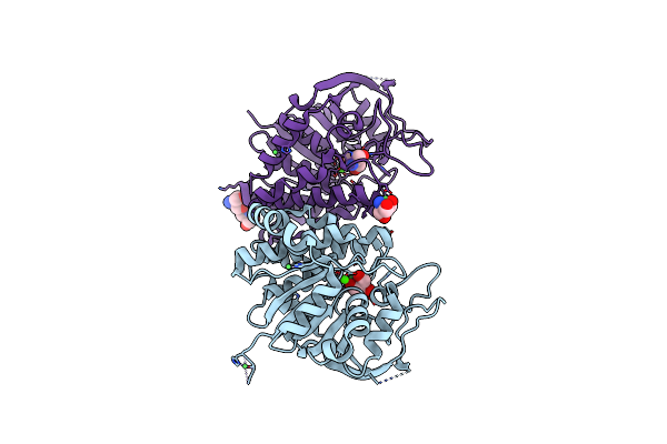
Crystal Structure Of The Trypanosoma Brucei Inosine-Adenosine-Guanosine Nucleoside Hydrolase In Complex With Compound Uamc-00312 And Allosterically Inhibited By A Ni2+ Ion
Organism: Trypanosoma brucei brucei
Method: X-RAY DIFFRACTION Resolution:1.68 Å Release Date: 2013-08-07 Classification: HYDROLASE/HYDROLASE INHIBITOR Ligands: CA, NI, MBY, TRS |
Organism: Trypanosoma brucei brucei
Method: X-RAY DIFFRACTION
Release Date: 2013-08-07
Ligands: CA, NI, MBY, TRS
 |
Crystal Structure Of The Trypanosoma Brucei Inosine-Adenosine-Guanosine Nucleoside Hydrolase In Complex With The Nitris Metalorganic Complex
Organism: Trypanosoma brucei brucei
Method: X-RAY DIFFRACTION Resolution:1.80 Å Release Date: 2013-08-07 Classification: HYDROLASE Ligands: CA, NI, TRS |
Organism: Trypanosoma brucei brucei
Method: X-RAY DIFFRACTION
Release Date: 2013-08-07
Ligands: CA, NI, TRS
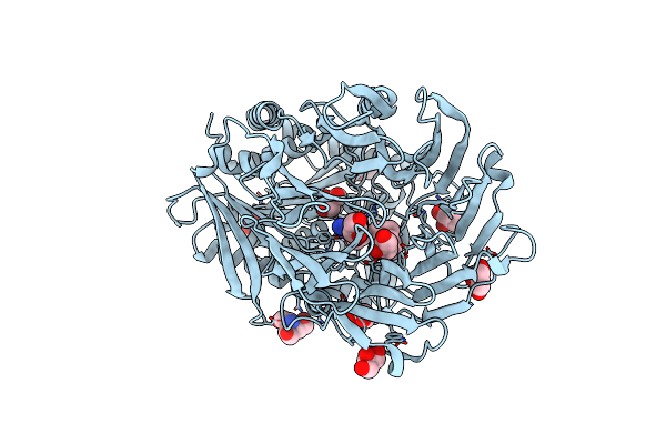 |
Prolyl Oligopeptidase From Porcine Brain With A Covalently Bound P2- Substituted N-Acyl-Prolylpyrrolidine Inhibitor
Organism: Sus scrofa
Method: X-RAY DIFFRACTION Resolution:1.70 Å Release Date: 2013-03-13 Classification: HYDROLASE Ligands: 4I4, TAM, GOL |
Organism: Sus scrofa
Method: X-RAY DIFFRACTION
Release Date: 2013-03-13
Ligands: 4I4, TAM, GOL
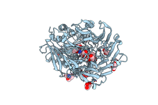 |
Prolyl Oligopeptidase From Porcine Brain With A Covalently Bound P2- Substituted N-Acyl-Prolylpyrrolidine Inhibitor
Organism: Sus scrofa
Method: X-RAY DIFFRACTION Resolution:1.65 Å Release Date: 2013-03-13 Classification: HYDROLASE Ligands: JKT, TAM, GOL |
Organism: Sus scrofa
Method: X-RAY DIFFRACTION
Release Date: 2013-03-13
Ligands: JKT, TAM, GOL
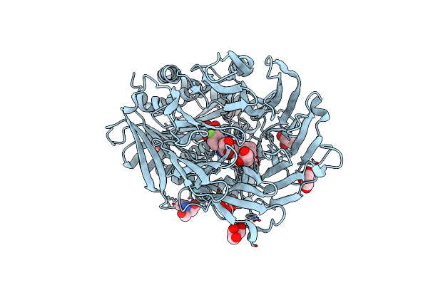 |
Prolyl Oligopeptidase From Porcine Brain With A Non-Covalently Bound P2-Substituted N-Acyl-Prolylpyrrolidine Inhibitor
Organism: Sus scrofa
Method: X-RAY DIFFRACTION Resolution:1.50 Å Release Date: 2013-03-13 Classification: HYDROLASE Ligands: TDV, TAM, GOL |
Organism: Sus scrofa
Method: X-RAY DIFFRACTION
Release Date: 2013-03-13
Ligands: TDV, TAM, GOL