

Search Count: 35
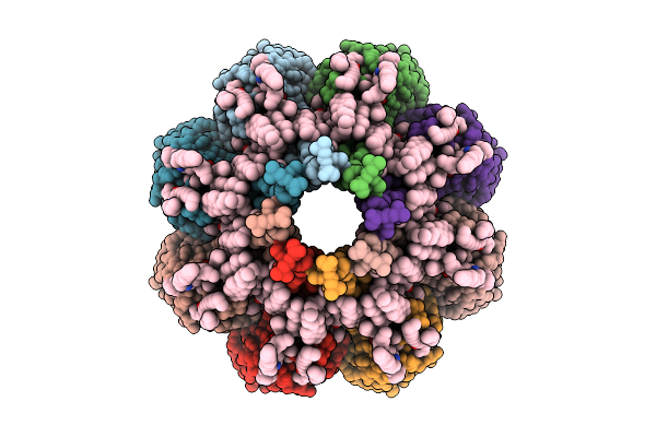 |
Structure Of Sticholisin Ii In Large Unilamellar Vesicles.
Organism: Stichodactyla helianthus
Method: ELECTRON MICROSCOPY Release Date: 2025-10-29 Classification: MEMBRANE PROTEIN Ligands: FO4, CLR |
Organism: Stichodactyla helianthus
Method: ELECTRON MICROSCOPY
Release Date: 2025-10-29
Ligands: FO4, CLR
 |
Structure Of Fragacetoxin C In Lipid Nanodiscs
Organism: Actinia fragacea
Method: ELECTRON MICROSCOPY Release Date: 2025-10-29 Classification: MEMBRANE PROTEIN Ligands: FO4, CLR |
Organism: Actinia fragacea
Method: ELECTRON MICROSCOPY
Release Date: 2025-10-29
Ligands: FO4, CLR
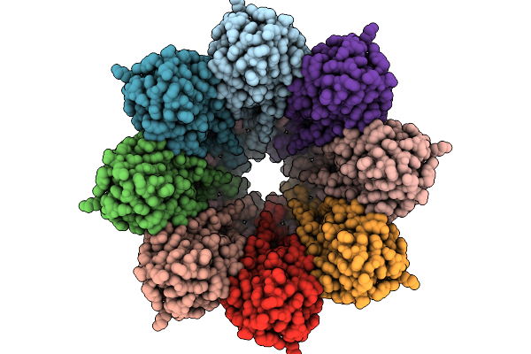 |
Structure Of The Octameric Pore Of Fragaceotxin C (Frac Or Delta-Actitoxin-Afr1A) In Large Unilamellar Vesicles.
Organism: Actinia fragacea
Method: ELECTRON MICROSCOPY Release Date: 2025-10-08 Classification: MEMBRANE PROTEIN Ligands: FO4, CLR |
Organism: Actinia fragacea
Method: ELECTRON MICROSCOPY
Release Date: 2025-10-08
Ligands: FO4, CLR
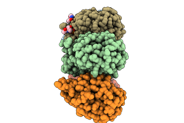 |
Structure Of 6Mer Pore Intermediate Of Sticholysin Ii (Stnii) Toxin In Lipid Nanodiscs
Organism: Stichodactyla helianthus
Method: ELECTRON MICROSCOPY Release Date: 2025-10-08 Classification: MEMBRANE PROTEIN Ligands: FO4 |
Organism: Stichodactyla helianthus
Method: ELECTRON MICROSCOPY
Release Date: 2025-10-08
Ligands: FO4
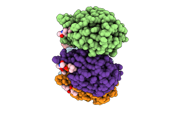 |
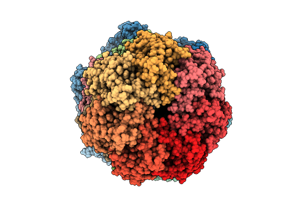
Structure Of 5Mer Pore Intermediate Of Sticholysin Ii (Stnii) Toxin In Lipid Nanodiscs
Organism: Stichodactyla helianthus
Method: ELECTRON MICROSCOPY Release Date: 2025-09-17 Classification: MEMBRANE PROTEIN Ligands: FO4 |
Organism: Stichodactyla helianthus
Method: ELECTRON MICROSCOPY
Release Date: 2025-09-17
Ligands: FO4
 |
Structure Of The Mycobacterium Tuberculosis Clpc1P1P2 Complex Bound To The Activator Bz-Leu-Leu
Organism: Mycobacterium tuberculosis, Synthetic construct
Method: ELECTRON MICROSCOPY Release Date: 2025-08-27 Classification: CHAPERONE Ligands: ADP, ATP |
Organism: Mycobacterium tuberculosis, Synthetic construct
Method: ELECTRON MICROSCOPY
Release Date: 2025-08-27
Ligands: ADP, ATP
 |
Sars-Cov-2 Spike In Complex With The 17T2 Neutralizing Antibody Fab Fragment (Local Refinement Of Rbd And Fab)
Organism: Severe acute respiratory syndrome coronavirus, Homo sapiens
Method: ELECTRON MICROSCOPY Release Date: 2024-01-10 Classification: VIRAL PROTEIN |
Organism: Severe acute respiratory syndrome coronavirus, Homo sapiens
Method: ELECTRON MICROSCOPY
Release Date: 2024-01-10
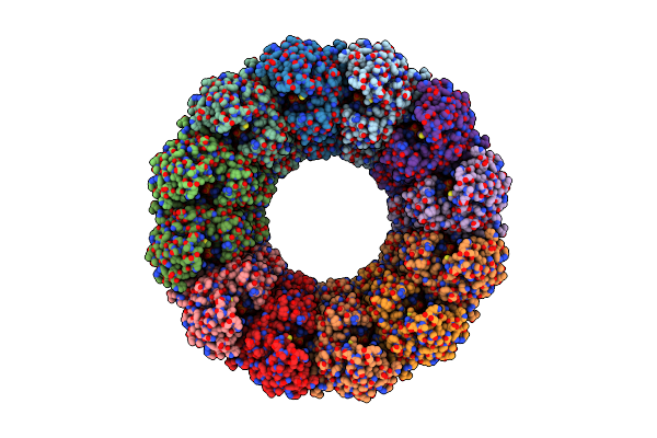 |
Cryoem Structure Of The Chikungunya Virus Nsp1 Capping Pores In Complex With Gtp
Organism: Chikungunya virus strain s27-african prototype
Method: ELECTRON MICROSCOPY Release Date: 2023-05-31 Classification: VIRAL PROTEIN Ligands: ZN, GTP |
Organism: Chikungunya virus strain s27-african prototype
Method: ELECTRON MICROSCOPY
Release Date: 2023-05-31
Ligands: ZN, GTP
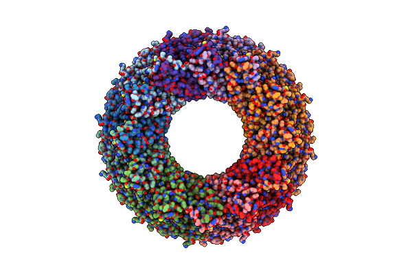 |
Cryoem Structure Of The Chikungunya Virus Nsp1 Capping Pores In Covalent Complex With A 7Gmp Cap Structure
Organism: Chikungunya virus strain s27-african prototype
Method: ELECTRON MICROSCOPY Release Date: 2023-05-03 Classification: VIRAL PROTEIN Ligands: ZN, SAH |
Organism: Chikungunya virus strain s27-african prototype
Method: ELECTRON MICROSCOPY
Release Date: 2023-05-03
Ligands: ZN, SAH
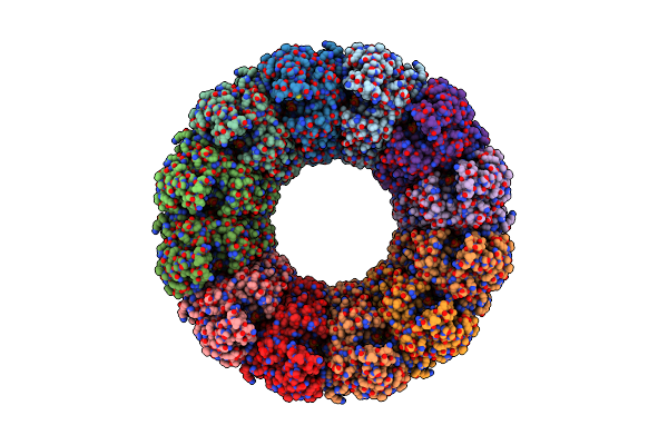 |
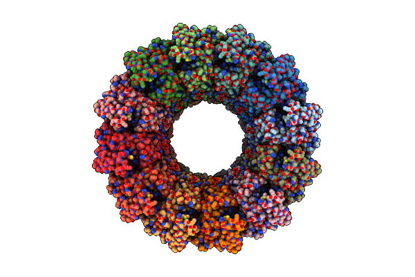
Cryoem Structure Of The Chikungunya Virus Nsp1 Capping Pores In Complex With M7Gtp And Sah Ligands
Organism: Chikungunya virus strain s27-african prototype
Method: ELECTRON MICROSCOPY Release Date: 2023-03-29 Classification: VIRAL PROTEIN Ligands: ZN, SAH, MGP, MG |
Organism: Chikungunya virus strain s27-african prototype
Method: ELECTRON MICROSCOPY
Release Date: 2023-03-29
Ligands: ZN, SAH, MGP, MG
 |
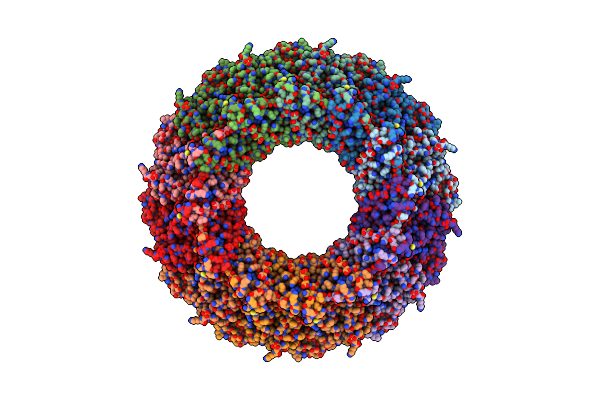
Cryoem Structure Of The Chikungunya Virus Nsp1 Capping Pores In Complex With Sam
Organism: Chikungunya virus strain s27-african prototype
Method: ELECTRON MICROSCOPY Release Date: 2023-03-29 Classification: VIRAL PROTEIN Ligands: ZN, SAM |
Organism: Chikungunya virus strain s27-african prototype
Method: ELECTRON MICROSCOPY
Release Date: 2023-03-29
Ligands: ZN, SAM
 |
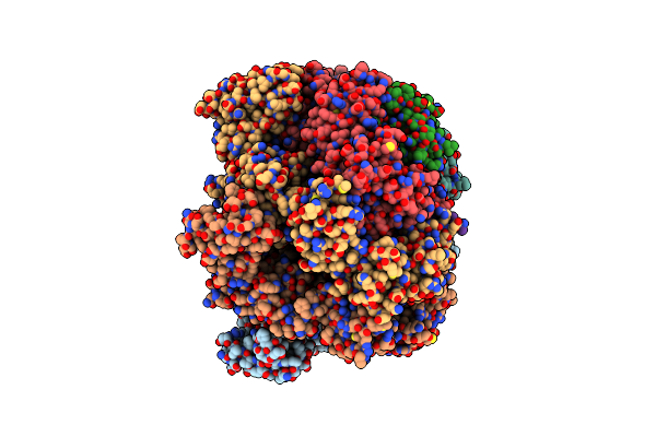
Structure Of An Open Form Of Chikv Nsp1 Capping Pores
Organism: Chikungunya virus strain s27-african prototype
Method: ELECTRON MICROSCOPY Release Date: 2023-03-29 Classification: VIRAL PROTEIN Ligands: ZN, MY6 |
Organism: Chikungunya virus strain s27-african prototype
Method: ELECTRON MICROSCOPY
Release Date: 2023-03-29
Ligands: ZN, MY6
 |
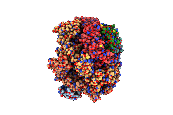
Mycobacterium Tuberculosis Clpc1 Hexamer Structure
Organism: Mycobacterium tuberculosis
Method: ELECTRON MICROSCOPY Release Date: 2022-10-26 Classification: CHAPERONE Ligands: ADP |
Organism: Mycobacterium tuberculosis
Method: ELECTRON MICROSCOPY
Release Date: 2022-10-26
Ligands: ADP
 |
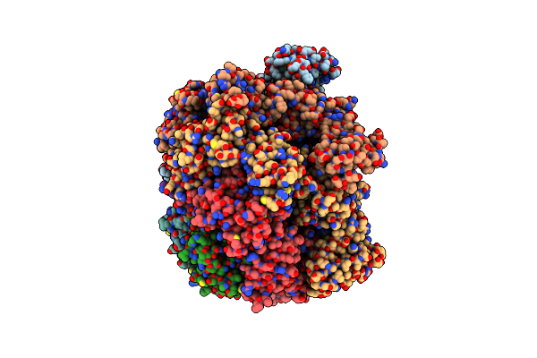
Mycobacterium Tuberculosis Clpc1 Hexamer Structure Bound To The Natural Product Antibiotic Cyclomarin
Organism: Mycobacterium tuberculosis
Method: ELECTRON MICROSCOPY Release Date: 2022-10-26 Classification: CHAPERONE Ligands: ADP |
Organism: Mycobacterium tuberculosis
Method: ELECTRON MICROSCOPY
Release Date: 2022-10-26
Ligands: ADP
 |
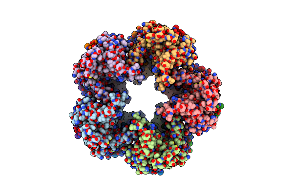
Mycobacterium Tuberculosis Clpc1 Hexamer Structure Bound To The Natural Product Antibiotic Ecumycin (Class 1)
Organism: Mycobacterium tuberculosis
Method: ELECTRON MICROSCOPY Release Date: 2022-10-26 Classification: CHAPERONE Ligands: ADP |
Organism: Mycobacterium tuberculosis
Method: ELECTRON MICROSCOPY
Release Date: 2022-10-26
Ligands: ADP
 |
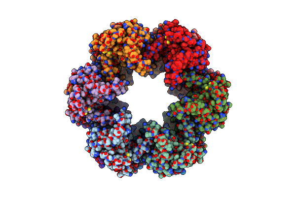
Cryo-Em Structure Of Mal De Rio Cuarto Virus P9-1 Viroplasm Protein (Decamer)
Organism: Mal de rio cuarto virus
Method: ELECTRON MICROSCOPY Release Date: 2022-06-15 Classification: VIRAL PROTEIN |
Organism: Mal de rio cuarto virus
Method: ELECTRON MICROSCOPY
Release Date: 2022-06-15
 |
Cryo-Em Structure Of Mal De Rio Cuarto Virus P9-1 Viroplasm Protein (Dodecamer)
Organism: Mal de rio cuarto virus
Method: ELECTRON MICROSCOPY Release Date: 2022-06-15 Classification: VIRAL PROTEIN |
Organism: Mal de rio cuarto virus
Method: ELECTRON MICROSCOPY
Release Date: 2022-06-15
 |
The Sars-Cov-2 Spike In Complex With The 2.15 Neutralizing Nanobody
Organism: Severe acute respiratory syndrome coronavirus 2, Camelus dromedarius
Method: ELECTRON MICROSCOPY Release Date: 2022-06-08 Classification: VIRAL PROTEIN Ligands: NAG |
Organism: Severe acute respiratory syndrome coronavirus 2, Camelus dromedarius
Method: ELECTRON MICROSCOPY
Release Date: 2022-06-08
Ligands: NAG
 |
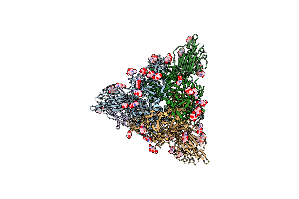
The Sars-Cov-2 Spike In Complex With The 1.29 Neutralizing Nanobody
Organism: Severe acute respiratory syndrome coronavirus 2, Camelus dromedarius
Method: ELECTRON MICROSCOPY Release Date: 2022-06-08 Classification: VIRAL PROTEIN Ligands: NAG |
Organism: Severe acute respiratory syndrome coronavirus 2, Camelus dromedarius
Method: ELECTRON MICROSCOPY
Release Date: 2022-06-08
Ligands: NAG
 |
The Sars-Cov-2 Spike In Complex With The 1.10 Neutralizing Nanobody
Organism: Severe acute respiratory syndrome coronavirus 2, Camelus dromedarius
Method: ELECTRON MICROSCOPY Release Date: 2022-06-08 Classification: VIRAL PROTEIN Ligands: NAG |
Organism: Severe acute respiratory syndrome coronavirus 2, Camelus dromedarius
Method: ELECTRON MICROSCOPY
Release Date: 2022-06-08
Ligands: NAG