

Search Count: 25
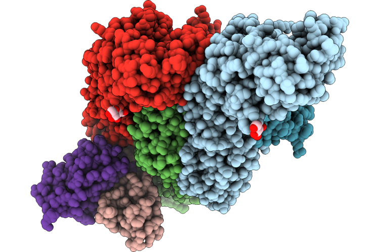 |
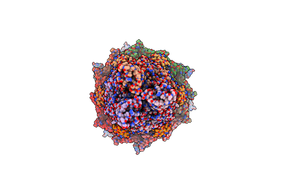
Cryoem Structure Of Wnv (Kunjin Strain) With The Fab Of Wnv-86 Antibody
Organism: Kunjin virus, Homo sapiens
Method: ELECTRON MICROSCOPY Release Date: 2026-07-08 Classification: VIRUS/IMMUNE SYSTEM Ligands: NAG |
Organism: Kunjin virus, Homo sapiens
Method: ELECTRON MICROSCOPY
Release Date: 2026-07-08
Ligands: NAG
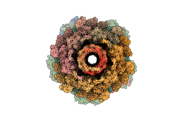 |
Structure Of The Bacteriophage T4 Portal-Neck-Tail Connector Complex
Organism: Escherichia phage t4
Method: ELECTRON MICROSCOPY Release Date: 2026-01-28 Classification: VIRAL PROTEIN |
Organism: Escherichia phage t4
Method: ELECTRON MICROSCOPY
Release Date: 2026-01-28
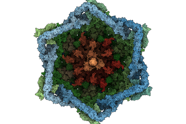 |
Structure Of The Distal Part Of The Bacteriophage T4 Tail
Organism: Escherichia phage t4
Method: ELECTRON MICROSCOPY Release Date: 2026-01-28 Classification: VIRAL PROTEIN Ligands: ZN |
Organism: Escherichia phage t4
Method: ELECTRON MICROSCOPY
Release Date: 2026-01-28
Ligands: ZN
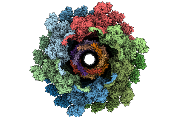 |
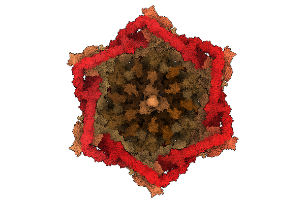
Structure Of The Middle Part Of The Bacteriophage T4 Tail
Organism: Escherichia phage t4
Method: ELECTRON MICROSCOPY Release Date: 2026-01-28 Classification: VIRAL PROTEIN |
Organism: Escherichia phage t4
Method: ELECTRON MICROSCOPY
Release Date: 2026-01-28
 |
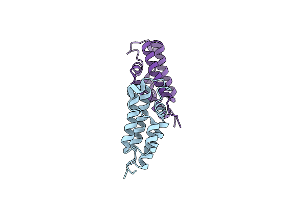
Structure Of The Bacteriophage T4 Portal-Neck-Tail Complex
Organism: Escherichia phage t4
Method: ELECTRON MICROSCOPY Release Date: 2025-12-24 Classification: VIRAL PROTEIN Ligands: ZN |
Organism: Escherichia phage t4
Method: ELECTRON MICROSCOPY
Release Date: 2025-12-24
Ligands: ZN
 |
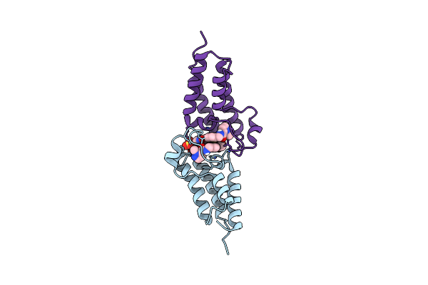
Crystal Structure Of Candida Glabrata Bdf1 Bromodomain 1 In The Unbound State
Organism: Nakaseomyces glabratus
Method: X-RAY DIFFRACTION Resolution:1.08 Å Release Date: 2024-10-30 Classification: TRANSCRIPTION |
Organism: Nakaseomyces glabratus
Method: X-RAY DIFFRACTION
Release Date: 2024-10-30
 |
Crystal Structure Of Candida Glabrata Bdf1 Bromodomain 1 Bound To A Pyrazole Ligand
Organism: Nakaseomyces glabratus
Method: X-RAY DIFFRACTION Resolution:1.82 Å Release Date: 2024-10-30 Classification: TRANSCRIPTION Ligands: Y7F, SO4 |
Organism: Nakaseomyces glabratus
Method: X-RAY DIFFRACTION
Release Date: 2024-10-30
Ligands: Y7F, SO4
 |
Crystal Structure Of Candida Glabrata Bdf1 Bromodomain 1 Bound To A Phenyltriazine Ligand
Organism: Nakaseomyces glabratus
Method: X-RAY DIFFRACTION Resolution:2.19 Å Release Date: 2024-10-30 Classification: TRANSCRIPTION Ligands: Y78, PO4, K |
Organism: Nakaseomyces glabratus
Method: X-RAY DIFFRACTION
Release Date: 2024-10-30
Ligands: Y78, PO4, K
 |
Crystal Structure Of Candida Glabrata Bdf1 Bromodomain 2 In The Unbound State
Organism: Nakaseomyces glabratus
Method: X-RAY DIFFRACTION Resolution:1.96 Å Release Date: 2024-10-30 Classification: TRANSCRIPTION Ligands: MPD, GOL, ACT |
Organism: Nakaseomyces glabratus
Method: X-RAY DIFFRACTION
Release Date: 2024-10-30
Ligands: MPD, GOL, ACT
 |
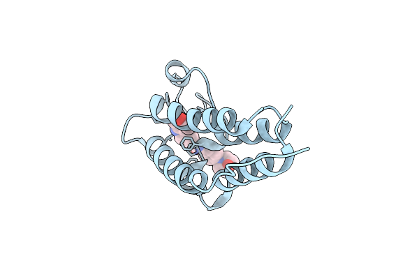
Crystal Structure Of Candida Glabrata Bdf1 Bromodomain 2 Bound To I-Bet151
Organism: Nakaseomyces glabratus
Method: X-RAY DIFFRACTION Resolution:1.95 Å Release Date: 2024-10-30 Classification: TRANSCRIPTION Ligands: GOL, SO4, 1GH |
Organism: Nakaseomyces glabratus
Method: X-RAY DIFFRACTION
Release Date: 2024-10-30
Ligands: GOL, SO4, 1GH
 |
Crystal Structure Of Candida Glabrata Bdf1 Bromodomain 2 Bound To A Pyridoindole Ligand
Organism: Nakaseomyces glabratus
Method: X-RAY DIFFRACTION Resolution:2.11 Å Release Date: 2024-10-30 Classification: TRANSCRIPTION Ligands: Y7B |
Organism: Nakaseomyces glabratus
Method: X-RAY DIFFRACTION
Release Date: 2024-10-30
Ligands: Y7B
 |
Tail Tip Of Siphophage T5 : Tip Proteins
Organism: Escherichia phage t5
Method: ELECTRON MICROSCOPY Release Date: 2023-02-08 Classification: VIRAL PROTEIN |
Organism: Escherichia phage t5
Method: ELECTRON MICROSCOPY
Release Date: 2023-02-08
 |
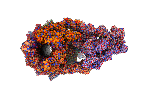
Tail Tip Of Siphophage T5 : Central Fibre Protein Pb4
Organism: Escherichia phage t5
Method: ELECTRON MICROSCOPY Release Date: 2023-02-08 Classification: VIRUS LIKE PARTICLE |
Organism: Escherichia phage t5
Method: ELECTRON MICROSCOPY
Release Date: 2023-02-08
 |
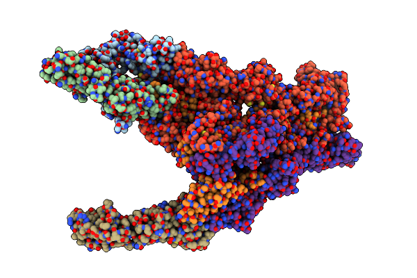
Tail Tip Of Siphophage T5 : Full Complex After Interaction With Its Bacterial Receptor Fhua
Organism: Escherichia phage t5
Method: ELECTRON MICROSCOPY Release Date: 2023-02-08 Classification: VIRAL PROTEIN |
Organism: Escherichia phage t5
Method: ELECTRON MICROSCOPY
Release Date: 2023-02-08
 |
Tail Tip Of Siphophage T5 : Bent Fibre After Interaction With Its Bacterial Receptor Fhua
Organism: Escherichia phage t5
Method: ELECTRON MICROSCOPY Release Date: 2023-02-08 Classification: VIRAL PROTEIN |
Organism: Escherichia phage t5
Method: ELECTRON MICROSCOPY
Release Date: 2023-02-08
 |
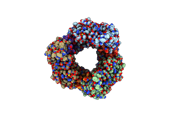
Tail Tip Of Siphophage T5 : Full Structure
Organism: Escherichia phage t5
Method: ELECTRON MICROSCOPY Release Date: 2023-02-08 Classification: VIRAL PROTEIN |
Organism: Escherichia phage t5
Method: ELECTRON MICROSCOPY
Release Date: 2023-02-08
 |
Tail Tip Of Siphophage T5 : Open Cone After Interaction With Bacterial Receptor Fhua
Organism: Escherichia phage t5
Method: ELECTRON MICROSCOPY Release Date: 2023-02-08 Classification: VIRAL PROTEIN |
Organism: Escherichia phage t5
Method: ELECTRON MICROSCOPY
Release Date: 2023-02-08
 |
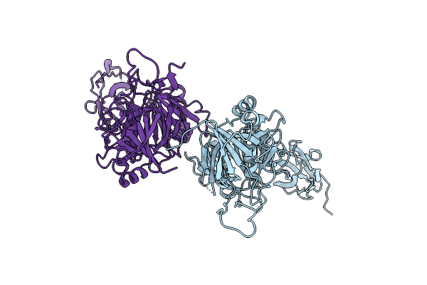
Tail Tip Of Siphophage T5 : Common Core Proteins
Organism: Escherichia phage t5
Method: ELECTRON MICROSCOPY Release Date: 2022-12-21 Classification: VIRAL PROTEIN |
Organism: Escherichia phage t5
Method: ELECTRON MICROSCOPY
Release Date: 2022-12-21
 |
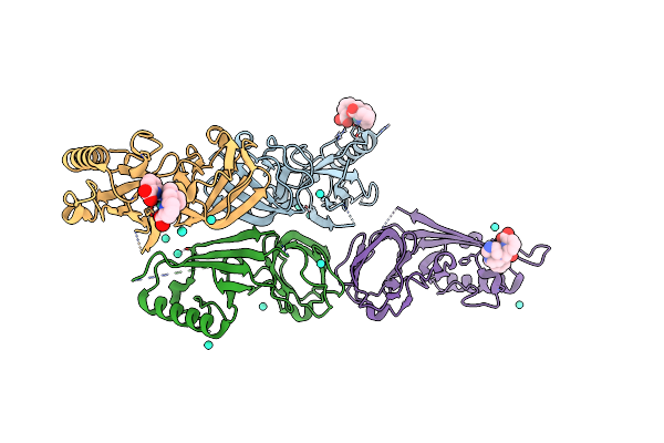
Crystal Structure Of Pb6, Major Tail Tube Protein Of Bacteriophage T5
Organism: Escherichia phage t5
Method: X-RAY DIFFRACTION Resolution:2.20 Å Release Date: 2018-01-03 Classification: VIRAL PROTEIN Ligands: CL |
Organism: Escherichia phage t5
Method: X-RAY DIFFRACTION
Release Date: 2018-01-03
Ligands: CL
 |
Bacteriophage T5 Distal Tail Protein Pb9 Co-Crystallized With Tb-Xo4
Organism: Escherichia phage t5
Method: X-RAY DIFFRACTION Resolution:2.00 Å Release Date: 2017-09-20 Classification: VIRAL PROTEIN Ligands: TB, 7MT, CL |
Organism: Escherichia phage t5
Method: X-RAY DIFFRACTION
Release Date: 2017-09-20
Ligands: TB, 7MT, CL