

Search Count: 24
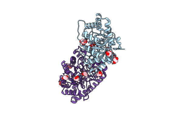 |
Crystal Structure Of Ssopox Asa6 Mutant (F46L-C258A-W263M-I280T) - Open Form
Organism: Sulfolobus solfataricus
Method: X-RAY DIFFRACTION Resolution:1.40 Å Release Date: 2018-01-10 Classification: HYDROLASE Ligands: FE2, CO, GOL, EDO |
Organism: Sulfolobus solfataricus
Method: X-RAY DIFFRACTION
Release Date: 2018-01-10
Ligands: FE2, CO, GOL, EDO
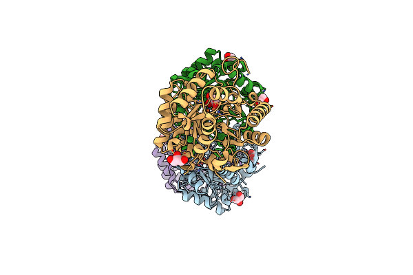 |
Crystal Structure Of Ssopox Asa1 Mutant (C258L-I261F-W263A)
Organism: Sulfolobus solfataricus
Method: X-RAY DIFFRACTION Resolution:2.00 Å Release Date: 2018-01-10 Classification: HYDROLASE Ligands: FE2, CO, GOL |
Organism: Sulfolobus solfataricus
Method: X-RAY DIFFRACTION
Release Date: 2018-01-10
Ligands: FE2, CO, GOL
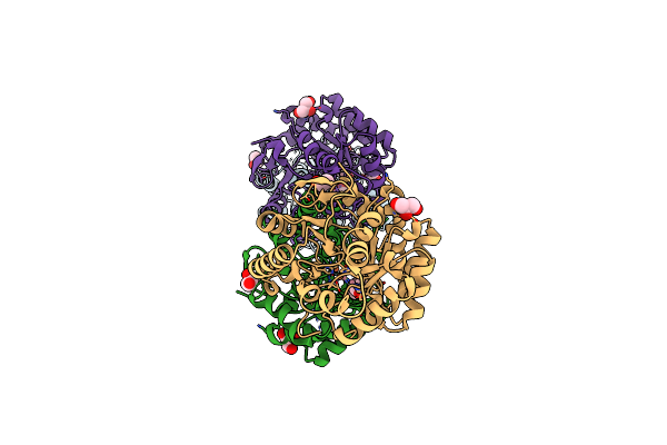 |
Crystal Structure Of Ssopox Asd6 Mutant (V27A-Y97W-L228M-W263M) - Open Form
Organism: Sulfolobus solfataricus
Method: X-RAY DIFFRACTION Resolution:2.95 Å Release Date: 2018-01-10 Classification: HYDROLASE Ligands: FE2, CO, EDO, GOL |
Organism: Sulfolobus solfataricus
Method: X-RAY DIFFRACTION
Release Date: 2018-01-10
Ligands: FE2, CO, EDO, GOL
 |
Crystal Structure Of Ssopox Asc6 Mutant (L72I-Y99F-I122L-L228M-F229S-W263L)
Organism: Sulfolobus solfataricus
Method: X-RAY DIFFRACTION Resolution:2.55 Å Release Date: 2018-01-10 Classification: HYDROLASE Ligands: FE2, CO, EDO, GOL |
Organism: Sulfolobus solfataricus
Method: X-RAY DIFFRACTION
Release Date: 2018-01-10
Ligands: FE2, CO, EDO, GOL
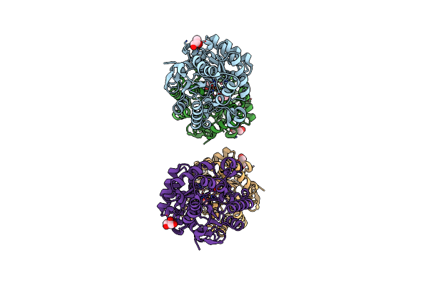 |
Crystal Structure Of Ssopox Asa6 Mutant (F46L-C258A-W263M-I280T) - Closed Form
Organism: Sulfolobus solfataricus
Method: X-RAY DIFFRACTION Resolution:2.15 Å Release Date: 2017-12-20 Classification: HYDROLASE Ligands: CO, FE2, EDO, GOL |
Organism: Sulfolobus solfataricus
Method: X-RAY DIFFRACTION
Release Date: 2017-12-20
Ligands: CO, FE2, EDO, GOL
 |
Crystal Structure Of Ssopox Asb5 Mutant (V27A-I76T-Y97W-Y99F-L130P-L226V)
Organism: Sulfolobus solfataricus
Method: X-RAY DIFFRACTION Resolution:2.50 Å Release Date: 2017-12-20 Classification: HYDROLASE Ligands: FE2, CO, GOL, EDO |
Organism: Sulfolobus solfataricus
Method: X-RAY DIFFRACTION
Release Date: 2017-12-20
Ligands: FE2, CO, GOL, EDO
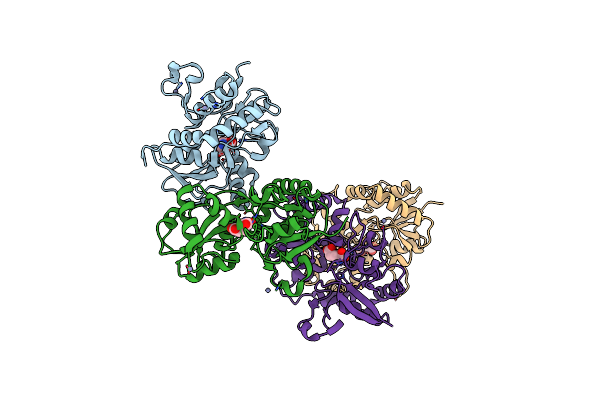 |
Measurement Of Conformational Changes Accompanying Desensitization In An Ionotropic Glutamate Receptor: Structure Of G725C Mutant
Organism: Rattus norvegicus
Method: X-RAY DIFFRACTION Resolution:2.40 Å Release Date: 2006-10-17 Classification: MEMBRANE PROTEIN Ligands: ZN, GLU |
Organism: Rattus norvegicus
Method: X-RAY DIFFRACTION
Release Date: 2006-10-17
Ligands: ZN, GLU
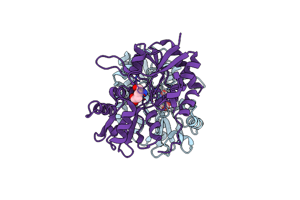 |
Measurement Of Conformational Changes Accompanying Desensitization In An Ionotropic Glutamate Receptor: Structure Of S729C Mutant
Organism: Rattus norvegicus
Method: X-RAY DIFFRACTION Resolution:2.30 Å Release Date: 2006-10-17 Classification: MEMBRANE PROTEIN Ligands: GLU |
Organism: Rattus norvegicus
Method: X-RAY DIFFRACTION
Release Date: 2006-10-17
Ligands: GLU
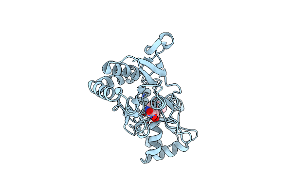 |
Glur2 Ligand Binding Core (S1S2J) Mutant L650T In Complex With Kainate
Organism: Rattus norvegicus
Method: X-RAY DIFFRACTION Resolution:1.60 Å Release Date: 2003-06-10 Classification: MEMBRANE PROTEIN Ligands: KAI |
Organism: Rattus norvegicus
Method: X-RAY DIFFRACTION
Release Date: 2003-06-10
Ligands: KAI
 |
Crystal Structure Of The Glur2 Ligand-Binding Core (S1S2J) Mutant L650T In Complex With Quisqualate
Organism: Rattus norvegicus
Method: X-RAY DIFFRACTION Resolution:1.60 Å Release Date: 2003-06-10 Classification: MEMBRANE PROTEIN Ligands: SO4, QUS |
Organism: Rattus norvegicus
Method: X-RAY DIFFRACTION
Release Date: 2003-06-10
Ligands: SO4, QUS
 |
Crystal Structure Of The Glur2 Ligand Binding Core (S1S2J) L650T Mutant In Complex With Ampa
Organism: Rattus norvegicus
Method: X-RAY DIFFRACTION Resolution:2.00 Å Release Date: 2003-06-10 Classification: MEMBRANE PROTEIN Ligands: ZN, AMQ |
Organism: Rattus norvegicus
Method: X-RAY DIFFRACTION
Release Date: 2003-06-10
Ligands: ZN, AMQ
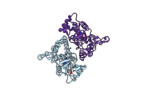 |
Crystal Structure Of The Glur2 Ligand-Binding Core (S1S2J) L650T Mutant In Complex With Ampa (Ammonium Sulfate Crystal Form)
Organism: Rattus norvegicus
Method: X-RAY DIFFRACTION Resolution:2.00 Å Release Date: 2003-06-10 Classification: MEMBRANE PROTEIN Ligands: SO4, AMQ |
Organism: Rattus norvegicus
Method: X-RAY DIFFRACTION
Release Date: 2003-06-10
Ligands: SO4, AMQ
 |
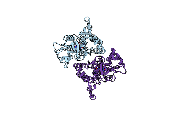
Crystal Structure Of The Glur2 Ligand-Binding Core (S1S2J) With The L483Y And L650T Mutations And In Complex With Ampa
Organism: Rattus norvegicus
Method: X-RAY DIFFRACTION Resolution:1.80 Å Release Date: 2003-06-10 Classification: MEMBRANE PROTEIN Ligands: SO4, AMQ |
Organism: Rattus norvegicus
Method: X-RAY DIFFRACTION
Release Date: 2003-06-10
Ligands: SO4, AMQ
 |
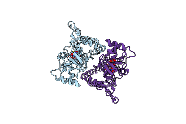
Crystal Structure Of The Non-Desensitizing Glur2 Ligand Binding Core Mutant (S1S2J-L483Y) In Complex With Ampa At 2.3 Resolution
Organism: Rattus norvegicus
Method: X-RAY DIFFRACTION Resolution:2.30 Å Release Date: 2002-06-05 Classification: MEMBRANE PROTEIN Ligands: AMQ |
Organism: Rattus norvegicus
Method: X-RAY DIFFRACTION
Release Date: 2002-06-05
Ligands: AMQ
 |
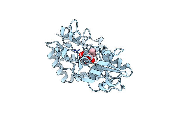
Crystal Structure Of The Non-Desensitizing Glur2 Ligand Binding Core Mutant (S1S2J-L483Y) In Complex With Antagonist Dnqx At 2.3 A Resolution
Organism: Rattus norvegicus
Method: X-RAY DIFFRACTION Resolution:2.30 Å Release Date: 2002-06-05 Classification: MEMBRANE PROTEIN Ligands: DNQ, SO4 |
Organism: Rattus norvegicus
Method: X-RAY DIFFRACTION
Release Date: 2002-06-05
Ligands: DNQ, SO4
 |
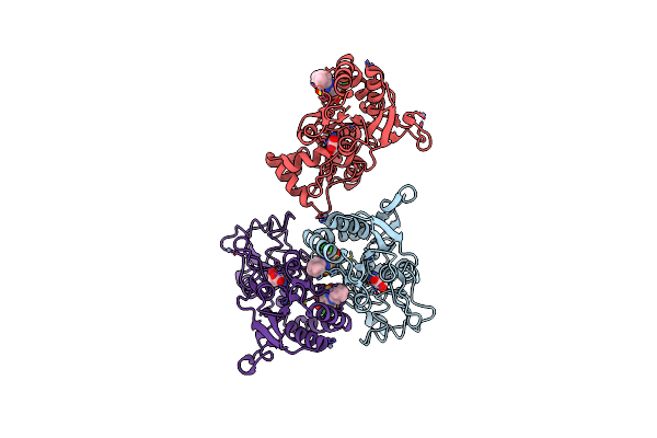
Crystal Structure Of The Glur2 Ligand Binding Domain Mutant (S1S2J-N754D) In Complex With Kainate At 2.1 A Resolution
Organism: Rattus norvegicus
Method: X-RAY DIFFRACTION Resolution:2.10 Å Release Date: 2002-06-05 Classification: MEMBRANE PROTEIN Ligands: KAI |
Organism: Rattus norvegicus
Method: X-RAY DIFFRACTION
Release Date: 2002-06-05
Ligands: KAI
 |
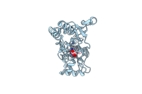
Crystal Structure Of Glur2 Ligand Binding Core (S1S2J-N775S) In Complex With Cyclothiazide (Ctz) As Well As Glutamate At 1.8 A Resolution
Organism: Rattus norvegicus
Method: X-RAY DIFFRACTION Resolution:1.80 Å Release Date: 2002-05-29 Classification: MEMBRANE PROTEIN Ligands: ZN, GLU, CYZ |
Organism: Rattus norvegicus
Method: X-RAY DIFFRACTION
Release Date: 2002-05-29
Ligands: ZN, GLU, CYZ
 |
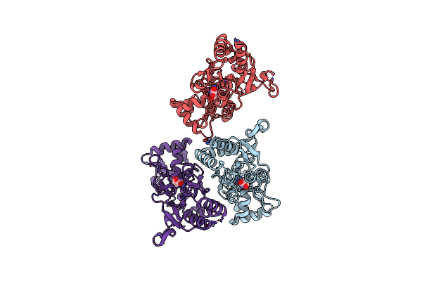
Crystal Structure Of The Glur2 Ligand Binding Core (S1S2J) In Complex With Kainate At 2.0 A Resolution
Organism: Rattus norvegicus
Method: X-RAY DIFFRACTION Resolution:1.90 Å Release Date: 2000-11-15 Classification: MEMBRANE PROTEIN Ligands: KAI |
Organism: Rattus norvegicus
Method: X-RAY DIFFRACTION
Release Date: 2000-11-15
Ligands: KAI
 |
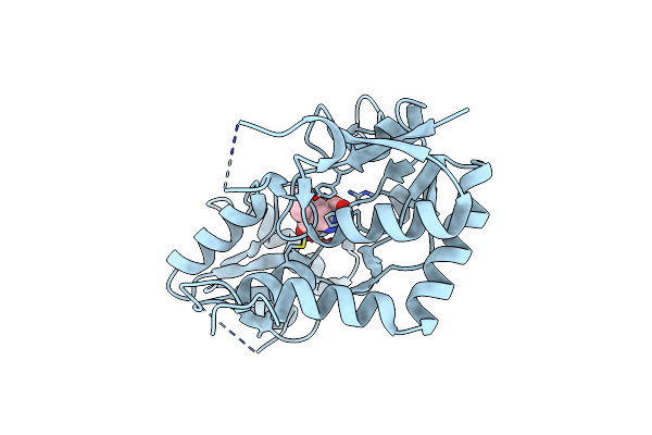
Crystal Structure Of The Glur2 Ligand Binding Core (S1S2J) In Complex With Glutamate At 1.9 Resolution
Organism: Rattus norvegicus
Method: X-RAY DIFFRACTION Resolution:1.90 Å Release Date: 2000-11-01 Classification: MEMBRANE PROTEIN Ligands: ZN, GLU |
Organism: Rattus norvegicus
Method: X-RAY DIFFRACTION
Release Date: 2000-11-01
Ligands: ZN, GLU
 |
Crystal Structure Of The Glur2 Ligand Binding Core (S1S2I) In Complex With Kainate At 1.6 A Resolution
Organism: Rattus norvegicus
Method: X-RAY DIFFRACTION Resolution:1.60 Å Release Date: 2000-11-01 Classification: MEMBRANE PROTEIN Ligands: KAI |
Organism: Rattus norvegicus
Method: X-RAY DIFFRACTION
Release Date: 2000-11-01
Ligands: KAI